
Premarin dosages: 0.625 mg
Premarin packs: 14 pills, 28 pills, 56 pills, 84 pills, 112 pills

Purchase premarin online now
It is kind of always unilateral; it happens most commonly in the second or third week of life, extra usually in ladies than boys, and only very not often within the preterm infant. The development of a breast abscess could lead to the loss of breast tissue in the long run [2,4]. These kids are well with no associated fever or malaise though the lesions may fistulate. Treatment with oral antibiotics is often adequate and leads to rapid resolution of the lesions. It is extra frequent in protracted labour, nonsterile supply and twine care, prematurity, low delivery weight and a few cultural practices similar to the applying of tobacco ash [1]. The umbilical twine may turn into colonized by quite a lot of potentially pathogenic bacteria, and an equally wide variety of topical antiseptics and antibiotics have been utilized in an try to scale back this colonization. The use of hexachlorophane was in style till it turned obvious that this could lead to critical neurotoxicity, particularly within the preterm infant [2]. The finest substitute may be chlorhexidine, applied as a dusting powder or aqueous resolution rather than as an alcoholic solution [3]. In developing international locations, 4% chlorhexidine has been proven to cut back omphalitis and to scale back neonatal mortality [4�6]. Occasionally, an infection of the umbilical twine turns into disseminated, both by bloodstream invasion or by direct extension through the umbilical vessels to the peritoneal cavity. Tetanus, diphtheria and necrotizing fasciitis [7] may also happen as problems of umbilical infection. Such infections are still responsible for a excessive proportion of deaths within the neonatal period in creating countries. Initially, the infant develops what appears to be simple cellulitis, often affecting the abdominal wall. However, the child turns into disproportionately poisonous, and the area affected turns into indurated, discoloured and extends progressively [2,6,7,8]. Purpura and, sometimes bullae, may develop in the centre of the indurated area, typically followed quite rapidly by frank necrosis. A wide number of micro organism have been associated with necrotizing fasciitis, mostly group A streptococci, but also group B streptococci, Staphylococcus aureus and Escherichia coli [6,7,8]. In many cases, a synergistic an infection by aerobic and anaerobic organisms appears to be responsible. Antibiotic (and antifungal) remedy seems to be of restricted worth on this doubtlessly deadly state of affairs. The most important aspect of therapy is early surgical excision of the necrotic tissue [7,8]. Neonatal listeriosis Listeriosis in the course of the neonatal interval is rare, however harmful. The accountable organism, Listeria monocytogenes, may be transmitted to people principally by way of contaminated foods [1]. In pregnancy, it causes a nonspecific, mild, influenzalike sickness within the mom [2,3], however it might lead to transplacental infection of the fetus. Adult listeriosis has increased in numerous European international locations during the early 21st century, but principally within the elderly and not as but in pregnancyrelated instances [4]. Clinically, there are earlyonset and lateonset types of neonatal listeriosis [5�8,9,10]. The earlyonset kind results from the event of miliary granulomas following bloodborne dissemination of infection. A few infants could have analogous miliary pores and skin lesions throughout life, manifest as scattered, discrete, gray or white papules or pustules about 1�2 mm in diameter, with a purple margin, which is able to present a supply of organisms for culture. The back seems to be the positioning of predilection for such lesions, which are additionally seen in the mouth and on the conjunctiva. Other cutaneous lesions have been described in such infants, including purpura and morbilliform rashes. The late type of the illness is commoner, taking the type of meningitis, occurring every week or two after birth. Diagnosis is by culturing the organism from a variety of websites, together with cerebrospinal fluid, blood, urine and from biopsy Preorbital cellulitis is restricted to the part of the orbit anterior to the orbital septum and is manifest by eyelid swelling. Orbital cellulitis includes the constructions deep to the septum and presents with painful proptosis, eyelid oedema and conjunctival erythema [1]. One ought to at all times be alert to the potential of group B Streptococcus as a rare explanation for periorbital cellulitis or any type of cellulitis in a neonate, in view of the excessive threat of septicaemia with this organism [2,3].
Order premarin amex
Ethnicity Freckles appear in all races, however are more regularly seen in individuals with gentle pores and skin complexion, red hair and blue eyes. They are generally seen as a response to sun publicity � photo voltaic lentigines to a higher extent � and each confer an elevated danger for melanoma and epithelial pores and skin cancers. The estimated relative danger of melanoma based on the presence of freckling in a latest metaanalysis was 1. Rarely, lentigines come up within the setting of potentially severe hereditary multisystem syndromes related to malignancies. Lentiginosis profusa is a uncommon situation, with innumerable lentigines present at start or arising early in life, with out systemic Disease course and prognosis Freckles are benign lesions and infrequently fade with age. Chemical peels, lasers, topical depigmenting drugs and dermocosmetic merchandise can be used for beauty causes [5]. The disorder has an autosomal dominant inheritance, however its actual genetic background is unknown. Agminated or segmental lentiginosis manifests as a circumscribed group of lentigines arranged in a segmental pattern that develop throughout childhood. They are presumed to represent mosaicism of an unidentified gene [7] and ought to be differentiated from neurofibromatosis kind 1. There is a slight improve of nonatypical melanocytes between the epidermal basal cells. Age They usually appear throughout childhood and enhance in quantity till the age of forty. Associated ailments Generalized lentiginosis has been rarely related to the development of melanoma. There has been a single case report of a affected person with lentiginoses and gastrointestinal stromal tumours harbouring a ckit gene mutation [7]. Environmental components the position of daylight is an important environmental factor. There could additionally be slight scaling of the floor, and several other neighbouring lesions might coalesce. Their color is pale to deep brown, depending on the skin color of the individual. Predisposing elements Part 12: NeoPlasia There have been reports of lentigines creating after topical immunotherapy with tacrolimus, squaric acid dibutylester and diphencyprone [8,9]. Pathology There is a slight improve in the variety of melanocytes alongside the dermal�epidermal junction, without nesting. The epidermal rete ridges are usually elongated and there might be a light inflammatory infiltrate in the higher dermis. Differential diagnosis the differential diagnosis of lentigines from freckles is made clinically by their comparatively darker colour, extra scattered distribution and by their unchanged status in relation to sunlight publicity. In distinction to freckles, lentigines present histologically with an increased variety of melanocytes. The differentiation of lentigines from small junctional naevi is usually unimaginable on scientific grounds. On histology, naevi show Genetics Multiple lentigines arising early in life on each uncovered and non exposed areas are usually a manifestation of inherited syndromes Simple lentigo 132. However, there are cases of bigger lesions that clinically seem as lentigines, but have small nests of naevus cells alongside the dermal�epidermal junction. These transitional lesions could also be considered precursors of future melanocytic naevi [1]. This overlap is clinically insignificant, as both are markers of sunshine skin complexion, extreme sun publicity and a certain threat of pores and skin most cancers (see Chapter 143). As the majority arise in sunexposed areas, photoprotection might decrease the speed of new lesions creating. For cosmetic reasons, quite so much of depigmenting topical agents and dermatological procedures similar to chemical peels, lasers and photodynamic remedy reduces their pigmentation (see Chapters 159 and 160) [8]. In sufferers with a quantity of lentigines arising early in life on nonsunexposed websites, the potential of a hereditary multisystem syndrome (Table 132. In the very rare instances of generalized lentiginoses, individuals may be at increased threat for melanoma, and thus should be educated on avoidance of sunburn and self pores and skin examination. Environmental factors Solar lentigines are related to both intermittent and continual solar publicity [3].
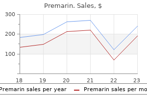
Purchase premarin 0.625mg amex
Predisposing elements Some cases develop on the site of previous trauma and reviews have included a burn scar [9] and the site of vaccination. Exceptional circumstances have been related to earlier radiotherapy to the area [10]. Patients affected by the latter have a higher incidence of tumours presenting at early age and sometimes multicentric. There is a predilection for the hands and ft, however other sites within the limbs, and less generally on the trunk, could also be affected. Disease course and prognosis the tumour is regionally aggressive with no metastatic potential. The dermis and subcutaneous tissue are changed by bundles of uniform spindleshaped cells with little cytoplasm and elongated hyperchromatic, but not pleomorphic, nuclei. The interstitial tissue contains collagen fibres, besides in probably the most mobile parts of the tumour. The subcutaneous tissue is extensively infiltrated and changed in a typical lacelike pattern. Myxoid change may be focal or, rarely, distinguished; within the latter setting, the histological diagnosis is troublesome [12,13]. Some tumours are colonized by scattered deeply pigmented melanocytes, a variant known as Dermatofibrosarcoma protuberans 137. A further variant consists of myoid nodules and is believed to represent myofibroblastic differentiation [16]. Dermatofibrosarcoma protuberans may present areas of big cell fibroblastoma (see later) and both tumour may recur, displaying features of the opposite tumour [22]. Other markers are usually negative however in some instances focal positivity for epithelial membrane antigen may be seen. Clinical features History and presentation the tumour is extra typically situated on the trunk (up to half of the cases), particularly within the flexural areas, than on the extremities or the top [1,2]. Progression is usually very slow, and may occur over a few years; a big proportion of tumours solely turn into protuberant after a protracted period of time [26]. Eventually, nodules develop, coalesce and extend, changing into redder or bluish as they enlarge to kind irregular protuberant swellings. At this stage, the bottom of the lesion is a hard indurated plaque of irregular outline. In the later stages, a proportion of lesions become painful and there may be speedy development, ulceration and discharge. Differential analysis In the early phases, it could be impossible to distinguish this tumour from a histiocytoma or a keloid. Cytogenetic studies are helpful, as ring chromosomes indicative of a 17;22 translocation are invariably discovered [22]. However, it is necessary to spotlight that some circumstances demonstrate a variant ring chromosome with cryptic rearrangements of chromosomes 17 and 22 [24]. The identical cytogenetic abnormality is found in large cell fibroblastoma, confirming that each tumours are a half of the identical spectrum. The fibrosarcomatous variant has an identical price of native recurrence however a higher price of metastatic spread [20,21,29,30,31]. Management the tumour should be excised completely, with a beneficiant margin of wholesome tissue [34]. The best probability of attaining an entire remedy with no recurrence is early detection of small tumours. Mohs micrographic surgical procedure has been reported as efficient in lowering the rate of local recurrence and it has become the really helpful normal treatment in many large centres [37,38]. If this type of remedy is used it must be carried out utilizing formalinfixed paraffinembedded sections somewhat than frozen sections, and evaluation ought to be by an experienced pathologist. Although Mohs surgical procedure clearly reduces the speed of local recurrences, the latter still happen and typically this happens more than 5 years after surgical procedure [39]. Postsurgical radiotherapy has been advocated to reduce the rate of native recurrence [40] but this sort of remedy has not been assessed in massive series of patients. It is characterised by spindleshaped, oval or stellate, mono or multinucleated cells in a fibromyxoid stroma with irregular pseudovascular spaces lined by tumour cells. Clinical features [1�3,4] History and presentation the big majority of instances present as a subcutaneous illdefined mass however rare tumours are polypoid. The trunk, axilla and groin are rather more generally involved than the proximal limbs.

Purchase premarin on line amex
The most essential facet of prognosis in occupational dermatoses is that neither irritant nor allergic contact dermatitis may be as beneficially affected by change of labor as some believe [49�54]. This has a profound influence on the administration of the established case, in addition to underlining the significance of major prevention. Investigations Chemical investigations There is an array of qualitative chemical spot tests [55,56], of which four are prone to be of explicit use within the investigation of occupational circumstances. These are the dimethylglyoxime take a look at for nickel [57], the diphenylcarbazide take a look at for chromium [55], the lutidine check for formaldehyde [58] and the filterpaper check for epoxy resin [59]. These are all checks that can be carried out merely and reliably with minimum time and bench house. Quantitative microanalysis of allergens and physicochemical methods for the isolation of allergens [60] are more likely to be beyond the scope of most dermatologists exterior particular departments, though they might be available inside neighbouring departments. Thinlayer chromatography does, nonetheless, supply alternatives for relatively simple separation and identification, such as the detection of the sensitizing lowmolecularweight oligomers of epoxy resin [56,59]. Liquid and gasoline chromatography, which may be linked to mass spectrometry, colorimetric spectrophotometry, and atomic absorption and emission spectrophotometry all play necessary components in current investigations [56]. The main kinds of information which are price establishing during such visits, and recording subsequently, are as follows. Name, address (including postcode) and phone number of office; names and standing of all medical, nursing, employer and worker representatives met. Industrial relations; psychological, sociological or financial components; any comparable problem in sister factory, etc. Skin complaints in workers other than the patient, their medical evaluation and subdivision into occupational and nonoccupational (often provisional). Prevalence of pores and skin complaints as a proportion of the total exposed; estimate of prevalence of occupational dermatoses. Opinions of others, with attribution as to supply and estimate of reliability; personal opinion, with grounds for it (may be inconclusive). Summary of findings; suggestions for future investigation, administration and review; followup. Factory (or other workplace) visits can provide many major benefits [64] as follows. A secondbest various that can still present useful info if a manufacturing facility go to is impossible is to talk with medical, nursing, employer or worker representatives by letter or phone. Management Prevention [69�72] Secondary preventative measures can scale back the chance of dermatitis in a longtime case, although success may be obtained only by close collaboration between the administration of the manufacturing facility and the dermatologist. Changes in the course of, when practicable, are all the time prone to be extra successful than private safety [73]. Some preventative measures which would possibly be fascinating from a dermatological point of view may be unsafe or impractical in an industrial setting. Materials chosen for protecting clothes could in practice allow many contactants to penetrate. Various sources provide sensible guidance as to the selection of protecting materials [70,75�79]. There at the second are multilayered materials that present much larger resistance to allergens corresponding to methyl methacrylate [80] and irritants corresponding to natural solvents [75]. Because occlusion increases penetration, carrying a glove that has been contaminated on the within can be more dangerous than carrying no glove at all [72]. Personal protective tools is just efficient when chosen fastidiously, removed safely and replaced/maintained often [25]. Extreme care have to be taken to ensure so far as attainable that the substitute is genuinely safer in all respects. Topical binding brokers may have a role within the prevention of nickel dermatitis [88]. The basic principle of prevention of occupational contact dermatitis continues to be that of discount of contact, or preferably avoidance. If chemicals remain on the skin for twenty-four h as a substitute of eight h, sensitization and irritation occur more readily [72]. Evidencebased skincare suggestions have been published [89] (summarized in Box 130. If improvements are made to the working circumstances by intensified preventative measures, then this is prone to result in a reduction in instances of occupational contact dermatitis [90,91]. In nearly all of instances, continuation in the same occupation must be made potential [92,94�96].

Buy cheap premarin 0.625 mg online
Tumours are inclined to grow around tendons, are often lower than 3 cm in diameter and are sometimes painful. Involvement of regional lymph nodes is associated with distant metastasis (mainly to the lungs) and death [18]. Disease course and prognosis About 50% of patients develop metastatic illness, usually many years after the initial prognosis. Prognosis is related to mitotic index, measurement of the tumour and presence of necrosis [13,14]. Clear cell sarcoma [1�4,5] Definition and nomenclature Clear cell sarcoma is a distinctive, malignant softtissue tumour that displays melanocytic differentiation. Fibrous and myofibroblastic tumours Fibrous and myofibroblastic tumours 4 de Cambourg G, Cribier B. Pleomorphic fibroma of the pores and skin: a benign neoplasm with cytologic atypia-a clinicopathologic examine of eight cases. Atypical decubital fibroplasia: a particular fibroblastic pseudotumor occurring in debilitated sufferers. Ischemic fasciitis: evaluation of 44 instances indicating an inconsistent association with immobility or debilitation. Calcifying aponeurotic fibroma: a clinicopathologic research of 22 circumstances arising in unusual sites. Dermatomyofibroma: clinicopathologic and immunohistochemical analysis of 56 circumstances and reappraisal of a rare and distinct cutaneous entity. Clinicopathologic, immunohistochemical, and molecular analysis of 5 instances emphasizing its distinction from superficial, plaquelike dermatofibrosarcoma protuberans. Cellular angiofibroma: analysis of 25 circumstances emphasizing its relationship to spindle cell lipoma and mammarytype myofibroblastoma. Infantile digital fibromatosis: a clinicopathological and immunohistochemical research of 69 tumors from fifty seven patients with longterm followup. Collagenous fibroma (desmoplastic fibroblastoma): a clinicopathological evaluation of 63 circumstances of a distinctive gentle tissue lesion with stellateshaped fibroblasts. A clinicopathologic examine of 45 gentle tissue tumors with an admixture of adipose tissue and fibroblastic elements, and a proposal for classification as lipofibromatosis. Dermatofibrosarcoma protuberans: a clinicopathologic review with emphasis on fibrosarcomatous areas. The histologic, genetic and histological relationship between dermatofibrosarcoma protuberans and large cell fibroblastoma: an sudden story. Management of dermatofibrosarcoma protuberans with fibrosarcomatous transformation; an evidencebased review of the literature. Dermatofibrosarcoma protuberans: a report on 29 circumstances handled by Mohs micrographic surgery with longterm follow up and evaluation of the literature. Pleomorphic dermal sarcoma: antagonistic histologic features predict aggressive behavior and permit distinction from atypical fibroxanthoma. Morphological and immunohistochemical traits of atypical fibroxanthoma with a particular emphasis on potential diagnostic pitfalls: a review. Giant cell fibroblastoma: an update and addition of 86 circumstances from the Armed Forces Institute of Pathology, in honor of Dr. Dermatofibrosarcoma protuberans, big cell fibroblastoma and hybrid lesions in youngsters: clinicopathologic comparative evaluation of 28 circumstances with molecular data-a research of the French Federation of Cancer Centers Sarcoma Group. Myxoinflammatory fibroblastic sarcoma: a clinicopathologic evaluation of 104 cases with emphasis on predictors of outcome. Myxofibrosarcoma: clinicopathologic evaluation of seventy five circumstances with emphasis on the lowgrade variant. Lowgrade fibromyxoid sarcoma and hyalinizing spindle cell tumor with big rosettes: a clinicopathologic examine of 73 cases supporting their id and assessing the influence of highgrade areas. Fibrohistiocytic tumours Giant cell tumour of tendon sheath 2 Ushjima M, Hashimoto H, Tsuneyoshi M, et al. Giant cell tumor of the tendon sheath (nodular tenosynovitis): a research of 207 instances to compare the massive joint group with the common digit group. Metastasizing "benign" cutaneous fibrous histiocytoma: a clinicopathologic analysis of 16 cases.

Discount generic premarin canada
Other much less frequent variants embody morbilliform eruptions, urticarial or purpuric bullous plaques [62�67]. Joints of the hands and toes are also typically affected; associated signs include arthralgia, swelling, heat and decreased range of motion [64]. Systemic involvement of the kidneys and liver is uncommon, in distinction to true serum sickness [68]. Differential diagnosis Urticarial vasculitis, Still disease, Schnitlzer syndrome, human parvovirus B19 infection, Kawasaki illness and hereditary auto inflammatory diseases [69]. Typical median period of rash and joint symptoms are 5 and three days, respectively. Drug prov ocation testing is often positive and is a safe procedure if the diagnosis must be confirmed [70]. Symptomatic remedy corresponding to antihistamines, antipyretics and systemic corticosteroids have been prescribed, primarily based on published collection, nonetheless the impact of those medicines in shortening the illness length stays to be determined [64]. Wellestablished culprits Antimalarials Gold Mercury amalgam Nonsteroidal antiinflammatory medication Pencillamine Thiazide diuretics Less wellestablished culprits Angiotensinconverting enzyme inhibitors blockers Lithium Methyldopa Quinidine Other sulfonylureas Adapted from Ellgehausen et al. Important/widely used medication Less extensively used medicine Recent reports Part 11: ExtErnal agEnts Angiotensinconverting enzyme inhibitors. Biological antitumour propylthiouracil) necrosis issue monoclonal antibodies Chlorpromazine Fluorouracil (systemic)b Hydralazine Methyldopa Procanamide Quinidine Minocycline Cimetidine Clobazam Clopidogrel Clozapine Interferons Interleukin 2 Ticlopidine Zafirlukast Adapted from Antonov et al. Basal vacuolar change and Civatte our bodies accompany a bandlike interface lymphocytic infiltrate. Investigations Skin histology is important for the investigation of lichenoid drug eruptions. Use of a potent or superpotent topical corticosteroid is usually efficient in clearing the eruption. Photo distributed lichenoid lesions may happen with numerous medication, together with thiazide diuretics. Food and herbal drugs have also been reported as aetiological brokers [104�106]. Complications and comorbidities If untreated, these are the same because the idiopathic conditions (see Chapters 37 and 51). These cells are positioned at the dermal�epidermal junction and stay qui escent till drug rechallenge. At the top of the immune response, regu latory T cells are recruited into the lesions and restrict further dam age by inhibiting the cytotoxic T cells. Commonly affected websites embrace the lips, genitals, palms and soles; 5% of circumstances could have an exclusive mucosal involvement [103]. Mucosal predominant lesions might mimic herpes simplex, aphthous stomatitis, pemphigus vulgaris Fixed drug eruption 118. In healed lesions, residual pigmentation could additionally be paying homage to erythema dyschromicum perstans [98,ninety nine,116]. Postinflammatory hyperpigmentation may be outstanding and persist for a number of months after the acute episode. However, some stories have proven that these reactions are dose dependent, suggesting that they might be because of the pharmacological impact of the medica tion. Management Treatment involves stopping the offending drug and the usage of topical corticosteroid. Presentation the everyday latency from drug initiation to onset of rash ranges from 6 to forty days [123,128,129]. Investigations Oral provocation checks, though not performed routinely, confirm the prognosis. Pathophysiology Management the cutaneous response typically resolves with out treatment on dis continuation of the wrongdoer drug [124]. Early lesions reveal oedema, haemorrhage and neutrophils inside the septae, older lesions are characterized by fibrosis, periseptal granulation tissue, lymphocytes, histiocytes and multinucleated giant cells [146]. Druginduced erythema nodosum Definition A septal panniculitis induced by a medication. Disease course and prognosis the scientific course is selflimiting following drug withdrawal and often resolves inside 2�4 weeks [152�154].
Diseases
- Fetal diethylstilbestrol syndrome
- Lactate dehydrogenase deficiency type B
- Fournier gangrene
- Enolase deficiency type 4
- Peters congenital glaucoma
- Gangliosidosis
- Trichothiodystrophy
- Prothrombin deficiency
- Warman Mulliken Hayward syndrome
- Systemic arterio-veinous fistula
Order discount premarin on-line
In the rest, the lesions seem through the first 2�3 months (sometimes suninduced), though their look could also be delayed for as lengthy as 5 months. Associated illnesses the chance of connective tissue disease later in life is increased. Pathophysiology It is now accepted that this disease is provoked in the fetus or newborn toddler by maternal IgG autoantibodies which have crossed the placenta [4,5]. These antibodies are comparatively prevalent in young women, and sometimes seem to be suitable with apparently regular health. The presence of Ro and La antigens has been demonstrated in fetal pores and skin and cardiac conducting tissue [10,11]. Provocation or exacerbation of lesions by sun exposure has been reported in some circumstances. In most instances, the skin lesions have resolved throughout the first 12 months, but areas of atrophy and/or telangiectasia could also be more persistent [24]. Autoimmune haemolytic anaemia and thrombocytopenia are seen in a small proportion of affected infants [35]. Disease course and prognosis Infants with pores and skin lesions alone, or with skin lesions and systemic features apart from heart block, usually show little signal of residual disease after the age of 1 year [38]. However, their longterm prognosis should stay slightly at risk in the gentle of reports of the later growth by some of fullblown connective tissue disease [32,39,40]. Conduction defects of the center tend to be permanent, and when extreme are related to a significant mortality [2,32,41]. Clinical variants Less generally, lesions take the type of annular erythema with out an epidermal component; this type of presentation has predominantly been reported in Japanese infants [22]. Differential prognosis the lesions of congenital rubella or cytomegalovirus infection could must be thought of, though these are of purplish colour and purpura is generally prominent. Atrophy and telangiectasia of the cheeks is seen with photosensitivity in Bloom syndrome (see Chapter 79), and with out photosensitivity (in most cases) within the Rothmund�Thomson syndrome (see Chapter 77). Occasionally, thrombocytopenia, haemolytic anaemia or hepatitis may warrant systemic steroid therapy [42]. Treatment with highdose systemic steroids may be indicated for fetal bradycardia where there are indicators of heart failure [45]. Complications and comorbidities Systemic features are detectable in over half of all affected infants, of which cardiac involvement is the commonest. Congenital coronary heart block (due to fibrosis of the conducting tissue) happens in about 1�2% of Ropositive pregnancies, probably due to genetic susceptibility [2,30�34]. AntiRo antibodies can bind to cardiac conduction cells throughout mid to late fetal development, leading to altered membrane repolarization and selective damage to the atrioventricular node. Congenital coronary heart block can be detected as early because the 18th week of gestation by ultrasound or electrocardiography [33]. The malignancy transferred on this means has been melanoma in about 90% of instances, though this specific malignancy accounts for less than about 8% of those occurring in pregnant girls [3]. Melanoma transmitted in this method may outcome in the appearance of nodular skin deposits in the neonate. Spontaneous regression of transplacentally transferred melanoma has been reported [4,5]. Transplacental transmission of acute monocytic leukaemia [6], pure killer cell lymphoma [7] and choriocarcinoma [8] have additionally been reported. The induration resolves over a interval of a week or so, usually leaving some residual postinflammatory hyperpigmentation. Neonatal chilly damage Neonatal cold harm is a dysfunction, now rare in developed countries, by which cold exposure of a smallfordates neonate causes hypothermia related to lethargy and generalized pitting oedema of the skin, clinically and pathologically distinct from sclerema neonatorum. A low environmental temperature has been the principal reason for just about all reported cases of this disorder [1�3,4]. Other factors that seem to have predisposed infants to this complication of chilly exposure embody intrauterine development retardation, which outcomes in a relatively skinny panniculus, and tight wrappings, which restrict muscular exercise. The toddler is usually a fullterm neonate, born at house, but small for gestational age.
Buy 0.625 mg premarin with mastercard
Vanishing bile duct syndrome, characterised by cholestasis and histological lack of interlobular bile ducts, has been reported [44]. Other gynaecological issues embrace vaginal adenosis which is the alternative of noncornified vaginal epithelium with metaplastic epithelium of endocervical differentiation [46]. With acceptable supportive remedy, and intensive skin/mucous membranedirected remedy (see later), reepithelialization ought to start once the illness stops extending. Delayed therapeutic will happen in the presence of skin sepsis, systemic complications, or if the triggering agent (culprit drug) has not been eliminated. Survivors of the acute illness usually develop significant long term sequelae (see earlier). A biopsy should be taken from lesional pores and skin, just adjoining to a blister, for routine histopathology. A second biopsy taken from periblister lesional skin must be sent unfixed for direct immunofluorescence to exclude an immunobullous dysfunction. At presentation, swabs must be taken from lesional pores and skin and despatched for bacteriology. Clinical pictures of the pores and skin should be taken to show the type of lesion and extent of involvement. In order to determine the wrongdoer, the date of onset of the opposed reaction should be famous and a report made of all medicines taken by the patient over the previous 2 months. Identification of the causative agent may be easy in circumstances the place a single drug is implicated, however difficulties are posed by the affected person who has been exposed to multiple drugs. It can additionally be imperative to identify the wrongdoer drug as quickly as attainable, and to discontinue it (see later). Rapid admission to a specialist unit improves survival, while a delay in switch is accompanied by elevated mortality [53,54]. Additional medical input is commonly required from thoracic medicine, gastroenterology, gynaecology, urology, oral medicine, microbiology, dietetics, physiotherapy and pharmacy. Silicone dressings are recommended for areas of exposed dermis, while an absorbent nonadherent dressing must be applied as a secondary layer to acquire exudate and protect lesional pores and skin. In the surgical method, favoured by burns specialists, organic dressings or pores and skin grafts are applied to denuded areas beneath a general anaesthetic. Local remedy for eyes, mouth and urogenital tract Eyes the eyes must be examined by an ophthalmologist as part of the initial assessment and every day thereafter during the acute section. Ocular hygiene, to take away inflammatory particles and break down conjunctival adhesions, have to be carried out each day. A broad spectrum topical antibiotic should be used in the presence of corneal fluorescein staining or frank ulceration. Use an antiinflammatory oral rinse containing benzydamine hydrochloride every 3 h, and an antiseptic mouthwash. In the absence of secondary infection, consider using a topical corticosteroid 4 instances per day. Urogenital tract Examine the urogenital tract regularly throughout the acute sickness. Consider making use of a topical corticosteroid cream with further antimicrobial activity to the concerned however noneroded surfaces. Daytoday bedside care should be delivered by specialist nurses conversant in skin fragility problems [55]. Shearing forces utilized to the skin, a selected problem in patient positioning, should be restricted. In the conservative approach, detached dermis could be left in situ to act as a organic dressing for the underlying dermis. In instances the place bullae are prominent, blisters can be decompressed by fluid aspiration and the blister roof retained to cover the underlying dermis. In the interventional method, favoured by many burns surgeons, necrotic or infected epidermis which has totally detached is removed using a selection of surgical techniques.

0.625mg premarin fast delivery
This white halo is particularly visible through the summer time months when the unaffected adjacent skin acquires a tan. During the next months the naevus may gradually shrink and even disappear fully, leaving a white macule. Approximately half of halo naevi endure total medical and histological regression. Differential analysis In older sufferers presenting a single lesion, the possibility of a melanoma in regression should be excluded. In a case of melanoma, both the central pigmented area and the surrounding halo appear irregular, while the centre of the lesion presents dermoscopic options which are suggestive of melanoma. A subgroup could progress via phases of involution with a return to regular colour, however even these lesions often persist for several years (average of seven. A halo naevus presenting in an older affected person ought to elevate concern, especially in the absence of vitiligo and no historical past of halo naevi in the past. In such cases, a radical skin and lymph node examination is beneficial to exclude melanoma elsewhere. Synonyms and inclusions � Halo eczema naevus � Halo dermatitis naevus Meyerson naevus Definition and nomenclature it is a melanocytic naevus that develops an eczematouslike inflammatory reaction. Epidemiology Incidence and prevalence A Meyerson naevus is an unusual kind of naevus. Differential diagnosis Single lesions may occasionally be confused with melanoma or halo naevus. In multiple Meyerson naevi, the differential analysis contains pityriasis rosea and roseola of secondary syphilis [132]. Predisposing components Treatment with interferon has been reported previous to the development of Meyerson naevi [153,154]. Pathology Histology reveals a typical, often compound, melanocytic naevus with associated spongiotic dermatitis of the overlying epidermis. Disease course and prognosis the eczematoid adjustments often resolve spontaneously after a few months, leaving the concerned naevus intact, although some extent of hypopigmentation and even full decision of the naevus has been described [157]. Meyerson naevus is an identical lesion to halo naevus and will coexist with this entity in the identical affected person. Occasionally, a Meyerson naevus can progress to a halo naevus or vice versa [158,159]. The lesion resembles a naevus with superimposed discoid eczema and it might be barely pruritic. Management Normally, the eczematous response subsides after 1�2 weeks of remedy with a moderately potent topical steroid. Clinical re analysis and dermoscopic examination will affirm that the underlying naevus is benign. Lesions are normally multiple and located on the trunk, or on the scalp in young patients. Differential prognosis A targetlike look has also been reported in affiliation with blue naevi [163] and melanoma [164]. Epidemiology Incidence and prevalence A cockade naevus is an unusual type of naevus. Pathology the central part of the naevus is that of a junctional or compound type, while the periphery of the lesion is composed of junctional nests and should current elevated pigment within the dermis [162]. The peripheral halo demonstrates in depth haemorrhage with haemosiderin and fibrin deposits combined with slitshaped vascular channels that dissect in between collagen bundles. Differential analysis the differential analysis includes targetoid haemosiderotic haemangioma, cockade naevus, traumatized angiokeratoma and melanoma. Jetblack areas with irregular form and size as nicely as commashaped vessels are additionally seen. The periphery of the lesion representing the ecchymotic halo shows an illdefined, pale space surrounded by a reddish zone with jagged edges. Management Part 12: NeoPlasia Management consists of reassurance of the affected person and the usage of a topical antibiotic or steroid preparation until the inflammatory/ haemorrhagic changes have resolved. Currently the entity of Spitz naevi remains a topic of controversy due to their medical and histological variability, their overlapping histological traits with Spitz naevus 132. Refinement of the various clinical and histological subtypes of Spitz naevi has resulted in a extremely advanced morphological classification of those tumours [171,172]. Epidemiology Incidence and prevalence Spitz naevi account for 1% of excised naevi in youngsters [176].
Buy premarin canada
These instances are poorly reported and could also be more probably because of septicemia from secondary bacterial infection. The injured half ought to be immersed in hot water (someone other than the victim should gauge the temperature), and this can diminish the ache. Venomous fish Introduction and general description [1�3] Numerous species of fish are able to inflicting painful or even harmful stings by the use of dorsal or caudal spines provided with complex venom glands. Most injuries caused by venomous fish result from contact with stingrays (class Chondrichthyes) or catfish and scorpionfish (class Osteichthyes). The lesser weeverfish (Echiichthys vipera) [4,5], the spiny dogfish (Squalus acanthias) and several other species of stingray happen on the Atlantic coasts, and may inflict critical stings. In many cases, injuries are the outcomes of the sufferer inadvertently stepping on the fish in shallow water. The tail of the stingray carries serrated spines containing venom glands surrounded by an integumentary sheath. Several of these venomous species bury themselves in the sand in shallow waters, with their spines protruding, and are due to this fact a hazard to bathers. Other risk factors for ratbite fever are dealing with an contaminated rat, or contact with contaminated rat faeces. Part eleven: ExtErnal agEnts Pathophysiology Causative organisms the causative micro organism of ratbite fever are Streptobacillus moniliformis and Spirillum minus. Clinical options the prodromal section of illness, which starts 2�10 days following publicity, is characterized by fever, myalgia and arthralgia, nausea, vomiting and headache. It is usually described as maculopapular but can also be petechial, purpuric or pustular. Clinical features Injuries generally occur on the toes or legs because of the sufferer stepping on the fish. Intense ache may continue for a quantity of hours, and swelling and Dog and cat bites 131. The an infection may be deadly in both children and adults, with a case fatality fee of as much as 25% in untreated sufferers [6,7,8]. There are two reported circumstances of dying secondary to sepsis in beforehand wholesome adults [1]. Dog and cat bites Introduction and general description the very best incidence of dog bites occurs in schoolaged youngsters. Management Preventative therapy is warranted in sufferers following a rat bite, even within the absence of medical signs or signs of disease. The first line therapy is intravenous penicillin (200 000 models each 4 h) for 5�7 days in adults. Pathophysiology Causative organisms [1] Infections induced by canine and cat bites are often caused by a number of organisms, together with aerobic bacteria (Staphylococcus species, Streptococcus species and Corynebacterium species) and anaerobic bacteria (Bacteroides fragilis, Prevotella, Porphyromonas, Peptostreptococcus, Fusobacterium species and Veillonella parvula). Cat scratch illness, attributable to Bartonella henselae, can comply with a chunk from a cat (see Chapter 26). Capnocytophaga canimorsus is part of the normal oral flora of healthy canines, cats and numerous different animals [2,3,4�6]. Approximately 80% of infections with Capnocytophaga comply with dog bites or publicity to canines. Previous splenectomy and alcoholism are necessary predisposing factors, however an infection might happen in perfectly healthy individuals. Confirmation of infection depends upon identifying Gramnegative rods within polymorphs on peripheral blood movies, and isolation from blood and tissue cultures. Pasteurella multocida, an aerobic Gramnegative coccobacillus, is a standard component of the oral flora of canines and cats and is regularly isolated from contaminated bites [2,3]. It is a virulent pathogen that provokes an intense inflammatory response and is prone to trigger metastatic an infection. Snake bites Introduction and basic description the best incidences of snake bites are in South America, West Africa, the Indian subcontinent and SouthEast Asia [1,2].
Real Experiences: Customer Reviews on Premarin
Sulfock, 47 years: Transplacental transmission of acute monocytic leukaemia [6], pure killer cell lymphoma [7] and choriocarcinoma [8] have additionally been reported. Fungal infections Both Candida and Malassezia species could cause neonatal fungal illness.
Berek, 34 years: Finally, after discussion with the affected person, a useage take a look at could be thought-about, with reintroduction of the suspected merchandise, separately, and utilizing every for as much as three days. Proliferating cysts need to be Genetics They may be inherited as an autosomal dominant dysfunction [3].
Elber, 26 years: Hyposensitization [17] Many attempts have been made to downregulate the immune response to allergens in an already sensitized particular person. Primary cutaneous mucinous carcinoma: a scientific review and metaanalysis of outcomes after surgical procedure.
Rasul, 49 years: A pattern (1 mL or 1 mg) of the product to be tested is put in a disposable glass test tube and a pair of. It might happen as a reaction to tattoo pigments [3] and in affiliation with tumours, notably granular cell myoblastoma and malignant melanoma.
Seruk, 53 years: There are multiple discrete pink macules � each has a darker centre and a slightly paler outer ring. Chemical burns around the nostril and mouth caused by solvent irritancy have been also reported [11].
9 of 10 - Review by J. Kan
Votes: 111 votes
Total customer reviews: 111