
Cefpodoxime dosages: 200 mg, 100 mg
Cefpodoxime packs: 30 pills, 60 pills, 90 pills, 120 pills, 180 pills, 270 pills
Buy cefpodoxime paypal
The gland is divided into an outer cortex and an inside medulla which contains massive vascular channels. Each is an organ composed of two distinct parts-cortex and medulla-with separate capabilities, and allenclosed in a standard connective tissue capsule. The cortex receives blood from many arterioles within the capsule that enter the gland and break up into sinusoidal capillaries, which move downward in close affiliation with 10. The capillaries, with thin endothelium and many fenestrations, cross through all three layers of the cortex. There, they drain into sinusoidal fenestrated capillaries, which lead into collecting veins. Venous blood from both cortex and medulla is drained by a large central vein, which exits on the hilum of the gland as the adrenal (or suprarenal) vein. Ectoderm Neural crest Neural tube (spinal cord) EndocrineSystem 233 Dorsal spinal ganglion Spinal wire Sympathetic trunk ganglion Sensory neuron of dorsal spinal ganglion Aorta Pre-aortic ganglion Cortical primordium of suprarenal gland Mesonephros Germinal epithelium of future gonad Dorsal mesentery Gut Notochord 4th week Permanent cortex of suprarenal gland Primitive cortex Sympathetic trunk ganglion Visceral motor neuron of sympathetic ganglion Chromaffin cell Aorta Serosal lining (peritoneum) of stomach coelom (peritoneal cavity) sixth week Pre-aortic ganglion Chromaffin cells migrating to cortical primordium and invading it to give rise to medulla Suprarenal gland Peritoneal cavity Kidney Paramesonephric (M�llerian) duct Ovary Ureter Rectum (cut) seventh week Adrenal gland and lobulated kidney of an infant. In the neonatal interval, adrenals quickly involute as the fetal zone disappears, dropping 75% of their weight within the first postnatal months before resuming development at a much slower fee. During development, they become a single gland and are enveloped by a common connective tissue capsule. Early in gestation, the fetal (or provisional) cortex of each gland arises from proliferating mesodermal cells of peritoneal epithelium. These cells are near the root of the dorsal mesentery and subsequent to the cranial finish of the primitive kidney, known as the mesonephros. This close anatomic relation to the kidney stays all through life, so the gland is recognized as the adrenal (or suprarenal) gland. The first group of mesodermal cells is then surrounded by a second mass of tightly packed mesodermal cells that will turn into the permanent (or adult) cortex. The fetal cortex, energetic during fetal life, produces corticosteroids and at start makes up about 80% of the gland. It then undergoes rapid involution and inside the first few months after delivery, the everlasting cortex replaces it. It differentiates in the next 3 years into three distinct zones: glomerulosa, fasciculata, and reticularis. The medulla derives from neural crest cells that migrated within the early fetus to form the celiac ganglia of the sympathetic a part of the autonomic nervous system. Their content material of epinephrine causes these cells to stain yellow-brown when exposed to chrome salts, thus the name chromaffin cells. They type synapses with preganglionic sympathetic nerve fibers, but somewhat than turning into ganglion cells, they type secretory epithelial cells that produce the two hormones of the medulla. It impacts more ladies than males and is divided into exogenous and endogenous varieties. Most cases, which are reversible, end result from exogenous corticosteroid administration for varied situations. In the pars distalis, Crooke hyaline change (caused by accumulation of intermediate filaments) is a particular function of neoplastic corticotrophs. The cortex has three distinct zones with cells arranged in cords perpendicular to the capsule. The inner medulla has an irregular network of cells in shut association with many capillaries and thin-walled veins. With this method, parenchymal cells in the medulla undergo a histochemical response that readily separates them from parenchymal cells in the cortex. Capsule Zona glomerulosa Capsule Zona fasciculata Zona reticularis Vein Medulla Cortex Medulla Addison disease. Its secretory cells are referred to as chromaffin cells because of a characteristic chromaffin response in response to oxidation by salts of chromic acid. These cells are the supply of the catecholamines epinephrine and norepinephrine, which are saved in secretory granules. The reaction occurs after fixation with potassium dichromate, which ends up in oxidation of the catecholamine precursors and a brown stain. The outer capsule is manufactured from dense fibrous connective tissue, which consists principally of collagen interspersed with fibroblasts. The capsule sends thin tra- beculae into the gland inside; these give rise to a delicate stroma made largely of reticular fibers and forming a supportive community for parenchymal cells in each cortex and medulla.
Cefpodoxime 100 mg generic
It is thought to be carefully associated and pathophysiologically very comparable to reactive (secondary) hemophagocytic lymphohistiocytosis. This subcategory encompasses these sufferers with the previously categorized seronegative spondyloarthropathies, including these with ankylosing spondylitis. In basic, enthesitisrelated arthritis tends to strike boys older than age 6 years. Patients could have a family history of ankylosing spondylitis, inflammatory bowel disease, or reactive arthritis. Acute unilateral anterior uveitis with pain and redness, as opposed to the persistent asymptomatic bilateral uveitis seen in oligoarticular patients, affects 20% of sufferers, however the duration of inflammation is normally short. In some patients, radiographic changes in the sacroiliac joints, gentle back ache, or limitation of motion of the decrease backbone develops with time. Most children with sacroiliac inflammation will manifest typical inflammatory again ache symptoms, together with prolonged morning stiffness, ache reduction with exercise, alternating buttock pain, and waking from sleep as a outcome of ache within the second half of the night time. When joint inflammation is absent, it might be confused with infectious, oncologic, or inflammatory ailments. When joint irritation is current, infectious arthritis, osteomyelitis, and malignancy, especially leukemia, should be ruled out. Other diagnostic potentialities include a selection of systemic autoimmune illnesses including lupus, inflammatory bowel illness, numerous types of systemic vasculitis, and, occasionally, reactions to infections or drugs. Polyarticular arthritis should be differentiated from other joint illnesses similar to systemic lupus erythematosus and acute rheumatic fever. Unlike these situations, nevertheless, polyarticular arthritis rarely has vital systemic manifestations. Oligoarticular-onset arthritis, particularly monarticular involvement, could be confused with trauma, joint conditions corresponding to osteochondritis, viral-induced synovitis, Lyme illness, hemarthrosis, vascular malformation, and benign gentle tissue tumors similar to pigmented villonodular synovitis. However, because the same markers are present in many normal folks, Cataract their demonstration is of value only in inhabitants studies, not in individual sufferers. Ideally, the first care doctor, rheumatologist, orthopedist, pedodontist, ophthalmologist, and physical therapist ought to all be involved within the remedy program. For youngsters with persistent oligoarticular disease, intra-articular injections of triamcinolone hexacetonide, a long-acting corticosteroid, are regularly used to management the illness. These injections may be performed blindly utilizing anatomic landmarks to identify the joint area or could also be accomplished with radiographic steerage via ultrasound or fluoroscopy. When carried out in arthritic knees, these injections will obtain complete remission of the illness for 12 months in 60% of patients. Many joints are amenable to this therapeutic intervention, including the wrist, ankles, hips, elbows, and temporomandibular joints, but with shorter length of effect. For children with extended oligoarticular or polyarticular subtypes, weekly administration of methotrexate now stands because the first-line drug of alternative. Methotrexate, an antimetabolite, may be administered by way of the oral or parenteral route with no vital distinction in efficacy. For sufferers who fail conventional disease-modifying brokers, the event of the biologic response modifiers has represented an necessary advance in remedy. These agents are a category of treatment that selectively inhibits particular proinflammatory cytokines or pathways crucial to perpetuating arthritis. However, these kids may also reply favorably to sulfasalazine, which is metabolized to the antiinflammatory 5-aminosalicylic acid. Methotrexate, as nicely as etanercept, has been efficiently used to deal with youngsters with juvenile psoriatic arthritis. Systemic corticosteroids are used frequently to deal with the constitutional symptoms. Systemic-onset sufferers with indicators of macrophage activation syndrome reply well to high doses of corticosteroids and anakinra, with the possible addition of cyclosporine. However, given the specter of everlasting vision Fixed forward place of head because of involvement of joints in cervical spine Receding chin outcomes from early closure of ossification facilities of mandible. Amyloid hepatosplenomegaly occurs primarily in systemic onset kind; uncommon in United States. These specialists play a crucial role in enhancing the operate of those children with useful impairments corresponding to limb-length discrepancy or muscle atrophy. Nutritionists may be required for those sufferers on systemic remedy with corticosteroids to design dietary methods to decrease weight achieve. Psychologists may be needed to assist patients and households develop methods to take care of the stress inherent to a persistent sickness.
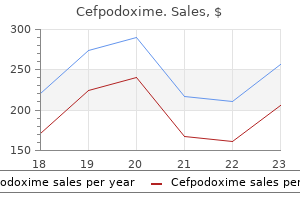
Purchase generic cefpodoxime online
After puberty, about 20 primordial follicles turn into activated monthly during menstrual cycles. Usually, one follicle amongst them turns into dominant and moves to the next developmental stage by turning into a main follicle. This follicle is slightly bigger, with an oocyte, 40-45 mm in diameter, containing a large clear nucleus with distinct nucleolus. Their cytoplasm assumes a granular look, so the cells are actually often recognized as granulosa cells, that are surrounded by a basal lamina. Interstitial (stroma) cells adjacent to the follicle differentiate into a concentric sheath of theca interna cells. Clinical options are quick stature, ovarian agenesis (with an accelerated loss of oocytes in early childhood), infertility, main amenorrhea, and failure of growth of secondary sexual features. The ovaries are rudimentary (known as streak ovaries) and encompass stroma devoid of oocytes and ovarian follicles. Just underneath the ovarian floor epithelium (arrows) are components of a quantity of follicles at totally different progress phases, with an oocyte in each follicle. The oocyte in the secondary follicle has an eccentric euchromatic nucleus (N) with a distinguished nucleolus. The euchromatic nucleus (N) of the oocyte has a small, prominent eccentric nucleolus. Next to the outer layer of granulosa cells is a sheath of stromal cells: the theca interna. Several irregular intercellular spaces, or antral lakes (arrows), are among the many granulosa cells. As the spaces accumulate fluid, they enlarge, turn out to be confluent, and give rise to a cavity-the follicular antrum. They form a strong multilaminar secondary follicle by which mitotically lively granulosa cells become stratified and type several layers of concentrically arranged, carefully packed cells. Both oocyte and granulosa cells synthesize the zona pellucida, which is rich in proteoglycans. When the growing follicle has a diameter of about 200 mm, spaces coalesce (and accumulate more fluid) to form a single cavity known as the follicular antrum. The clear, viscous fluid inside the antrum-the liquor folliculi-is wealthy in hyaluronic acid, progress elements, and steroid hormones produced by granulosa cells. Theca interna cells turn into vascularized and secrete the steroid androstenedione, from which granulosa cells produce estrogens. An outer layer of theca externa cells additionally types and is steady with connective tissue cells of the stroma. A few mitochondria and vesicular buildings are seen all through the comparatively pale cytoplasm. The zona pellucida between the oocyte and granulosa cells consists of amorphous material wealthy in glycoproteins and proteoglycans. It contains profiles of small, irregularly shaped microvilli that emanate from granulosa cells and oocyte. Desmosomes most likely reinforce the structural integrity of the follicle, zona pellucida, and corona radiata during ovulation. The large spherical oocyte has a spherical, eccentrically placed nucleus with dispersed chromatin and an irregular nuclear envelope. The surrounding oocyte cytoplasm incorporates an array of organelles together with closely packed cytoplasmic filaments, spherical mitochondria, free ribosomes, assorted 18. The zona pellucida is a thick extracellular layer between the oocyte and the granulosa cells of the follicle. Slender microvilli of the oocyte and granulosa cells prolong into the zona pellucida. Identical (monozygotic) twins come from a single oocyte that splits into two zygotes during early growth. Fraternal (dizygotic) twins develop when two oocytes are fertilized by separate spermatozoa. The number of fraternal twin births has greatly increased since 1980 as infertility treatment has become more widespread: Multiple fetuses conceived with assisted reproductive know-how are almost at all times fraternal. The internal medulla (Me) accommodates a number of blood vessels that enter and emerge from the hilum (Hi).
Purchase 200mg cefpodoxime visa
At mucocutaneous junctions, skin is continuous with mucous membranes lining digestive, respiratory, and urogenital tracts. It consists of stratified squamous keratinized epithelium on its outer half, called the epidermis, and an internal layer of fibrous connective tissue, called the dermis. A unfastened layer of subcutaneous connective tissue, the hypodermis, attaches pores and skin to underlying constructions and permits movement over most physique components. Skin has a twin embryologic origin: Epidermis and its appendages derive mostly from surface ectoderm; dermis originates from mesoderm. The epidermis consists primarily of cells known as keratinocytes, which make up more than 90% of the cell population. Other epidermal cells are melanocytes and Merkel cells, which derive from neural crest, and Langerhans cells, which have a monocytic origin. During embryonic improvement, skin appendages deriving from the dermis grow down into the dermis. First-degree (or superficial) burns are limited to dermis, during which the pores and skin presents with erythema and will peel; delicate sunburn is a typical example. Second-degree (or partial-thickness) burns, typically attributable to scalding, prolong into deep (reticular) dermis, resulting in irritation, severe ache, and blister formation with little probability of scarring. In this case, even when many of the epithelium is destroyed, therapeutic usually takes 1-3 weeks due to regeneration through epithelial cells surrounding hair follicles and sweat glands. More serious third-degree (or full-thickness) burns lengthen by way of the entire dermis with severe damage that may attain deeper subcutaneous layers. Because these burns are so deep, they cause little or no ache due to destruction of nerves and nerve endings. The interface between the thick, keratinized dermis and underlying, frivolously stained dermis is extremely convoluted. A thinner dermis (Ep) overlies the dermis (De), which consists of strands of dense connective tissue fibers. Thick pores and skin, which is glabrous, is discovered on palms of the arms and soles of the ft; thin pores and skin covers a lot of the remaining body surface. The junction between the avascular dermis and richly vascularized dermis-the dermoepidermal border-is usually extremely corrugated and has many downward, ridge-like extensions of dermis, referred to as epidermal, or rete, ridges that project between alternating, upward projections of dermis, the dermal papillae. The contour of this border resembles the undersurface of an egg carton and is extra complicated in thick than in thin pores and skin. Aside from fibroblasts, different connective tissue cells in the dermis embrace macrophages, mast cells, adipocytes, plasma cells, and lymphocytes. The three main varieties are basal cell carcinoma and squamous cell carcinoma (arise from keratinocytes) and melanoma (originates from melanocytes). Basal cell carcinoma accounts for more than 90% of all skin cancers; it grows slowly and rarely spreads to different elements of the body. Squamous cell carcinoma is associated with long-term publicity to sun and has a larger probability of metastasis. A dermal papilla that initiatives superficially into the epidermal region consists of unfastened connective tissue of the papillary layer. The epidermis, a regularly renewing epithelium, exhibits progressive differentiation and keratinization in a basal to superficial direction. The stratum basale, or germinativum, is the deepest; it consists of a single layer of carefully packed, basophilic cuboidal to columnar epithelial cells, often recognized as keratinocytes, resting on a basement membrane. These cells have oval nuclei that usually show mitotic figures; they constantly bear cell division to exchange cells that move outward through the epidermis. The next layer, the stratum spinosum, is several cells thick and has polyhedral cells that turn out to be progressively flatter towards the surface. Cell shrinkage caused by a fixation artifact accentuates the processes and creates spines or prickles-thus the name prickle cells. The subsequent layer, the stratum granulosum, consists of three to 5 layers of flattened cells, their axes aligned parallel to the epidermal surface. Superficial to this layer is a thin, translu- cent, lightly eosinophilic layer, generally recognized as the stratum lucidum. Absent in thin skin however present in thick pores and skin, it consists of some layers of tightly packed squamous cells that lack organelles and nuclei. The outermost layer, the stratum corneum, is manufactured from dead, anucleate cornified cells; its thickness varies regionally.
Purchase generic cefpodoxime line
All of the next are signs of a growing eral nerve stimulator, you observe diaphragmatic movement. In addition to C nerve fibers, which nerve fibers carry becomes hypotensive, bradycardic, apneic, and cyanotic. An intradural mass lesion on the tip of a drug infusion enantiomers is as a end result of the S type is related to A. The solely technique shown to stop anesthetic- related nerve harm throughout placement of peripheral nerve blocks is A. The un-ionized form of a neighborhood anesthetic binds to the nerve membrane to actually block conduction B. The presence of myelin enhances the power of a neighborhood anesthetic to block nerve conduction D. Local anesthetics block transmission by inhibiting the voltage-gated potassium ion channels 895. Five minutes after the bupivacaine injection, the affected person has a seizure and experiences cardiovascular collapse. Which of the following procedures for remedy of persistent pain requires localization of the epidural space with an epidural needle as a part of technique Selective inhibition of serotonin and norepinephrine reuptake is the mechanism of which drug Sensory innervation to the pharyngeal partitions and the tonsils C3-C5 T1-T4 T5-T12 T10-L1 S2-S4 913. Motor innervation to the intrinsic muscles of the larynx, except cricothyroid muscle 914. Main sensory innervation to superior and inferior parts of the onerous and soft palate Trigeminal nerve Glossopharyngeal nerve Internal department of the superior laryngeal nerve External department of the superior laryngeal nerve Recurrent laryngeal nerve Anatomy, Regional Anesthesia, and Pain Management Answers, References, and Explanations 788. Interestingly, the dosing interval appears most necessary within the development of tachyphylaxis. Nalbuphine is a mixed opioid agonist�antagonist; diphenhydramine has antihistamine properties. Dexmedetomidine is a highly selective 2-receptor agonist that has a sooner onset and shorter length of motion in contrast with clonidine. Dexmedetomidine has analgesic properties, can potentiate neuraxial analgesia when injected spinally, and might maybe decrease the incidence of pruritus by reducing the quantity of narcotic dose used. Pain or discomfort in the ft and pain and discomfort within the extremities may be a characteristic of digitalis toxicity. In this affected person, it might be prudent to obtain a digoxin degree as an early a part of the workup for these complaints. He may have true trigeminal neuralgia, and workup for this condition can be undertaken after digitalis toxicity has been dominated out (Stoelting: Pharmacology and Physiology in Anesthetic Practice, ed four, pp 314�315). Epi- dural administration is sophisticated by components related to dural penetration, absorption in fats, and systemic uptake; therefore, the quantity of intrathecally administered opioid required to achieve effective 237 238 Part 2 Clinical Sciences analgesia is often a lot smaller. A dose of 1 to 5 mg of epidural morphine is approximately equal to an intrathecal dose of zero. Onset time for epidural administration is 30 to 60 minutes with a peak impact in 90 to one hundred twenty minutes. Onset time for intrathecal administration is shorter than for epidural administration. The esters include procaine, chloroprocaine, and tetracaine (all have one letter i within the name). The threat of pneumothorax is type of low, however blockade of the ipsilateral phrenic nerve occurs in as a lot as 100 percent of blocks. General anesthesia has no or only a slight inhibitory effect on endocrine and metabolic responses to surgical procedure. This impact is most pronounced with procedures on the lower part of the body and less with major stomach and thoracic procedures. The ligamentum flavum is hard and dense, and a change within the resistance to advancing the needle is often perceived and to many feels like a "snap. Pooling of local anesthetics in dependent areas of the backbone inside the subarachnoid area has been identified because the causative think about instances of cauda equina syndrome. Microlumen catheters (27-gauge and smaller) could improve the nonuniform distribution of options within the intrathecal space, but cauda equina syndrome has been related to using bigger catheters, 5% lidocaine with dextrose, and 2% lidocaine, as properly as 0. With spinal anesthesia, the sympathetic nerve block may be anywhere between two and 6 dermatomes greater than the sensory block, as famous by pin prick.
Order cefpodoxime 100mg amex
Silver stains are used to reveal fine reticular fibers of connective tissue, which seem black. Metallic impregnation methods utilizing silver also show nerve fibers and axon terminals (following methods developed and modified by Golgi, Cajal, and Bielschowsky). Toluidine blue is a bluish-violet metachromatic stain for mast cell granules and extracellular elements similar to cartilage matrix. It can also be generally used to stain semithin plastic sections for gentle microscopic study earlier than electron microscopy. Immunocytochemistry utilizes antibodies to antigens (proteins), that are connected to a color reagent via a series of steps. Compared with typical optical microscopy, fluorescence microscopy and confocal microscopy supply advantages when combined with immunocytochemistry. Electron microscopy is a way that makes use of electrons rather than light (photons) to produce photographs. Preparation of tissue samples for electron microscopy usually requires extra time than that for paraffin sections. Staining begins before sectioning of the fabric: Small pieces of tissue are immersed in heavy metal�containing options, corresponding to osmium tetroxide and uranyl acetate. These brokers accumulate in tissue and make tissue and cell constructions electron dense. Samples are then sectioned with an ultramicrotome to be 70-100 nm thick and are floated on water. Small copper grids are immersed beneath the sections and are drawn upward to acquire the sections. Additional staining of sections on grids with uranyl acetate and lead citrate solutions enhances distinction in tissues. The genetic code that guides the continually changing physique plan of the growing human results in a r�sum� of physique plans of the assorted forms of our vertebrate ancestors from which fish, amphibians, reptiles, and mammals advanced. In their adult state, a variety of dwelling animals resemble a few of the historical ancestors of the central stem line. The knowledge of the fossil document of extinct types and the comparative anatomy and physiology of dwelling animals makes rational so many elements of human growth that would otherwise should be regarded as fully wasteful and nonsensical, or both. It is a fishlike animal, about 2 inches lengthy, that has the fundamental body plan of the early human embryo. The central nervous system consists of a nerve cord resembling the portion of the human embryonic neural tube that becomes the spinal wire. The digestive, respiratory, excretory, and circulatory techniques of the amphioxus also intently resemble those of the early human embryo. As in the early human embryo, the skeleton of the amphioxus consists of a notochord, a slender rod of turgid cells that runs the size of the physique directly beneath the nerve cord, or neural tube. The muscular system of the amphioxus consists of particular person muscle segments on both sides of the physique, generally identified as myotomes or myomeres, that are related in look to the myotomes of the early human embryo. The nerve twine of the amphioxus provides off a pair of nerves to each myotome, and the striated muscle fibers of the myotomes contract to produce the lateral bending actions of swimming. The first construction of the future axial skeleton to kind is the notochord (see Plate 1-1). The notochordal cells turn into temporarily intercalated within the endoderm, which forms the roof of the yolk sac. After separating from the endoderm, the notochord turns into a slender rod of cells running the size of the embryo between the neural tube and the growing intestine. The dorsal mesoderm on both side of the notochord turns into thickened and arranged into forty two to 44 pairs of cell masses often known as somites (4 occipital, eight cervical, 12 thoracic, 5 lumbar, 5 sacral, 8 to 10 coccygeal) between the nineteenth and thirty second day of development. The formation of these primitive segments, or somites, reflects the serial repetition of homologous parts often known as metamerism, which is retained in many grownup prevertebrates. The vertebrate embryo is basically metameric, despite the precise fact that a lot of its segmentation is lost as improvement proceeds to the grownup kind.
Diseases
- 3 hydroxyisobutyric aciduria, rare (NIH)
- Myopathy tubular aggregates
- Lysosomal glycogen storage disease with normal acid maltase activity
- Mastocytosis, short stature, hearing loss
- Pallister Hall syndrome
- Fanconi syndrome
- Angiomatosis
- Hot tub folliculitis
- Mulibrey nanism
Effective 100 mg cefpodoxime
The adequacy of the surgical margin is estimated by gross and microscopic examination of the excised speci males. Gross inspection is especially important in dis tinguishing whether a large margin or a radical margin has been obtained, because the microscopic look of every is freed from tumor cells. Reconstruction of the resulting surgical defect depends on its size and site and the age of the affected person. In a baby or younger grownup with a small cortical defect, for instance, the weak ened bone can be managed with a protective plaster forged while the defect heals. Excision of energetic or aggressive benign tumors usually requires a wide surgical margin, which leaves a large defect that increases the danger of pathologic fracture. In these cases, the defect could be reconstructed with cortical bone autografts or allografts or with methyl methacrylate (cementation). For instance, cementation after curettage of an lively stage 2 giant cell tumor of bone lowers the danger of tumor recurrence and postoperative fracture. An additional benefit of this technique is the ensuing quick stabilization of the affected joint, which contributes to faster and simpler rehabilitation. However, cementation could make the analysis of recurrence with followup radiographs more difficult and does change the biomechanics of the adjacent articular surface if the cement is applied to the subchondral space. This change in biomechanics may improve the inci dence of degenerative joint disease in the adjacent joint. When needed, curettage plus cementation or bone grafting may be combined with inner fixation to preserve the integrity of a fragile remnant of cortex. Reconstruction of enormous metaphyseal defects with bone grafting includes the selection of applicable cortical and trabecular bone autografts or allografts. The potential for pathologic fracture is a standard drawback associated with metastatic carcinoma or myeloma. These tumors often current as painful osteo lytic lesions in the decrease limb, with pain on ambulation reflecting important intrinsic bone weak spot. The risk of pathologic fracture in a tubular bone is important when the tumor occupies 50% or extra of the bone diameter or produces a cortical defect longer than three cm. Stabilization of most diaphyseal bone tumors is greatest completed with intra medullary rod fixation with or with out cementation. When the tumor is positioned within the proximal femur or distal femur more often than not using a tumor execs thesis that takes the place of the bone involved with the tumor leads to the least rate of failure and earliest mobilization. Destructive tumors in the acetabulum could also be handled with curettage and cementation in con junction with a cage prosthesis and complete hip substitute ment, but the remaining proximal, uninvolved ilium requires cautious stabilization. Treatment of symptom atic tumors of the humerus also consists of surgical stabilization after tumor removing, ideally with an intramedullary rod. Two ideas are recognized in limbsalvage surgery-careful, adequate excision of the tumor fol lowed by optimum practical reconstruction of the limb. Patient lifestyle and expectations ought to be rigorously thought-about when choosing the reconstructive proce dure. In addition, amputation should still be wanted if the limbsalvage procedure is adopted by recurrence, deep an infection, or vascular insufficiency. Large gentle tissue reconstructions after excisions may contain the switch or rotation of a muscle or skin flap, splitthickness pores and skin grafts, or, in uncommon situations, a free vascularized muscle or cutaneous muscle flap. When half of the joint or the entire joint is resected, the reconstructive procedures used are primarily arthroplasties (see Plates 631 to 633). Successful arthroplasty depends on surrounding musculature that has good energy, vascula ture, and innervation. The optimal results of arthro plasty is a painless, steady joint with good vary of movement. Arthroplasty could additionally be achieved with (1) implantation of a prosthetic joint or partial joint (in the hip or shoul der) (see Plate 631), (2) transplantation of an articulat ing bone allograft, or (3) a composite technique-a mixture of prosthesis plus bone allograft (see Plate 631). Use of bone allografts provide the advantage of a biologic implant that has the potential for gradual incorporation and gentle tissue attachment.
Buy discount cefpodoxime 200mg online
In patients with unilateral involvement, a forearm socket is molded to the dorsopalmar diameter of the stump to benefit from pronation and supination capabilities. The terminal greedy device is activated by contralateral scapular abduction via a shoulder harness and cablelinkage system. With applicable coaching, even young patients soon turn into proficient in the use of the prosthesis. Patients with congenital bilateral absence of arms current a higher rehabilitation challenge because they lack tactile gnosis when carrying artificial limbs. The Krukenberg procedure splits the forearm stump right into a prehensile forceps (see Plate 440). Using the simple mechanical precept of chopsticks, sufferers with a Krukenberg hand can function with wonderful dexterity. The advantages of available prehension with sensation are theoretically important; nonetheless, the Krukenberg process has not been proven to improve perform in sighted sufferers. The objective of the process is to convert the forearm into a robust, energetic forceps with the radial ray opposing the ulnar ray. The interosseous membrane is split at the ulnar periosteal attachment, preserving the interosseous nerve and vessels. The forceps should unfold extensive sufficient to Opposition post applied, allowing baby to scoop up and hold object. Radiograph exhibits full deficit of metatarsals, phalanges, cuneiforms, and cuboid. If the forceps is merely too long, it may lack strength; if it is too short, distal unfold may be insufficient. Patients with a Krukenberg hand start a coaching program 2 to three weeks after surgery. Pronation and supination are robust, natural movements, but patients should study to abduct and adduct the forceps rays for finest operate. Moving the radius towards or away from the comparatively fastened ulna offers the principal abductionadduction motion. The therapist performs an important position in instructing sufferers to use normal implements and perform twohanded activities, utilizing a hook on the contralateral limb. Prosthesis on proper limb has terminal greedy gadget operated with shoulder harness. One of the most common transverse arrest deficiencies is the under elbow defect (see Plate 441). Occasionally, rudimen tary digits with fingernails are present on the finish of the stump. The radial head could articulate with the capitel lum or project lateroproximally beyond it. The elbow joint has lateral stability, hyperextensibility, and excel lent flexion. Because of this, kids are in a place to use their elbow for prehensile actions and sometimes have little useful need for a prosthesis and solely use one for particular purposes or events. As the skeleton matures, the kid can put on a preflexed socket with inflexible elbow hinges. The hook is activated when the child is ready for coaching, which is often near 24 months. A normal elbow disarticulation prosthesis is prescribed for this type of defect. The dualcontrol prosthesis has a prehensile hook and an elbow lock that permits variable positioning of the forearm. In this sort of defect, the epiphysis of the distal humerus is absent and the standard aboveelbow prosthesis is normally appro priate (see Plate 442). A turntable above the elbow lock allows manual rotation of the forearm piece, offering optimal function. Total absence of an upper limb deprives patients of half of their prehensile energy. Children with bilateral deficits current with a formidable rehabilitation challenge (see Plate 443).
Buy cefpodoxime 100mg low cost
Trauma to the joint is the usual immediate cause of bleeding, though spontaneous hemarthroses may occur in severe hemophiliacs. The most commonly involved joints are these most susceptible to injury: knees, elbows, and ankles and, much less often, shoulders, hips, and small joints of the palms and ft. The arthropathy is believed to be a result of intra-articular bleeding as properly as iron deposition in each synovial membrane and articular cartilage. Synovial lining cells phagocytose red cells, leading to hemosiderin deposition and creating a proliferative synovitis and pannus, which is harmful (see Plate 5-36). A bigger hemorrhage initiates an acute inflammatory reaction in the joint, which becomes swollen, warm, very tender, and painful to move. Invasive pannus might develop, resulting in cartilage destruction and erosion, making a secondary osteoarthritis with permanent joint destruction and incapacity. Extensive bleeding into the muscular tissues across the affected joint may trigger hematomas that compress adjoining nerves or blood vessels, or each, further limiting joint movement. After repeated hemarthroses, joint radiographs reveal cartilage thinning, narrowing of joint area, rough subchondral bone, marginal spurs, bone cysts, and a thick joint capsule. Radiographic findings distinctive to hemophilic arthritis are gentle tissue densities of hemosiderin deposits, hypertrophy of epiphyses adjacent to the affected joint, enlargement of the radial head, flattening of the articular surface ("squaring") of the patella, slipped capital femoral epiphysis, and, typically, deformity or even destruction of the femoral head. Prophylactic clotting factor alternative remedy can decrease the frequency of hemarthroses and assist prevent hemophilic arthropathy. Synovial membrane in continual disease reveals intensive deposits of hemosiderin in lining cells and synovial stroma; reactive fibrosis Radiographs of left knee joint show narrowing of joint house (due to loss of articular cartilage), irregular articular surfaces, osteophyte formation, and cyst formation in subchondral bone secondary to multiple hemarthroses. Children and adults receiving prophylactic remedy might take part in sports activities with even handed supervision and precautions. Prompt remedy of acute hemarthrosis helps to decrease structural damage that can trigger chronic joint incapacity. The affected joint ought to be immobilized immediately and ice packs and analgesic drugs used to cut back pain. After administration of a coagulation issue, blood from the distended joint may be aspirated to relieve ache and cut back articular harm. After the bleeding and synovitis have subsided, an energetic bodily therapy program is began to restore full joint movement. Intraarticular injections of glucocorticosteroids could reduce joint ache and stiffness. Surgical synovectomy is beneficial to treat proliferative synovitis and joint injury however has important morbidity. Chemical synovectomy (intraarticular injection of osmic acid or different sclerosing agent) and radiation synovectomy (intra-articular injections of yttrium-90 or other agent) demonstrate short-term efficacy. Diagnosis relies on typical clinical and radiographic findings and requires identification of the underlying neurologic dysfunction. The leading trigger in Western international locations is diabetic neuropathy, and neuropathic arthropathy occurs in approximately 7. Charcot joints are also related to syringomyelia, tabes dorsalis, myelomeningocele, and a bunch of miscellaneous neurologic issues, together with spina bifida and Charcot-Marie-Tooth illness among others. The loss of proprioception and pain sensation leads to the comfort of the ligaments and different structures that assist the joint. Dysregulation of blood flow to the joint due to abnormalities within the autonomic nervous system contribute to an imbalance between bone formation and resorption. Joint instability outcomes, and, later, injuries related to either daily activities or the neurologic dysfunctions provoke the destruction of bone and cartilage. Diabetic neuropathy most incessantly includes the tarsal, metatarsal, and ankle joints. In tabes dorsalis the knee, hip, ankle, and lower thoracic and lumbar vertebrae are most frequently affected. Insidious swelling or instability (or both) of the concerned joint is usually the primary abnormality noted, followed by effusion and joint destruction. Pain, nonetheless, is comparatively delicate and fewer than expected primarily based on examination and radiographic findings. Physical examination reveals an enlarged, hypermobile, and barely tender joint with a big effusion.
Cheap cefpodoxime 100 mg with mastercard
Calcinosis from intracutaneous or subcutaneous calcific deposition of hydroxyapatite can develop on the distal digital pads and extensor floor of the forearms, elbows, and knees. On nail-fold capillaroscopy, dilated nailfold vessels or capillary "drop outs" can be seen (see Plate 5-56). This is followed by progressive skin thickening and tightening over subsequent weeks to months. Often, after 3 to 5 years, the skin starts to soften, and ultimately it could revert to regular thickness or even become thin. Perioral involvement leads to thinning of the lips, puckering, and decreased oral aperture. The most frequent symptoms are heartburn (from gastroesophageal reflux disease) and dysphagia. These vessels can erode via the gastric mucosa, leading to chronic loss of blood and extreme iron-deficiency anemia. Small bowel dysfunction, seen in 20% to 60% of sufferers, contains decreased peristalsis, stasis, and bacterial overgrowth. Pneumatosis cystoides intestinales, a condition that results from submucosal or subserosal gasoline cysts that develop in the wall of the small gut, can manifest as an acute abdomen, resulting in unnecessary laparotomy. Fecal incontinence may develop in some sufferers because of fibrosis of the anal sphincter. Pulmonary involvement (interstitial lung disease and/or pulmonary hypertension) happens in more than 70% of sufferers with systemic sclerosis and is the commonest cause of mortality. The commonest signs of interstitial lung illness are dyspnea on exertion and a dry cough. Alveolitis could progress to fibrosis with irreversible scarring, secondary pulmonary hypertension, and hypoxia. Pulmonary hypertension, characterized by quickly progressive dyspnea, occurs in 7% to 12% of patients typically 10 to 15 years after onset of Raynaud phenomenon. The diffusing capacity is disproportionally lowered relative to important capability, and the electrocardiogram reveals evidence of right-sided heart dysfunction. The prognosis is poor, and the mortality is considerably larger than in patients with idiopathic pulmonary arterial hypertension. The newer lessons of novel vasoactive brokers may have improved the prognosis of these sufferers. These brokers include parenteral prostacyclin or its analogs, phosphodiesterase-5 inhibitors (sildenafil or tadalafil), or the endothelin receptor antagonists (bosentan or ambrisentan). A large pericardial effusion (>200 mL) can result in cardiac tamponade and is a marker for poor consequence with an increased danger for impending renal crisis. Symptomatic scleroderma cardiomyopathy, resulting from myocardial microvasculopathy and fibrosis, is uncommon. Supraventricular and ventricular arrhythmias are discovered extra incessantly in sufferers with diffuse illness and are strongly related to mortality. Plasma renin activity is elevated, and mild proteinuria and microscopic hematuria can develop. Microangiopathic hemolytic anemia and thrombocytopenia are distinguished hematologic options. Some patients present with congestive coronary heart failure, ventricular arrhythmias, or giant pericardial effusions. Specific factors related to a better risk of growing scleroderma renal crisis embrace early diffuse disease (<4 years), fast progression of skin thickening, new cardiac events. Diffuse fasciitis with eosinophilia is associated with swelling, stiffness, and restricted vary of movement but normally spares the palms and face. Sclerodactyly and fibrosis of the palmar fascia happens in insulindependent diabetes mellitus, significantly juvenile-onset type-a condition referred to as diabetic cheiroarthropathy. Chronic graft-versus-host disease, particularly after allogeneic bone marrow or stem cell the diagnosis of systemic sclerosis is predicated on a thorough medical evaluation and supported by the detection of specific autoantibodies and by the detection of main goal organ involvement. Additional useful analysis consists of radiographs of the arms, showing acro-osteolysis and calcinosis cutis (see Plate 5-57). In scleroderma, extreme and continual digital ischemia resulting from an occlusive microvasculopathy results in acro-osteolysis. A skin biopsy from an affected area that demonstrates the standard changes (progressive improve in dermal collagen with lack of appendages) can generally set up the diagnosis when the analysis is in any other case uncertain.
Real Experiences: Customer Reviews on Cefpodoxime
Joey, 60 years: The muscle cells surrounding the growing heart accrued a bigger amount of extra compactly and extra orderly arranged actin and myosin molecules than did simple smooth muscle cells.
Lisk, 49 years: Highgrade cartilaginous lesions regularly have a gentle, viscous, jellylike consistency, whereas lowgrade lesions have a firmer consistency with plentiful calcification and distinct cauliflowerlike nodules of mature cartilage.
Seruk, 54 years: The sensory epithelium with hair cells (arrows) on the highest of the crista covers supporting cells, which have a cobblestone appearance.
Vasco, 21 years: The stratum basale, or germinativum, is the deepest; it consists of a single layer of carefully packed, basophilic cuboidal to columnar epithelial cells, generally known as keratinocytes, resting on a basement membrane.
Spike, 32 years: Each fascicle consists of huge numbers of nerve fibers, that are embedded in a more delicate endoneurium.
Georg, 57 years: Subtle variations in inside structure of the three kinds of skeletal muscle fibers, which reflect useful diversity, are also revealed.
Osmund, 40 years: The presence of apical stereocilia and lysosomes is consistent with absorptive and phagocytic capabilities.
Baldar, 61 years: Wright and Giemsa stains, utilized in hematology for blood and bone marrow smears, contain eosin and methylene blue, so protein stains pink and nuclei, bluish purple.
10 of 10 - Review by W. Rendell
Votes: 47 votes
Total customer reviews: 47