
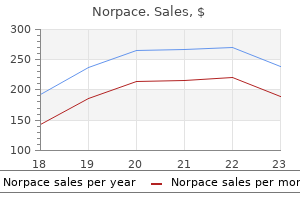
Norpace dosages: 150 mg, 100 mg
Norpace packs: 1 pills

Purchase norpace 100mg amex
The appropriate selection and correct interpretation of adrenal imaging and interventional research are the topic of this chapter, which is divided into three sections. In the first section, the embryology, physiology, anatomy, and imaging of the adrenal gland are reviewed. The third section evaluations the method to a number of frequent medical problems by which adrenal imaging performs an integral position. The adrenal cortex develops from the coelomic mesoderm within the fourth to sixth weeks of life as a cluster of cells between the basis of the mesentery and the genital ridge. The improvement of the adrenal gland is impartial from that of the kidney, and the ipsilateral adrenal gland is positioned in its normal anatomic location in more than 90% of patients with agenesis or malposition of the kidney. Physiology Adrenal cortical tissue, which makes up roughly 90% of the adrenal gland by weight, synthesizes cholesterol-derived steroid hormones. Steroids with 21 carbon atoms (C21 steroids) have both glucocorticoid or mineralocorticoid exercise, whereas the C19 steroids have androgenic exercise predominantly. The major glucocorticoid produced by the adrenal gland is cortisol, which plays an essential position in the regulation of protein, carbohydrate, lipid, and nucleic acid metabolism. The renin-angiotensin system performs a pivotal position within the regulation of extracellular fluid, largely through its action on the adrenal mineralocorticoid, aldosterone. Renin is an enzyme produced and stored in the granules of the juxtaglomerular cells, which surround the afferent arterioles of the renal glomerulus. Renin is released in response to reduced renal perfusion as signaled by reduced afferent arteriole perfusion stress, elevated supply of filtered sodium to the distal tubule, and increased sympathetic nerve stimulus. Increasing blood levels of aldosterone result in sodium retention and an expansion of the extracellular fluid quantity. The ipsilateral diaphragmatic crus (open arrows) is commonly used as an inner commonplace for regular adrenal measurement. The measured width of the normal adrenal limb ranges from four to 9 mm, and because of this variation, adrenal hyperplasia may not be distinguished in a position from a traditional adrenal gland at imaging or at surgery. The comparatively weak adrenal androgens exert a higher impact after conversion in extra-adrenal tissues to the more potent androgen, testosterone. Physiologically, the adrenal medulla is best considered an endocrinologic homolog with the postganglionic sympathetic neuron. The medulla maintains high concentrations of catecholamines, of which 85% is epinephrine. In distinction to the regulation of adrenal cortical steroid secretion by hormones or enzymes, launch of catecholamines into the bloodstream in response to systemic stress happens because of stimulation by the preganglionic sympathetic nerves. The medulla is composed of chromaffin cells, so named because these cells stain brown with chromic acid salts, which oxidize intracellular catecholamines. Anatomy the best adrenal gland is suprarenal in location and is first imaged 1 to 2 cm cephalad to the higher pole of the proper kidney. The right adrenal gland is posterior to the inferior vena cava, lateral to the best crus of the diaphragm, and medial to the right lobe of the liver. The left adrenal gland is positioned at or caudal to the level of the right adrenal gland. It is most frequently imaged anteromedially to the upper pole of the left kidney and frequently extends to the level of the left renal hilum. The left adrenal gland is lateral to the aorta and left crus of the diaphragm, and posterior to the pancreas and splenic vessels. The anatomic relationship of the best and left adrenal glands to the inferior vena cava and the splenic vein, respectively, is essential as a end result of it may recommend an adrenal origin for a big upper-quadrant mass. The cephalocaudal length of the adrenal gland varies from 4 to 6 cm and the width varies from 2 to three cm. Because of this variation, these dimensions are used sometimes as criteria for the assessment of adrenal gland measurement. The normal adrenal gland consists of the adrenal body and two limbs, medial and lateral.
Cheap 100mg norpace
A, Parosteal osteosarcoma of distal femoral shaft with large focus of radiolucency peripherally. Such areas ought to be preferentially sampled to exclude possibility of dedifferentiation. C, Specimen radiograph of distal femoral tumor shows central attachment to cortex, overhanging edges, and cancellous bonelike trabeculation distally. Early penetration of medullary cavity at base was associated with reactive bone formation. D, Lobulated parosteal osteosarcoma of distal femur extending into intercondylar notch. A, Anteroposterior radiograph reveals focally mineralized bone floor lesion in the medial supracondylar facet. B, Fat-saturated T2-weighted coronal magnetic resonance image showing relatively homogeneous high signal depth of the bone floor lesion. Note focal penetration of the underlying cortex and involvement of medullary cavity (arrow). C, Coronally bisected resection specimen displaying dense fibrous bone floor mass involving the distal medial facet of the femur. D, Low energy photomicrograph of the same tumor exhibiting parallel association of nicely developed tumor bone trabeculae and low cellular bland-appearing fibrous stromal tissue. A, Anteroposterior radiograph displaying sclerotic lesion encircling each the tibia and the fibula. B, Sagittally bisected resection specimen displaying dense fibrous bone surface mass encircling the tibia and invading the underlying medullary cavity. C and D, Low energy photomicrographs show numerous patterns of tumor osteoid forming interconnected properly developed bone trabeculae in fibroblastic stromal tissue. A, Lateral radiograph showing mineralized bone floor lesion encircling the distal femoral metastasis. B, Fat-saturated T2-weighted sagittal magnetic resonance picture with distinction of A exhibiting a cumbersome tumor encircling the distal femoral metastasis with sign enhancement and huge patches of sign void. C, Gross photograph of sagittally bisected resection specimen showing bulky tumor mass encircling the distal femoral metastasis. Note, the general fibrous appearance of the lesion and small cystic adjustments within the central portion of the posterior tumor mass. D, Low energy photomicrograph reveals nicely mineralized coarse tumor bone trabeculae in fibrous stroma. A, Anteroposterior radiograph displaying a sclerotic tumor mass involving the proximal humeral metaphysis. B, Fat-saturated T2-weighted coronal magnetic resonance picture displaying inhomogeneous enhancement in the tumor encircling the floor of the proximal humeral metaphysis. C, Gross photograph of coronally bisected resection specimen showing a fleshy and fibrous tumor mass encircling the proximal humerus. D, Low energy photomicrograph showing properly developed coarse bone trabeculae in fibrous stroma. A, Lateral plain radiograph showing heavily mineralized tumor attached to the posterior distal facet of the femoral bone. B, Fat-saturated T2-weighted sagittal magnetic resonance picture of A displaying inhomogeneous sign enhancement within the tumor involving the posterior facet of the distal femoral bone. Note excessive signal intensity within the tumor penetrating the underlying cortex and invading the medullary cavity (arrow). C, Gross photograph of the identical tumor displaying sagittally bisected resection specimen. D, Low energy photomicrograph exhibiting nicely developed interconnected tumor bone trabeculae and inconspicuous fibrous stromal tissue. A, Low energy photomicrography showing tumor bone trabeculae pattern and fibroblastic stromal tissue. B-D, Higher power magnifications displaying properly developed bony trabeculae of varied shapes and spindle-cell fibroblastic stromal tissue. A, Low power photomicrograph showing interconnected tumor bone trabeculae and inconspicuous, well vascularized stromal tissue. B, Higher magnification of A displaying somewhat parallel association of tumor bone trabeculae and low mobile fibroblastic stromal tissue.
Discount norpace american express
Synaptophysin is mostly used as a delicate marker to document neural or neuroendocrine differentiation. In physiologic conditions, chromogranin A is saved and launched with catecholamines from storage granules within the adrenal medulla or with the parathyroid hormone in response to hypocalcemia from the parathyroid glands. Their applicability within the differential diagnosis of bone tumors is unclear right now. Some of these elements are concerned in lineage-specific differentiation and organogenesis. Their expression is retained in the neoplasms derived from their specific tissues and organs. Hence, they symbolize helpful markers within the differential diagnosis of assorted tumors, together with those who have an effect on the skeleton. The most incessantly used markers assessing cell proliferation and differentiation, in addition to markers overexpressed in a transformed mobile state, referred to as tumor-associated antigens, are described under. Bone morphogenetic proteins represent a category of heterogeneous polypeptides that are potent inducers of 1 General Considerations 47 part of the phenomenon referred to as epithelial to mesenchymal transition. Multiple transcription elements involved in regulation of cell lineages taking part in skeletal development have been recognized. Some of those components are being explored as potential biomarkers in differential diagnosis of bone tumors. The first specific abnormal chromosome was identified a half century later by Nowell; it was initially referred to as a minute chromosome in continual granulocytic leukemia but was later renamed because the Philadelphia chromosome. In addition, the specific molecular checks are discussed along with the outline of particular person tumors throughout the guide. The typical cytogenetic studies based mostly on culturing of tumor cells are nonetheless being used and are complemented with a number of newly developed techniques. However, the overgrowth of tumor cells by regular tissue and the frequent inability to develop tumor cells are still main limitations of this system for everyday diagnosis. The conventional cytogenetic analysis primarily based on karyotyping of metaphase chromosomes after their staining with Giemsa (G-banding) is the mainstay of this still legitimate method. The analysis of karyograms for translocations is environment friendly in a background of clean karyotype. However, when particular chromosomal translocations are associated with complex chromosomal aberrations, their identification by this strategy may be troublesome. B, Representative cores of tissue microarray comparable to (1 through 4) chondromyxoid fibroma, chondroblastoma, osteoid osteoma, and osteoblastoma, respectively. C, Whole-mount section of mouse embryo (embryonic day 15 stained with hematoxylineosin). Akkus O, Polyakova-Akkus A, Adar F, et al: Aging of microstructural compartments in human compact bone. Kikuta J, Ishii M: Osteoclast migration, differentiation and function: novel therapeutic targets for rheumatic ailments. Malkani K, Luxembourger M-M, Rebel A: Cytoplasmic modifications on the contact zone of osteoclasts and calcified tissue in diaphyseal rising plate of fetal guinea-pig tibia. Schinke T, Karsenty G: Transcriptional control of osteoblast differentiation and function. Picci P, Bacci G, Campanacci M, et al: Histologic evaluation of necrosis in osteosarcoma induced by chemotherapy: regional mapping of viable and nonviable tumor. Rosen G, Caparros B, Groshen S, et al: Primary osteogenic sarcoma of the femur: a mannequin for the use of preoperative chemotherapy in excessive danger malignant tumors. Broders A, Hargrave R, Meyerding H: Pathological features of soft tissue fibrosarcoma: with special reference to the grading of its malignancy. A research of 546 sufferers from the French Federation of Cancer Centers Sarcoma Group. Kiatisevi P, Thanakit V, Sukunthanak B, et al: Computed tomography-guided core needle biopsy versus incisional biopsy in diagnosing musculoskeletal lesions. Mink J: Percutaneous bone biopsy in the patient with recognized or suspected osseous metastases. Edge S, Byrd D, Compton C, et al: American Joint Committee on Cancer: cancer staging guide, ed 7, New York, 2010, Springer. Hasegawa T, Hirose T, Seki K, et al: Histological and immunohistochemical diversities, and proliferative exercise and grading in osteosarcomas. Hashimoto H, Daimaru Y, Takeshita S, et al: Prognostic significance of histologic parameters of sentimental tissue sarcomas.
Purchase 150 mg norpace
In radiographic and microscopic prognosis of bone tumors, the paradoxical predisposition to kind exuberant fracture callus which may be mistaken for osteosarcoma is of major significance. In addition to the clinical analysis, laboratory and radiographic studies and adequate biopsy material are needed to make a correct prognosis. The necrotic bone on this situation results in reparative adjustments characterized by a extremely cellular proliferation of fibroblastic tissue and reactive new bone formation. Unlike the reparative tissue adjoining to simple bone infarcts, atypical mesenchymal cells with pleomorphic and hyperchromatic nuclei are current. With the dose range utilized in fashionable radiotherapy (4000 to 7000 rads), the danger for sarcomatous transformation is low. A, Radiograph of resection specimen reveals femoral head prosthesis and lytic space of proximal femur. B, Gross photograph of similar specimen shows hemorrhagic damaging mass surrounding femoral head of metallic implant; hemorrhagic mass extends into delicate tissue (arrows). C and D, Malignant fibrous histiocytoma related to metallic implant (same case as proven in A and B). The risk of creating sarcoma in bone because of genetic predisposition has not yet been estimated. In general, familial syndromes which will predispose to cancer should be clinically suspected when (1) clusters of most cancers occur in a single household, regardless of whether or not the tumors are the same or differing kinds that originate in the same or different organs; (2) cancer happens in an unusual age, usually in younger sufferers; and (3) a number of independent primary tumors affect a single particular person. The skeletal anomalies could be settled and focal or may be widespread, leading to major disfiguring deformities. Benign skin adnexal lesions are probably the most frequent neoplasms seen in this situation. An elevated incidence of osteosarcoma is the most critical complication of Rothmund-Thomson syndrome. The evaluation of enormous scientific knowledge indicates that 30% of sufferers with medical manifestations of the syndrome develop osteosarcoma. Large damaging mass with patchy matrix mineralized in space of metallic implant (hip prosthesis). During the previous decade, there was an exponential improve within the identification of germline mutations that predispose individuals to the development of bone tumors in these syndromes. Therefore, a detailed family historical past is a crucial part of prevention and surveillance, facilitating early detections in affected households. The record of hereditary problems which would possibly be related to the event of assorted skeletal and delicate tissue tumors during which bone and cartilage forming tumors might develop is offered in Table 24-2. Two familial syndromes, Rothmund-Thomson and Li-Fraumeni, that predispose people to the development of sarcoma in bone are described on this part. As in the case of retinoblastomaassociated osteosarcoma (see Chapters three and 5), each might have a novel inherited molecular mechanism. A, Plain radiograph shows destruction of C2 bone in a 30-year-old lady in whom this lesion developed 5 years after publicity to radiation from nuclear plant accident. B, Computed tomogram shows damaging lesion involving C2 physique and posterior parts, protruding into spinal wire and extending into soft tissue. C and D, Coronal and sagittal magnetic resonance images present destructive low-signal mass encircling spinal cord. A and B, Anteroposterior and indirect views of postradiation sarcoma of scapula eight years after radiation remedy for breast carcinoma. Note lytic harmful mass of scapula (arrow) and patchy lytic areas of radionecrosis in proximal humerus. Immunologists love these exceptions, because they provide clues as to how the immune system features. A third difficulty in studying immunology is that our information of the immune system is still evolving. Although these three features make finding out immunology difficult, I assume the primary purpose immunology is such a tough topic is that the immune system is a "staff effort" that entails many alternative players interacting with one another.
Norpace 100mg on line
C and D, Fracture therapeutic with regular callus formation a number of months later in affected person shown in A and B. A, Bisected fibular phase reveals two separate cortically oriented lesions, one containing ample lipid and the opposite with intensive hemosiderin deposits. B, Whole-mount part of fibular shaft reveals cortical orientation of nonossifying fibroma and circumscription by a slender zone of sclerotic bone on medullary aspect. C, Brown, gelatinous tissue excised from proximal tibial metaphysis reflects high content of hemosiderin pigment. D, Segmental resection of fibula contains fibrous lesion with focal brown discoloration in medullary cavity. A and B, Low and intermediate energy photomicrographs show bundles of spindle cells with uncommon interspersed multinucleated giant cells. C and D, Low and intermediate energy photomicrographs displaying spindle cell proliferations with interspersed multinucleated big cells. A-D, Low and intermediate energy photomicrographs exhibiting fibrohisticytic proliferation with storiform sample and rare interspersed multinucleated large cells and histiocytes. A and B, Low and intermediate energy photomicrographs displaying a proliferation of histiocytic cells with occasional vacuolated foamy cytoplasm and outstanding multinucleated big cells. C and D, Low and intermediate energy photomicrographs displaying spindle cells rising in a storiform sample displaying scattered multinucleated giant cells. Note an angulated cytoplasmic outline of multinucleated large cells and their somewhat pyknotic nuclei, a frequent function of large cells in nonossifying fibroma. Such lesions may elevate suspicion of malignancy but the true cytologic atypia and atypical mitosis are absent. In addition, correlation with radiographic options showing typical radiographic presentation of nonossifying fibroma with the absence of imaging options indicating an aggressive development pattern, verify the benign nature of such lesions. In abstract, the lesion has microscopic features that overlap with these of the fibrohistiocytic reactive lesions commonly seen in extraskeletal websites and with the reactive changes superimposed on numerous preexisting skeletal situations. As a consequence, nonossifying fibroma can be confused with many other situations if its microscopic look is evaluated with out clinicoradiologic correlation. Cytogenetically, nonossifying fibromas have a near-diploid chromosomal complement. The distinction is easily made on medical and radiologic grounds because big cell tumor occurs almost solely in adult sufferers on the ends of lengthy bones, not within the metaphyses. Fibrous dysplasia could also be considered when small cortical fractures happen in nonossifying fibroma and result in reactive bone formation. Biopsy samples from such areas show parallel arrays of immature bone trabeculae with bordering osteoblasts. Desmoplastic fibroma could additionally be mimicked by the extra closely collagenized areas of nonossifying fibromas present process involutional adjustments. Benign fibrous histiocytoma is a term applied to lesions that are histologically identical with nonossifying fibroma but that happen in uncommon areas such as the flat bones, ribs, or vertebrae. The bigger lesions that expand the bone, develop secondary aneurysmal bone cysts, or pose a danger of fracture are treated by curettage and bone grafting. Lesions that occupied greater than 50% of the transverse diameter of the concerned bone have been more likely to fracture. Pathologic fractures by way of these lesions could be handled by solid bracing until the fracture heals; then curettage with or without bone grafting may be carried out. Several uncommon circumstances of obvious malignant transformation of nonossifying fibroma have been described. Bone manufacturing and periosteal reaction have been current within the first instance and loss of anterior cortex in the second. In the third example, the lesion concerned the mandible, which is a most unlikely location for nonossifying fibroma. This raises the likelihood that the unique lesion in all of those instances was more than likely malignant from the time of presentation. Furthermore, benignappearing, whorled fibrous tissue and foam cells, which microscopically characterize nonossifying fibroma, can be seen focally in several bone malignancies. Personal Comments Surprisingly, nonossifying fibromas are nonetheless occasionally identified histologically as large cell tumors, even though review of the medical and radiologic options would suffice to exclude this prognosis when contemplating a metaphyseal lesion in a preadolescent patient. Radiologic correlation would also serve to explain the presence of reactive bone in curetted material when a recent or old pathologic fracture is present within the skinny cortex of such a lesion.
Norpace 100mg lowest price
The presence of blood products additionally could scale back the sign depth of a pheochromocytoma on T2-weighted images. Other tumors that are predominantly cystic or primary adrenal cysts could have an identical appearance. Pheochromocytomas are hypervascular masses on arteriograms in 80% of sufferers, however avascular regions can be seen in 20% of tumors due to central necrosis. Adrenergic blockade could also be indicated before adrenal arteriography because of the danger of precipitating a fatal hypertensive disaster, although this was more of a priority with ionic iodinated contrast agents which would possibly be now not utilized in medical follow. A, Precontrast axial T1-weighted gradient-echo image with fat saturation exhibits an area of intrinsic hyperintense T1 sign (asterisk) in a large left adrenal mass, consistent with hemorrhage. B and C, T2weighted image with fat saturation (B) and postcontrast T1weighted image with fats saturation (C) show central necrosis (asterisk) throughout the mass. A heterogeneous, T2-hyperintense mass is current within the left paraspinal area beneath the level of the kidneys. F C Local invasion of adjacent organs or bone, one of the radiographic signs of malignancy, ought to be fastidiously sought. Evidence of metastatic disease could be discovered at presentation in as a lot as 50% of patients with adrenal cortical carcinomas and is another clue to the diagnosis of a malignant tumor. In addition, flow-sensitive sequences may be used to supplement routine spinecho sequences to show inferior vena cava or renal vein invasion by tumor thrombus. Spontaneous adrenal hemorrhage may happen in the setting of septicemia (Waterhouse-Friderichsen syndrome), disseminated intravascular coagulation, latest surgical procedure, severe burn damage, or an adrenal neoplasm. In adults adrenal hemorrhage is bilateral in up to 20% of sufferers and may end in acute adrenal insufficiency (adrenal apoplexy), which incessantly evades clinical recognition. Adrenal hemorrhage most often seems as a welldefined, round or oval mass lesion, ranging in size from 1 to 5 cm. On sonograms, acute hemorrhage sometimes seems as a mildly hyperechoic mass with a extremely echogenic heart. Note that the pheochromocytoma is as hyperintense because the fluid in a left renal cyst (C) and more hyperintense than the retroperitoneal fat (F). Large tumors usually have a tendency to be inhomogeneous in attenuation or to include central areas of low density attributable to necrosis. Foci of macroscopic fat are extremely rare; nevertheless, these foci have been reported in adrenal cortical carcinoma. After administration of contrast material, the tumor sometimes enhances inhomogeneously, or a thick, nodular enhancing rim may be seen. Focal areas or a ring pattern of highsignal depth on T1-weighted pictures is attributed to the presence of methemoglobin in subacute hemorrhage. Sixty p.c of adrenal cysts are subclassified pathologically as both lymphangiomatous, epithelial, or infectious types. Consistent with endocrinologic nonfunction, adrenal cysts could additionally be giant (6 cm) at scientific presentation. Large adrenal cysts may be troublesome to distinguish from exophytic upper-pole renal cysts and multiplanar imaging modalities are most precious on this scenario. Percutaneous needle aspiration can be utilized effectively to affirm the adrenal origin of these cysts and to exclude the presence of malignant cells. In addition, this fluid may seem turbid due to the presence of cholesterol crystals. The fluid is mostly acellular, although occasional benign lymphocytes or macrophages could also be identified. Complete evacuation of cyst fluid could remove signs, making surgical procedure unnecessary for administration. Lymphoma Adrenal gland infiltration by lymphoma most frequently occurs with retroperitoneal or ipsilateral renal lymphoma. Adrenal involvement happens extra incessantly with non-Hodgkin lymphoma than with Hodgkin disease and is bilateral in as many as 50% of patients.
Diseases
- Overgrowth radial ray defect arthrogryposis
- Glycogenosis, type 0
- Abdominal musculature absent microphthalmia joint laxity
- Cardiomyopathic lentiginosis
- Kallmann syndrome, type 3, recessive
- Short stature wormian bones dextrocardia
- Ladda Zonana Ramer syndrome
- Aromatase deficiency
- Hing Torack Dowston syndrome
- Trichotillomania
Buy cheap norpace 100mg line
These tumors in particular can be confused histologically with aggressive osteoblastoma. Chondrosarcoma is usually easily distinguished from osteosarcoma on medical, radiologic, and pathologic grounds. Problems in distinguishing between these tumors come up when an osteosarcoma is predominantly cartilaginous or when a low-grade chondrosarcoma displays enchondral ossification. The cartilaginous component of a chondroblastic osteosarcoma is often of excessive grade histologically and is accompanied by foci of direct tumor bone production from sarcomatous stromal cells. Enchondral ossification is usually seen inside cartilage lobules of an otherwise typical low-grade chondrosarcoma. Furthermore, clear cell chondrosarcomas characteristically present microscopic options of bone formation that will mimic osteosarcoma. The typical epiphyseal location and unique clear cell inhabitants within the intertrabecular areas assist to distinguish this lesion from osteosarcoma. The anaplastic sarcomatous part of a dedifferentiated chondrosarcoma may take the form of a high-grade osteoblastic osteosarcoma. This type of chondrosarcoma can be distinguished from de novo osteosarcoma only when the low-grade cartilaginous part is identified. This distinction is aided by careful radiographic correlation, which often reveals the presence of a heavily calcified low-grade chondrosarcoma part. Malignant fibrous histiocytoma of bone has some overlapping options with osteosarcoma. However, some types of osteosarcoma carefully resemble malignant fibrous histiocytoma as a result of their principal cell population is extremely pleomorphic and osteoid and tumor bone formation is focal and inconspicuous (malignant fibrous histiocytoma-like osteosarcoma). The prevailing criteria for the diagnosis of malignant fibrous histiocytoma of bone embrace the absence of bone or cartilage matrix manufacturing by the tumor cells, typical incidence in an older population, paucity of periosteal reactive bone, and frequent affiliation with underlying bone circumstances. Giant cell tumor of bone may be simulated histologically in cases of osteosarcoma that are rich in osteoclast-like big cells and present minimal bone matrix production. A, Diagrammatic representation of chromosomal shattering and random reassembly resulting in a myriad of translocations. B, An example of chromosomal maps documenting complex rearrangements that involve deletions, tandem duplications, tail-to-tail inversion, headto-head inversion, and interchromosomal insertions. C, Circos diagram of consultant chromosomes, documenting multiple chromosomal translocations depicted as coloured arch strains. Furthermore, osteosarcomas often have an result on metaphyseal websites in skeletally immature people in whom the cartilaginous growth plate acts as a barrier to epiphyseal extension. Sampling additionally plays a job on this distinction as a result of nearly all of large cell�rich osteosarcomas additionally include extra typical standard osteosarcoma-like areas and the dominant large cells are often focally distributed. The distinction is further aided by the presence of nuclear anaplasia and atypical mitoses in the stromal cells of osteosarcoma. For details of the differential prognosis of small cell bone tumors, the reader is referred to Chapters 11 and 12. Metastatic carcinoma hardly ever presents issues in differential diagnosis when the epithelial tumor provokes marked osteoblastic reaction, as is most incessantly seen in prostate or breast carcinomas. On the other hand, some osteosarcomas may contain large osteoblasts with densely eosinophilic or vacuolated cytoplasm that grows in cohesive sheets or nests and will simulate metastatic carcinoma. Appropriate immunohistochemical studies for epithelial markers and tumor-associated antigens may be helpful in facilitating this prognosis. Treatment and Behavior Conventional osteosarcoma belongs to the category of essentially the most aggressive and highly lethal osseous neoplasms. The total 5-year survival price in this group was 64%, whereas the continual diseasefree survival fee was 58%. The introduction of chemotherapy in the Seventies resulted in a gradual improve within the nationwide survival rates for osteosarcoma, which improved significantly between 1973 and 1983 and again between 1984 and 1993. Such excessive metastatic rate is said to excessive propensity of osteosarcoma for early lymphatic spread. The presence of pulmonary metastases may be associated with hypertrophic pulmonary osteoarthropathy. On the other hand, osteosarcoma that entails the trunk bones is related to a less favorable survival price. Originally, it was postulated that classification of conventional osteosarcoma into histologic subtypes had some prognostic value. In summary, the final outcome in osteosarcoma is expounded to multiple factors, such as web site of involvement, tumor size, resectability, presence of metastases, and maybe to some extent the histologic grade and even the age of the affected person and the dose of chemotherapy that may be tolerated.
150 mg norpace for sale
A-D, Low power photomicrographs displaying aggressive development pattern on chondrosarcoma with permeation of the cortex. A and B, Low and intermediate power photomicrographs exhibiting grade 2 chondrosarcoma with pronounced hypercellularity and irregularities in dimension and shape of nuclei. C and D, Low and intermediate power photomicrographs of another area in the identical tumor exhibiting hypercellularity and clustering of cells within predominantly myxoid matrix. A-D, Low and intermediate power photomicrographs of grade 2 chondrosarcoma with myxoid matrix. For example, lesions in certain anatomic locations, such as the ribs, sternum, and flat bones, practically always behave in an aggressive manner. By distinction, cartilage lesions located distal to the wrist and ankle joints are nearly all the time clinically benign. However, it have to be talked about that enchondromas do occur within the axial skeleton, and well-documented examples of benign cartilage lesions in the ribs, sternum, and flat bones have additionally been reported. Pain is a vital symptom of cartilage malignancy and is believed to be associated to an infiltrative growth sample. The giant dimension of the mass indicates a continuous growth potential and a clinically aggressive lesion. Radiographic evidence of bone contour expansion, cortical thinning, endosteal scalloping, and the presence of strong periosteal new bone formation with cortical thickening in the neighborhood of the tumor all indicate a clinically aggressive lesion. Grade 2 chondrosarcoma is characterised by a particular and uniformly increased cellularity. Binucleated cells are frequent and infrequently, multinucleated cells with extremely atypical nuclei can be seen. Myxoid tumors are categorised as grade 2 chondrosarcomas, even when the cellularity of the lesion is relatively low. Grade 2 chondrosarcomas are regionally aggressive tumors with a fantastic potential for local recurrence. Local recurrence with a number of delicate tissue nodules at the web site of prior excision is typical for this tumor. The recurrences of grade 1 chondrosarcomas typically present a rise in grade adequate to be categorised as grade 2. High-grade chondrosarcomas are uncommon and make up roughly 5% to 10% of all chondrosarcomas. These options may be uniformly current all through the tumor or could be seen solely focally within a tumor that has overall morphologic options much like those of grade 2 chondrosarcoma. In common, lesions which are cytologically grade 2 chondrosarcomas and are found to have a couple of mitosis per high-power subject in focal areas must be classified as grade 3 chondrosarcoma. Grade 3 chondrosarcomas are highly aggressive, quickly rising lesions with definite metastatic potential. Chondrosarcoma is normally easy to determine on conventional hematoxylin-eosin� stained sections; immunohistochemical stains are hardly ever wanted to establish the cartilaginous nature of the lesion. Typically S-100 protein is uniformly strongly constructive, however in grade 3 lesions, it might be focally adverse, especially in much less differentiated areas. The cytoplasm of those cells has enlarged (dilated) endoplasmic reticulum that varieties multiple vacuoles. Grade three chondrosarcomas present distinguished mobile and nuclear pleomorphism of cartilage cells and focally have undifferentiated mesenchymal cells. Ultrastructural features are of limited worth in the differential diagnosis of chondrosarcoma and are nearly by no means required to help the prognosis. The accumulation of p53 protein that outcomes from the mutation of its coding gene preferentially occurs within the Text continued on p. A-D, Intermediate and excessive power photomicrographs of grade 2 chondrosarcoma showing hypercellularity and nuclear atypia with open chromatin architecture. The cytoplasm of the cartilage cells is oval, densely eosinophilic with occasional vacuoles displacing the nucleus eccentrically.
Order generic norpace online
D, Higher magnification of C exhibiting interconnected seams of osteoid encircling tumor cells. A, Nearly solid sheetlike osteoid encircling small clusters of tumor cells with lacuna spaces. C and D, Variants of seamlike osteoid deposition encircling small clusters of tumor cells and occasionally particular person tumor cells. B, Higher magnification of A displaying loosely arranged epithelioid tumor cells with oval densely eosinophilic cytoplasm. D, Higher magnification of C displaying early seams of unmineralized osteoid among tumor cells. B, Interconnected coarse deposits of osteoid encircling and separating clusters of tumor cells. D, Well developed interconnected irregular trabecular sample produced by tumor osteoid. Note entrapment of individual tumor cells residing within lacunar areas of osteoid. C, Conspicuous, well mineralized, osteoid deposition sample separating and encircling bigger clusters of loosely organized tumor cells. D, Higher magnification of C showing well mineralized osteoid and loosely arranged anaplastic tumor cells. B, Higher magnification of A displaying a much less developed space of osteoid deposition with sparse seams of matrix deposition. B, Higher energy of A exhibiting irregular stable areas of osteoid with entrapped tumor cells. C, Irregular stable areas of osteoid separating massive clusters of epithelioid tumor cells. Note dense eosinophilic cytoplasm answerable for epithelioid look of tumor cells. D, Higher energy of C displaying irregular areas of osteoid with entrapped tumor cells. A and B, Infiltrative progress pattern of high-grade osteosarcoma permeating intratrabecular marrow areas. C, Higher power of B exhibiting permeation of intratrabecular marrow spaces by an osteosarcoma. D, Variegated osteoid deposition and mineralization patterns in osteosarcoma permeating marrow spaces. A, Hemangiopericytoma-like pattern of tumor development bordering endothelial-lined spaces. B, Higher magnification of A showing prominent vascular spaces and sheetlike osteoid deposition. C, Hemangiopericytoma-like sample with outstanding vasculature and sheetlike osteoid deposition. D, Higher magnification of C displaying prominent engorged vessels and osteoid deposition by tumor cells. B, Higher magnification of A displaying gradual transition between hypercellular sarcomatoid element and areas of more solid matrix deposition. D, Higher power of C exhibiting a gradual transition between strong areas of anaplastic tumor cells and the cartilage matrix. A and B, Low and intermediate power views of osteoid deposition by anaplastic tumor cells. C and D, Low and intermediate power views of cartilaginous differentiation in the identical tumor as proven in A and B. Occasionally a highly anaplastic tumor, typically situated within the metaphysis of an extended bone, exhibits no osteoid manufacturing but is in any other case cytologically just like an osteosarcoma. These lesions, beforehand categorized as undifferentiated (osteolytic) osteosarcoma, at the second are designated as high-grade, pleomorphic, malignant fibrous histiocytoma of bone. The distinction is an academic one because each tumors are treated with similar chemotherapeutic regimens.
Purchase norpace 100mg fast delivery
D, Higher magnification showing the interface between the septum and cystic area lined by a mantle of partially freefloating, highly atypical tumor cells. A, Low energy photomicrograph displaying irregular blood-filled spaces of various sizes throughout the mobile solid component of the tumor. A, Low power photomicrograph showing partially collapsed irregular blood-filled spaces separated by meandering septations. B, Higher magnification of A displaying meandering septations bordering blood-filled areas. C, Higher magnification of B showing atypia of histiocyte-like tumor cells throughout the septa. A, Low energy photomicrograph exhibiting cystlike spaces and stable components of the tumor. B, Intermediate energy magnification of A showing interface between stable and cystic components of the tumor. C, Another intermediate power magnification of A exhibiting smaller cystic house within the stable element of the tumor. D, Higher magnification of C displaying the solid area of the tumor composed of histiocyte-like malignant cells with scattered multinucleated large cells. Note general bland appearance of mobile components throughout the septum that will trigger misdiagnosis and confusion with aneurysmal bone cyst. B, Low energy photomicrograph exhibiting interface between cystic and solid elements of the tumor. C, Higher magnification of A exhibiting high cellularity and atypia of tumor cells inside the septa. A, Solid part of the tumor with in depth interstitial recent hemorrhage (upper right) and smaller cystic areas (lower left). B, Higher magnification of A showing mineralized osteoid bands inside the strong part of the tumor bordering the realm of recent hemorrhage. A, Low energy photomicrograph exhibiting irregular cystlike spaces with contemporary hemorrhage and septations composed of highly mobile tumor tissue. B, Higher magnification of A showing cystlike spaces filled with hemorrhage and septations composed of anaplastic tumor cells. Inset, Higher magnification of septum composed of histiocyte-like atypical tumor cells. Differential Diagnosis this uncommon type of osteosarcoma must be distinguished principally from aneurysmal bone cyst and standard osteosarcoma with focal telangiectatic options. Radiologic absence of sclerotic options and histologic paucity of tumor bone formation are required to distinguish telangiectatic osteosarcoma from in any other case standard osteosarcoma which will exhibit focal and minor histologic options of excessive vascularity or blood-filled channels. The most necessary facet of differential diagnosis is the mimicry of the benign aneurysmal bone cyst. Clinical Behavior Originally, it was thought that telangiectatic osteosarcoma had a very doleful prognosis, a lot worse than typical osteosarcoma. Its sample of metastatic spread is just like that seen in standard osteosarcoma. The original Mayo Clinic knowledge indicated that greater than 80% of patients with this type of osteosarcoma die of the illness within 2 years of analysis. On the opposite hand, the expertise with trendy mixed modality remedy signifies that the notably unhealthy prognosis previously related to telangiectatic osteosarcoma in all probability not applies. Survival rates with present chemotherapy protocols are within the vary of 65% after 5 years. Incidence and Location this very uncommon variant of osteosarcoma accounts for approximately 1% of all osteosarcomas. Individual circumstances are recognized through the second decade of life and in folks older than age 50 years. Most circumstances occur within the major long bones of the lower extremities, the femur being essentially the most frequently involved (approximately 50%). As in other types of osteosarcoma, the knee area is concerned within the majority of cases. Intraosseous well-differentiated osteosarcoma has also been reported within the bones of the trunk and the craniofacial bones. This tumor usually involves the metaphyseal parts of the long bones of skeletally mature patients. Rare examples of intracortical or multicentric welldifferentiated osteosarcoma have been described.
Real Experiences: Customer Reviews on Norpace
Mojok, 34 years: C, Osteoid manufacturing in other areas of the same tumor, disclosing osteoblastic lineage differentiation in dedifferentiated component. The general architecture of neurofibroma is incessantly described as being patternless as opposed to neurilemmoma, which consists of bundles of palisaded cells. Data from the Surveillance, Epidemiology, and End Results study point out that less than 2% of primary bone sarcomas have been classified as angiosarcomas. Normal adrenal tissue is normally barely lower in sign depth than that of normal liver and renal cortex on T1-weighted images.
Hassan, 54 years: In distinction to cartilage cells, notochord cells produce a thick basement membrane and retain hydrated materials in massive vacuoles. Therefore one can expect intermediate cases exhibiting features of each subsets of adamantinoma. C and D, Irregular anastomosing vascular areas and solid areas of extremely atypical spin cell proliferations. Normal Langerhans cells come up from fetal macrophages that differentiate after migrating to the pores and skin.
8 of 10 - Review by Q. Owen
Votes: 275 votes
Total customer reviews: 275