Eldepryl dosages: 5 mg
Eldepryl packs: 60 pills, 90 pills, 120 pills, 180 pills, 270 pills, 360 pills
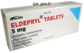
Discount eldepryl 5 mg without a prescription
Cell dying could end result from unintended cell damage or mechanisms that cause cells to self-destruct. It happens when cells are uncovered to an unfavorable bodily or chemical surroundings. Under physiologic conditions, harm to the plasma membrane can also be initiated by viruses, or proteins known as perforins. Today, the term programmed cell demise is applied extra broadly to any type of cell demise mediated by an intracellular death program, irrespective of the set off mechanism. During apoptosis, cells which may be now not needed are eliminated from the organism. This course of might happen during regular embryologic growth or different normal physiologic processes, similar to follicular atresia within the ovaries. Cells can provoke their very own dying by way of activation of an internally encoded suicide program. Apoptosis is characterized by managed autodigestion, which maintains cell membrane integrity; thus, the cell "dies with dignity" with out spilling its contents and damaging its neighbors. As a result of cell injury, injury to the cell membrane leads to an inflow of water and extracellular ions. As a result of the ultimate word breakdown of the plasma membrane, the cytoplasmic contents, together with lysosomal enzymes, are launched into the extracellular space. In apoptosis, the cell is an energetic participant in its own demise ("mobile suicide"). For instance, cell dying mediated by cytotoxic T lymphocytes combines some elements of both necrosis and apoptosis. Nuclear chromatin then aggregates, and the nucleus could divide into a number of discrete fragments bounded by the nuclear envelope. The cytoskeletal elements turn into reorganized in bundles parallel to the cell surface. Loss of mitochondrial perform is caused by changes in the permeability of the mitochondrial membrane channels. The integrity of the mitochondrion is breached, the mitochondrial transmembrane potential drops, and the electron-transport chain is disrupted. Thus, many researchers view mitochondria both as the "headquarters for the chief of a crack suicide squad" or as a "high-security jail for the leaders of a navy coup. These membrane-bounded vesicles originate from the cytoplasmic bleb containing organelles and nuclear material. The removing of apoptotic bodies is so environment friendly that no inflammatory response is elicited. In necrosis (left side), breakdown of the cell membrane leads to an inflow of water and extracellular ions, inflicting the organelles to endure irreversible modifications. Lysosomal enzymes are released into the extracellular area, causing injury to neighboring tissue and an intense inflammatory response. Apoptotic our bodies are later eliminated by phagocytotic cells without inflammatory reactions. Apoptosis can also be inhibited by indicators from different cells and the encircling environment through so-called survival elements. These include progress elements, hormones similar to estrogen and androgens, neutral amino acids, zinc, and interactions with extracellular matrix proteins. However, an important regulatory perform in apoptosis is ascribed to inner signals from the Bcl-2 (B-cell lymphoma 2) family of proteins. Members of this family consist of antiapoptotic and proapoptotic members that determine the life or dying of a cell. The proapoptotic members of the Bcl-2 household of proteins embody Bad (Bcl-2�associated demise promoter), Bax (Bcl-2�associated X protein), Bid (Bcl-2�interacting domain) and Bim (Bcl-2�interacting mediator of cell death). These proteins work together with one another to suppress or propagate their own activity by appearing on the downstream activation of varied executional steps of apoptosis. Note the areas containing condensed heterochromatin adjacent to the nuclear envelope. The heterochromatin in one of the nuclear fragments (left) begins to bud outward through the envelope, initiating a model new round of nuclear fragmentation. Note the reorganization of the cytoplasm and budding of the cytoplasm to produce apoptotic our bodies.
Purchase eldepryl 5 mg mastercard
When the osteoprogenitor cells come in apposition to the remaining calcified cartilage spicules, they turn out to be osteoblasts and start to lay down bone matrix (osteoid) on the spicule framework. The mixture of bone, which is initially solely a thin layer, and the underlying calcified cartilage is described as a combined spicule. Histologically, mixed spicules can be recognized by their staining traits. Calcified cartilage tends to be basophilic, whereas bone is distinctly eosinophilic. Also, calcified cartilage now not accommodates cells, whereas the newly produced bone could reveal osteocytes within the bone matrix. Such spicules persist for a short time before the calcified cartilage part is removed. The remaining bone element of the spicule may continue to develop by appositional growth, thus changing into bigger and stronger, or it could endure resorption as new spicules are fashioned. Growth of Endochondral Bone Endochondral bone progress begins within the second trimester of fetal life and continues into early maturity. As the chondrocytes enlarge, their surrounding cartilage matrix is resorbed, forming thin irregular cartilage plates between the hypertrophic cells. The hypertrophic cells begin to synthesize alkaline phosphatase; concomitantly, the encircling cartilage matrix undergoes calcification the occasions described beforehand represent the early stage of endochondral bone formation that happens within the fetus, starting at in regards to the 12th week of gestation. The continuing progress process that lasts into early adulthood is described within the following part. The process begins with the formation of a cartilage mannequin (1); subsequent, a periosteal (perichondrial) collar of bone forms around the diaphysis (shaft) of the cartilage mannequin (2); then, the cartilaginous matrix in the shaft begins to calcify (3). Blood vessels and connective tissue cells then erode and invade the calcified cartilage (4), making a primitive marrow cavity by which remnant spicules of calcified cartilage remain at the two ends of the cavity. As a primary heart of ossification develops, the endochondral bone is formed on spicules of calcified cartilage. Periosteal bone continues to form (5); the periosteal bone is formed as the outcome of intramembranous ossification. Blood vessels and perivascular cells invade the proximal epiphyseal cartilage (6), and a secondary heart of ossification is established in the proximal epiphysis (7). A comparable epiphyseal (secondary) ossification center types at the distal end of the bone (8), and an epiphyseal cartilage is thus shaped between every epiphysis and the diaphysis. With continued growth of the lengthy bone, the distal epiphyseal cartilage disappears (9), and at last, with cessation of progress, the proximal epiphyseal cartilage disappears (10). During endochondral bone formation, the avascular cartilage is progressively changed by vascularized bone tissue. The zones within the epiphyseal cartilage, starting with calcified cartilage � calcified. The calcified cartilage then serves as an initial scaffold for deposition of new bone. The calcified cartilage right here is in direct contact with the connective tissue of the marrow cavity. In this zone, small blood vessels and accompanying osteoprogenitor cells invade the area previously occupied by the dying chondrocytes. They kind a series of spearheads, leaving the calcified cartilage as longitudinal spicules. In a cross-section, the calcified cartilage appears as a honeycomb due to the absence of the cartilage cells. The invading blood vessels are the supply of osteoprogenitor cells, which can differentiate into osteoblasts, the bone-producing cells. In this Mallory-Azan� stained part, bone has been deposited on calcified cartilage spicules. In the middle of the photomicrograph, the spicules have already grown to create an anastomosing trabecula. The initial trabecula still incorporates remnants of calcified cartilage, as shown by the light-blue staining of the calcified matrix compared with the dark-blue staining of the bone.
Buy discount eldepryl 5 mg on-line
Only one cell type, tall columnar cells, is current in the epithelial layer (see upper right figure). This is said to its absorptive function and is in distinction to the staining of cells that are engaged within the manufacturing of protein. Lastly, with respect to its absorptive function, the epithelial cells regularly exhibit distended intercellular areas at their basal aspect (see upper proper figure arrows). This is a characteristic associated with the transport of fluid throughout the epithelium and, as noted above, commonly seen in intestinal absorptive cells. These are readily apparent in figure on high left and part of the wall of the Rokitansky-Aschoff sinus is shown at higher magnification in figure below. This specimen was taken from a website near the neck of the gallbladder the place mucous glands are often current. Note the attribute flattened nuclei on the base of the cell and the flippantly stained appearance of the cytoplasm, features attribute of mucin-secreting cells. It is a mixed gland containing both an exocrine part and an endocrine part that have distinctive characteristics. The exocrine element is a compound tubuloacinar gland with a branching community of ducts that convey the exocrine secretions to the duodenum. The endocrine part is isolated as highly vascularized islets of epithelioid tissue (islets of Langerhans). The islet cells secrete quite a lot of polypeptide and protein hormones, most notably insulin and glucagon, which regulate glucose metabolism all through the other tissues of the body. Other hormones secreted by islet cells include somatostatin, pancreatic polypeptide, vasoactive intestinal peptide, secretin, motilin, and substance P. All of those substances, aside from insulin, are additionally secreted by the population of enteroendocrine cells within the intestine, the organ from which the pancreas is derived throughout embryonic growth. While insulin and glucagon act primarily in endocrine regulation of distant cells, the opposite hormones (and glucagon) have vital roles in the paracrine regulation of the insulin-secreting B cells of the pancreatic islet. The pancreas is surrounded by a fragile capsule of reasonably dense connective tissue. Also inside the lobule are the small blood vessels and the connective tissue serving as a stroma for the parenchymal parts of the gland. The lumen of the acinus is small, and solely in fortuitous sections by way of an acinus is the lumen included (asterisks). Some acini reveal a centrally positioned cell with cytoplasm that exhibits no particular staining characteristics in H&E�stained paraffin sections. This determine demonstrates particularly well the morphology and relationships of the intercalated ducts. Note, first, the cross-sectioned intralobular duct (InD) consisting of cuboidal epithelium. This is because the plane of part cuts mainly by way of the cells somewhat than the lumen. As a consequence, this determine offers a good view of the nuclei of the duct cells. In addition, they display a staining sample much like that of centroacinar cells and different from that of nuclei of the parenchymal cells. Three principal functions are carried out by this method: air conduction, air filtration, and gasoline trade (respiration). In addition, air passing by way of the larynx is used to produce speech, and air passing over the olfactory mucosa within the nasal cavities carries stimuli for the sense of scent. The respiratory system also participates to a lesser diploma in endocrine functions (hormone manufacturing and secretion), as nicely as regulation of immune responses to inhaled antigens. Lungs develop from the laryngotracheal diverticulum of the foregut endoderm and its surrounding thoracic splanchnic mesenchyme. This preliminary diverticulum grows into the thoracic splanchnic mesenchyme surrounding the foregut. This lung bud divides into the left and proper bronchial buds, which enlarge to type the primordium of the left and proper main bronchi. Bronchial buds along with the encircling thoracic mesenchyme differentiate into lobar bronchi with subsequent progressive divisions into segmental bronchi. Each segmental bronchus with its surrounding mesenchyme additional differentiates and divides to form the bronchopulmonary segments of the lung.
Buy 5mg eldepryl amex
Brown adipocytes are also derived from mesenchymal stem cells but from a different cellular lineage than these differentiating into white adipocytes. Photomicrograph of brown adipose tissue from a new child in an H&E�stained paraffin preparation. This photomicrograph, obtained at a higher magnification, reveals the brown adipose cells with spherical and infrequently centrally located nuclei. For occasion, the conventional lipoma consists of mature white adipocytes, whereas a fibrolipoma has adipocytes surrounded by an excess of fibrous tissue and an angiolipoma incorporates adipocytes separated by an unusually giant variety of vascular channels. The majority of lipomas present structural chromosome aberrations that embrace balanced rearrangements, often involving chromosome 12. Lipomas are often found in subcutaneous tissues in middle-aged and aged people. They are characterized as well-defined, delicate, and painless lots of mature adipocytes normally found in the subcutaneous fascia of the back, thorax, and proximal elements of the upper and lower limbs. They are usually detected in older people and are mainly discovered within the deep adipose tissues of the decrease limbs, stomach, and the shoulder area. Tumors containing more cells in earlier phases of differentiation are more aggressive and more regularly metastasize. This photomicrograph was obtained from a tumor surgically removed from the retroperitoneal space of the stomach. Well-differentiated liposarcoma is characterized by a predominance of mature adipocytes that change in size and form. They are interspersed between broad fibrous septa of connective tissue containing cells (the majority of them are fibroblasts) with atypical hyperchromatic nuclei. A comparatively few scattered spindle cells with hyperchromatic and pleomorphic nuclei are found within connective tissue. Although the time period lipoma relates primarily to white adipose tissue tumors, tumors of brown adipose tissue are additionally found. They are uncommon, benign, and slow-growing gentle tissue tumors of brown fat most commonly arising in the periscapular area, axillary fossa, neck, or mediastinum. Most hibernomas comprise a mixture of white and brown adipose tissue; pure hibernomas are very rare. In contrast to white adipocytes, differentiation of brown adipocytes is beneath the influence of a unique pair of transcription elements. Clinical observations affirm that underneath regular situations, brown adipose tissue can expand in response to elevated blood levels of norepinephrine. This becomes evident in patients with pheochromocytoma, an endocrine tumor of adrenal medulla secreting excessive quantities of epinephrine and norepinephrine. In the past, it was thought that uncoupling proteins have been expressed only in brown adipose tissue. Recently, several comparable uncoupling proteins have been discovered in different tissues. Note that moderate improve of radioactive tracer uptake can also be detectable within the myocardium (yellow color). Regions of in depth metabolic exercise correlate with the distribution pattern of low-density brown adipose tissue. When oxidized, it produces heat to heat the blood flowing by way of the brown fats on arousal from hibernation and within the maintenance of body temperature in the cold. Brown adipose tissue can additionally be present in nonhibernating animals and humans and once more serves as a supply of warmth. Therefore, usually present brown adipose tissue can probably be induced and function in the context of human adaptive thermogenesis. Future research is being directed towards finding mechanisms for increased brown fat differentiation, which may potentially be an the mitochondria in eukaryotic cells produce and retailer vitality as an electrochemical proton gradient throughout the inner mitochondrial membrane. The power produced by the mitochondria is then dissipated as heat in a process generally known as thermogenesis. The metabolic activity of brown adipose tissue is regulated by the sympathetic nerve system and is related to ambient outside temperature. In addition, cold stimulates glucose utilization in brown adipocytes by overexpression of glucose transporters (Glut-4). An enhance within the quantity of brown adipose tissue has been reported on the neck and supraclavicular areas in the course of the winter months, particularly in lean individuals. This is supported by post-mortem findings of larger quantities of brown fats in outside employees exposed to chilly.
Order line eldepryl
Actin-capping proteins block additional addition of actin molecules by binding to the free end of an actin filament. An example is tropomodulin, which can be isolated from skeletal and cardiac muscle cells. Tropomodulin binds to the free end of actin myofilaments, regulating the size of the filaments in a sarcomere. Actin cross-linking proteins are liable for cross-linking actin filaments with one another. Immunofluorescence micrograph of a chick cardiac myocyte stained for actin (green) to show the thin filaments and for tropomodulin (red) to show the situation of the slow-growing ends of the skinny filaments. Tropomodulin seems as common striations due to the uniform lengths and alignment of the thin filaments in sarcomeres. The polarity of the skinny filament is indicated by the fast-growing finish and the slow-growing finish. The troponin complex binds to every tropomyosin molecule every seven actin monomers along the length of the skinny filament. Extensive research have revealed the presence of a wide selection of different nonmuscle myosin isoforms which may be responsible for motor capabilities in many specialized cells, corresponding to melanocytes, kidney and intestinal absorptive cells, nerve progress cones, and internal ear hair cells. As in lamellipodia, these protrusions comprise loose aggregations of 10 to 20 actin filaments organized in the identical direction, once more with their plus ends directed towards the plasma membrane. In listeriosis, an an infection attributable to Listeria monocytogenes, the actin polymerization machinery of the cell could be hijacked by the invading pathogen and utilized for its intracellular motion and dissemination throughout the tissue. Actin polymerization permits micro organism to cross into a neighboring cell by forming protrusions in the host plasma membrane. Intermediate Filaments Intermediate filaments play a supporting or common structural position. These rope-like filaments are called intermediate because their diameter of eight to 10 nm is between these of actin filaments and microtubules. Nearly all intermediate filaments encompass subunits with a molecular weight of about 50 kDa. Some proof means that many of the stable structural proteins in intermediate filaments developed from extremely conserved enzymes, with only minor genetic modification. Intermediate filaments are shaped from nonpolar and extremely variable intermediate filament subunits. Locomotion is achieved by the pressure exerted by actin filaments by polymerization at their growing ends. This mechanism is used in many migrating cells-in particular, on remodeled cells of invasive tumors. As a result of actin polymerization at their vanguard, cells prolong processes from their floor by pushing the plasma membrane forward of the rising actin filaments. The modern extensions of a crawling cell are referred to as lamellipodia; they contain elongating organized bundles of actin filaments with their plus ends directed toward the plasma membrane. These processes can be noticed in many other cells that exhibit small protrusions Unlike these of microfilaments and microtubules, the protein subunits of intermediate filaments present considerable variety and tissue specificity. The long, straight actin filament cores or rootlets (R) extending from the microvilli are cross-linked by a dense community of actin filaments containing quite a few actin-binding proteins. The community of intermediate filaments can be seen beneath the terminal net anchoring the actin filaments of the microvilli. Mechanism of brush border contractility studied by the quick-freeze, deep-etch methodology. Intermediate filaments are assembled from a pair of helical monomers that twist round each other to type coiled-coil dimers. Each tetramer, appearing as an individual unit, is aligned along the axis of the filament. The ends of the tetramers are sure together to type the free ends of the filament. This assembly process offers a stable, staggered, helical array during which filaments are packed collectively and additionally stabilized by lateral binding interactions between adjacent tetramers. Intermediate filaments are a heterogeneous group of cytoskeletal parts present in varied cell varieties. Intermediate filaments are self-assembled from a pair of monomers that twist round one another in parallel trend to type a steady dimer.
Chinese Red Ginseng (Ginseng, Panax). Eldepryl.
- Diabetes.
- How does Ginseng, Panax work?
- Thinking and memory.
- Hot flashes associated with menopause.
- Male impotence (erectile dysfunction).
- Are there any interactions with medications?
Source: http://www.rxlist.com/script/main/art.asp?articlekey=96961
Purchase eldepryl 5 mg online
The astrocyte membrane becomes depolarized, and the cost is dissipated over a big space by the extensive community of astrocyte processes. Instead, every tongue-like process appears to spiral across the axon, all the time staying in proximity to it, till the myelin sheath is shaped. Oligodendrocytes appear in specifically stained light microscopic preparations as small cells with comparatively few processes compared with astrocytes. Each oligodendrocyte provides off a number of tongue-like processes that find their way to the axons, where every course of wraps itself round a portion of an axon, forming an internodal section of myelin. The nucleus-containing area of the oligodendrocyte may be at far from the axons it myelinates. Thus, the place myelin sheaths of adjacent axons contact, they may share an intraperiod line. Cytoplasmic processes from the oligodendrocyte cell physique kind flattened cytoplasmic sheaths that wrap around every of the axons. The relationship of cytoplasm and myelin is essentially the identical as that of Schwann cells. Microglial cells are thought of a half of the mononuclear phagocytotic system (see Folder 6. Recent evidence suggests that microglia play a crucial role in protection in opposition to invading microorganisms and neoplastic cells. They remove bacteria, injured cells, and the debris of cells that endure apoptosis. They also mediate neuroimmune reactions, similar to those occurring in chronic pain circumstances. Photomicrograph of microglial cells (arrows) exhibiting their attribute elongated nuclei. In this condition, the microglial cells are current in giant numbers and are readily visible in a routine H&E preparation. The spikes could be the equivalent of the ruffled border seen on other phagocytotic cells. Ependymal cells type the epithelial-like lining of the ventricles of the brain and spinal canal. The cell body of tanycytes gives rise to an extended course of that initiatives into the mind parenchyma. Tanycytes are delicate to glucose concentration; subsequently, they might be concerned in detecting and responding to adjustments in energy balance as nicely as in monitoring different circulating metabolites within the cerebrospinal fluid. Photomicrograph of the central region of the spinal twine stained with toluidine blue. At greater magnification, ependymal cells, which line the central canal, may be seen to consist of a single layer of columnar cells. Transmission electron micrograph showing a portion of the apical area of two columnar ependymal cells. The modified ependymal cells and related capillaries are called the choroid plexus. Impulse Conduction An action potential is an electrochemical process triggered by impulses carried to the axon hillock after different impulses are acquired on the dendrites or the cell physique itself. In response to a stimulus, voltage-gated Na channels in the initial segment of the axon membrane open, inflicting an influx of Na into the axoplasm. This inflow of Na briefly reverses (depolarizes) the negative membrane potential of the resting membrane (70 mV) to optimistic (30 mV). After depolarization, the voltage-gated Na channels close and voltagegated K channels open. Depolarization of 1 part of the membrane sends electrical current to neighboring parts of unstimulated membrane, which is still positively charged. After a really temporary (refractory) interval, the neuron can repeat the process of producing an motion potential as quickly as once more. Physiologists describe the nerve impulse as "leaping" from node to node along the myelinated axon. However, the voltage reversal can solely happen at the nodes of Ranvier, the place the axolemma lacks a myelin sheath.
Discount eldepryl online visa
The nuclear envelope, fashioned by two membranes with a perinuclear cisternal area between them, separates the nucleoplasm from the cytoplasm. The nuclear lamina is composed of nuclear lamins, a specialised sort of intermediate filaments, and lamin-associated proteins. The G1 section is usually the longest and essentially the most variable section of the cell cycle; it begins at the finish of mitosis (M phase). This phase additionally contains the most important checkpoint within the cell cycle, the restriction level, at which the cell evaluates its personal replicative potential. Mitosis happens within the M section and is managed by the spindle-assembly and chromosome-segregation checkpoints. Passage via the cell cycle is driven by a two-protein complex consisting of cyclin and cyclin-dependent kinase (Cdk). These proteins are synthesized and degraded at common intervals throughout every cycle. Mitosis follows the S section of the cell cycle and incorporates 4 phases: prophase, during which chromosomes condense and become visible, the nuclear envelope disassembles, and the mitotic spindle develops from microtubules; metaphase, which involves the alignment of chromosomes in the equatorial plate; anaphase, during which the sister chromatids start to separate and are pulled to opposite poles of the cell; and telophase, which includes the reconstruction of the nuclear envelope and the division of cytoplasm. During the prophase of meiosis I (reductional division) homologous chromosomes are paired and the recombination of genetic materials occurs between maternal and paternal pairs. Apoptosis happens under regular physiologic circumstances to remove faulty or senescent cells with out inflammatory response of the tissue. At the sunshine microscope stage, the cells and extracellular parts of the assorted organs of the body exhibit a recognizable and infrequently distinctive sample of organization. This organized association reflects the cooperative effort of cells performing a selected function. Therefore, an organized aggregation of cells that operate in a collective manner known as a tissue [Fr. Cells inside tissues are related to each other by specialized anchoring junctions (cell-to-cell attachments, page 98). Cells additionally sense their surrounding extracellular setting and communicate with one another by specialized intercellular junctions (gap junctions, page 98); facilitating this collaborative effort permits the cells to operate as a functional unit. Other mechanisms that allow the cells of a given tissue to perform in a unified method embody specific membrane receptors that generate responses to various stimuli. Despite their disparate structure and physiologic properties, all organs are made up of solely four primary tissue varieties. Connective tissue underlies or supports the other three basic tissues, both structurally and functionally. Nerve tissue receives, transmits, and integrates information from inside and outside the body to control the activities of the body. The tissue concept supplies a foundation for understanding and recognizing the numerous cell varieties inside the body and the way they interrelate. Despite the variations in general look, structural organization, and physiologic properties of the Each basic tissue is outlined by a set of common morphologic traits or practical properties. Each type could additionally be further subdivided based on specific characteristics of its numerous cell populations and any particular extracellular substances which could be present. Another kind of contractile tissue, myoepithelium, capabilities as muscle tissue but is usually designated epithelium due to its location. Rather, college students are suggested to study the options or characteristics of the different cell aggregations that outline the four primary tissues and their subclasses. Epithelial cells, whether or not arranged in a single layer or in Tissues: Concept and Classification a quantity of layers, are always contiguous with each other. The intercellular area between epithelial cells is minimal and devoid of any construction except the place junctional attachments are present. Free surfaces are attribute of the outside of the physique, the outer surface of many inside organs, and the liner of the physique cavities, tubes, and ducts, each those that finally talk with the outside of the physique and those which would possibly be enclosed. The enclosed physique cavities and tubes embrace the pleural, pericardial, and peritoneal cavities as well as the cardiovascular system.
Buy eldepryl canada
The easiest way for many viruses, micro organism, and parasites to efficiently compromise the protective capabilities of the epithelial layer is to destroy the junctional complexes between epithelial cells. Several proteins found in junctional specializations of the cell membrane are affected by molecules produced or expressed by these pathogenic brokers. The oncogenic effect of those interactions is attributed, partly, to the sequestration and degradation of the zonula occludens and the tumor-suppressor proteins associated with the viruses. Bacteria A widespread bacterium that causes meals poisoning, Clostridium perfringens, attacks the zonula occludens junction. This microorganism is widely distributed within the external environment and is found within the intestinal flora of people and many domestic animals. Food poisoning symptoms are characterized by intense stomach ache and diarrhea that begins eight to 22 hours after consuming meals contaminated by these bacteria. Binding to claudins prevents their incorporation into the zonula occludens strands and leads to malfunction and breakdown of the junction. Dehydration that occurs with this sort of food poisoning is a results of a large movement of fluids by way of paracellular pathways into the lumen of the intestines. Helicobacter pylori, another bacterium, resides throughout the abdomen and binds to the extracellular domains of zonula occludens proteins. The loss of the protecting epithelial barrier in the lung exposes the lung to inhaled allergens and initiates an immune response that may lead to extreme asthma assaults. The cytoplasmic domains are linked via quite a lot of intracellular proteins to components of the cell cytoskeleton. Through this interplay, cadherins convey signals that regulate mechanisms of development and cell differentiation. Cadherins control cell-to-cell interactions and take part in cell recognition and embryonic cell migration. E-cadherin, probably the most studied member of this family, maintains the zonula adherens junction between epithelial cells. Integrins are represented by two transmembrane glycoprotein subunits consisting of 15 and 9 chains. This composition allows for the formation of various mixtures of integrin molecules which might be capable of work together with numerous proteins (heterotypic interactions). Integrins interact with extracellular matrix molecules (such as collagens, laminin, and fibronectin) and with actin and intermediate filaments of the cell cytoskeleton. Through these interactions, integrins regulate cell adhesion, management cell motion and shape, and participate in cell growth and differentiation. Selectins are expressed on white blood cells (leukocytes) and endothelial cells and mediate neutrophil� endothelial cell recognition. This heterotypic binding initiates neutrophil migration by way of the endothelium of blood vessels into the extracellular matrix. Selectins are additionally concerned in directing lymphocytes into accumulations of lymphatic tissue (homing procedure). Many molecules involved in immune reactions share a standard precursor element of their construction. However, several other molecules with no identified immunologic operate additionally share this identical repeat component. Together, the genes encoding these related molecules have been outlined because the immunoglobulin gene superfamily. It is likely considered one of the largest gene families in the human genome, and its glycoproteins carry out all kinds of essential biologic capabilities. These proteins play key roles in cell adhesion and differentiation, most cancers and tumor metastasis, angiogenesis (new vessel formation), inflammation, immune responses, and microbial attachment, in addition to many different functions. At these websites, cadherins preserve homotypic interactions with similar proteins from the neighboring cell. They are related to a group of intracellular proteins (catenins) the integrity of epithelial surfaces depends largely on the lateral adhesion of the cells with each other and their capacity to resist separation. Although the zonula occludens entails a fusion of adjoining cell membranes, their resistance to mechanical stress is proscribed. Reinforcement of this area is decided by a powerful bonding website beneath the zonula occludens.
Real Experiences: Customer Reviews on Eldepryl
Ortega, 64 years: The free or apical domain is at all times directed towards the exterior floor or the lumen of an enclosed cavity or tube. Both endothelium and endocardium, as well as mesothelium, are almost always easy squamous epithelia. Glycoproteins of the glycocalyx embrace terminal digestive enzymes similar to dipeptidases and disaccharidases. A large marrow cavity has shaped throughout the cartilage construction and is seen within the center of the micrograph.
Grobock, 22 years: The response usually develops about 15 to 30 minutes from the time of publicity to the antigen (allergen) and may cause a selection of symptoms involving skin (urticaria and eczema), eyes (conjunctivitis), nasal cavities (rhinorrhea, rhinitis), lungs (asthma), and alimentary tract (gastritis). Development of the upper part of the respiratory system containing nasal cavities, paranasal sinuses, nasopharynx, and oropharynx is associated with improvement of the oral cavity. Capillaries in these larger portal canals return the blood to the interlobular veins before they empty into the sinusoid. In older adults, the tunica intima may be expanded by lipid deposits, typically in the type of irregular "fatty streaks.
Copper, 41 years: The asterisk marks an artifact where epithelium separated during specimen preparation. Erythrocyte formation and launch are regulated by erythropoietin, a 34 kDa glycoprotein hormone synthesized and secreted by the kidney in response to decreased blood oxygen concentration. Clearly, the lymphocytes of the nodule are on both sides of the muscularis mucosae and, thus, inside both the mucosa and the submucosa. This photomicrograph reveals a cross-section (on the left) and a longitudinal section (on the right) of creating skeletal muscle fibers in the stage of secondary myotubes.
Nafalem, 47 years: Hard callus is progressively replaced by the motion of osteoclasts and osteoblasts that restores bone to its original form. This diagram shows the gross view of the diaphragmatic and visceral surfaces of the liver, with labeled anatomic landmarks discovered on each surfaces. Hepatic stellate cells (Ito cells) reside in perisinusoidal spaces and are loaded with lipid droplets for storage of vitamin A. Motor Innervation Skeletal muscle fibers are richly innervated by motor neurons that originate within the spinal cord or brainstem.
9 of 10 - Review by O. Rasarus
Votes: 282 votes
Total customer reviews: 282

