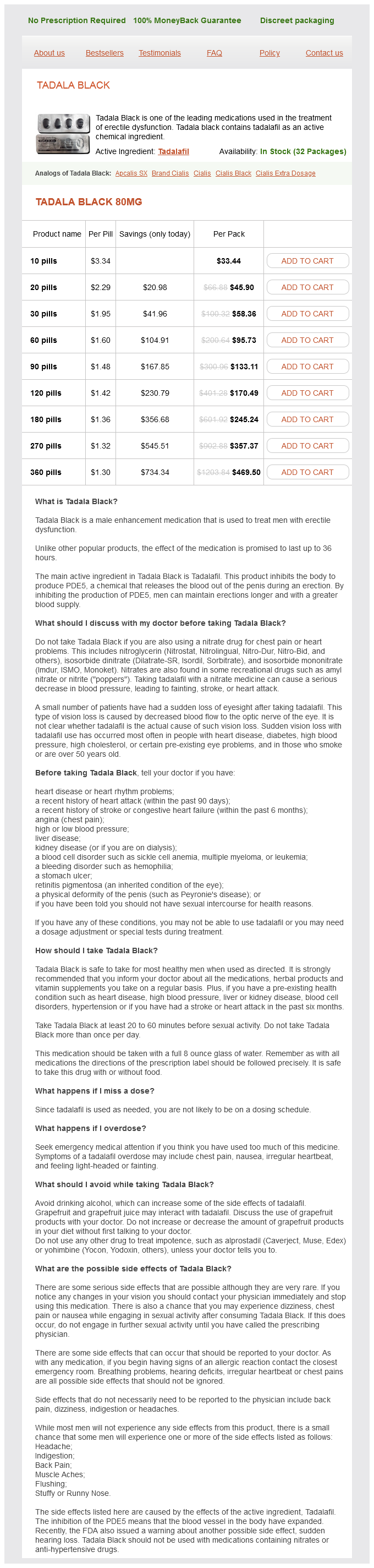
Tadala Black dosages: 80 mg
Tadala Black packs: 10 pills, 20 pills, 30 pills, 60 pills, 90 pills, 120 pills, 180 pills, 270 pills, 360 pills
Buy 80mg tadala black
Sometimes they envelop clusters of the epithelioid cells and ganglion cells, in a way resembling sustentacular cells. Those exterior the ampulla are normally small and lack infiltrative development [97], whereas these on the ampulla have produced clinically obstructive effects [96,98]. Gangliocytic paraganglioma this rare tumour has been the topic of a lot interest due to its uncertain histogenesis, its peculiar histological appearance and its virtually unique location in the periampullary portion of the duodenum. The tumour has been reported in patients aged 17�84 years, with a slight female predominance (1. Gangliocytic paragangliomas are usually solitary, sporadic lesions, though multiple tumours have been recorded [100]. Rare instances of gangliocytic paraganglioma of the jejunum have been reported, one arising in association with pancreatic heterotopia [105]. Macroscopically, gangliocytic paragangliomas are normally polypoid, exophytic lesions that protrude in to the lumen of the intestine. They measure up to 70 mm in diameter and present with haemorrhage, anaemia and/or duodenal or biliary obstruction. Grossly, the tumour is centred on the submucosa and there could additionally be focal ulceration. Amyloid may be present in relation to the epithelioid cells and concentrically laminated psammoma bodies may also be found, particularly if the endocrine cells comprise somatostatin [109]. They are often organized in well-demarcated nests, reminiscent of the Zellballen of traditional paragangliomas. Mitotic figures and necrosis are nearly by no means seen, although a average degree of pleomorphism may be present. The three cellular elements intermingle with the normal smooth muscle and small pancreatic ducts at the ampulla to produce a posh lesion. Two broad theories exist with regard to histogenesis: the primary proposes that they come up because of pancreatic maldevelopment [107]. This is supported by the frequent presence, within the tumour, of misplaced pancreatic tissue and the high incidence of immunoreactivity for pancreatic polypeptide and somatostatin. The second concept suggests a neoplastic lesion [108], with a complex triphasic progress pattern. Rare stories of local lymph node deposits composed of the endocrine cell component, both alone [111] or with sparse spindle cell or ganglion cell components [110], assist this. An equivalent tumour occurs within the cauda equina area [112], the origin of which is tough to explain on the idea of pancreatic maldevelopment. Poorly differentiated endocrine carcinoma/small cell carcinoma Fewer than 30 circumstances of this uncommon and highly aggressive periampullary tumour have been recorded within the literature, most presenting with obstructive jaundice, abdominal ache and weight loss, and followed by a rapidly fatal course [114,115]. The tumours are small (20�30 mm), ulcerated or protuberant lesions which microscopically reveal sheets or nests of small, mitotically lively cells with spherical or oval hyperchromatic nuclei, scanty cytoplasm and foci of necrosis. Using haematoxylin and eosin (H&E) staining, they resemble either small cell carcinomas or giant cell endocrine carcinomas. Malignant lymphoma must be excluded by immunocytochemistry and metastatic tumours by scientific examination and radiology. Localised tumours could additionally be resectable and metastatic tumours may respond to chemotherapy [72,117]. Goblet cell carcinoid A few instances of this rare tumour have been reported throughout the duodenum, both at the ampulla of Vater [118,119] or within the duodenal bulb [120]. Morphologically, these were small tumours that appeared identical histologically to their extra common counterparts in the appendix (see Chapter 30), being composed of small infiltrative nests of cytologically bland goblet cells, signet-ring cells and endocrine cells. By analogy with appendiceal goblet cell carcinoids, their prognosis should be intermediate between those of nicely differentiated endocrine tumours and adenocarcinomas of the duodenum. The neoplasm has an aggressive behaviour and demise normally results from widespread metastases [125]. One study confirmed loss of expression of p14, p15 and p16 in the 9p21 gene cluster [139]. Small tumours are sometimes found by the way submit mortem or at laparotomy whereas bigger neoplasms current with abdominal pain, intestinal obstruction or ischaemia from the local results of the tumour. In addition, these tumours incessantly end in a dense desmoplastic reaction of the mesentery, often at a distance from the primary tumour; this will cause subacute or frank obstruction. Multiple tumours are associated with a younger age of onset, an increased danger of carcinoid syndrome and a poorer prognosis [141].
Syndromes
- Adhesive dyes
- Sunken eyes
- How the condition or defect may be treated during or after the pregnancy
- Spastic arterial disease (arterial contractions brought on by cold or emotion)
- The name of the product or the object you think had lead in it
- Low-fat milk
- Bleeding from the lung tissue
- When did the weight loss begin?
- Fluids through a vein (by IV)
Purchase tadala black no prescription
There is extreme patchy replacement of the muscularis propria by comparatively acellular fibrous tissue. Patchy fibrosis of the muscle coats accounts for the attribute macroscopic appearances of scleroderma affecting the colon. Large, multiple sacculations appear which are confined to the anti-mesenteric border, presumably as a end result of the tissues of the mesenteric facet continue to give help to the weakened bowel wall, or possibly as a result of the blood provide is extra precarious on the anti-mesenteric border. Abnormal colonic motility, especially constipation, is frequent in sufferers with myotonic dystrophy [110] and progressive muscular dystrophy [111], in whom atrophy of the colonic clean muscle fibres without fibrosis could also be found. There could also be a stunning degree of lymphocytic infiltration within the muscularis propria, to not be confused with a myositis. Colonic motor dysfunction additionally occurs rarely in systemic amyloidosis, presumably due to a combination of smooth muscle infiltration, autonomic neuropathy and vascular insufficiency [112]. The inclusion is smooth-surfaced, spherical and stains darkish pink with haematoxylin and eosin. Defects of colonic innervation might produce numerous totally different clinical and pathological effects. This occasionally causes diarrhoea, however way more commonly leads to constipation with colicky stomach ache due to a failure of colonic transit. These inconspicuous our bodies appear to be markers of denervation, quite than particular products of a degenerative myopathy. Neurological disorders that result in chronic colonic pseudo-obstruction may be congenital or acquired. On uncommon events, nevertheless, the analysis can be delayed and so must be thought-about in any adolescent or grownup presenting with chronic constipation and megacolon [89]. Unusually for diseases inflicting colonic pseudo-obstruction, these problems are can be identified on rectal suction biopsy, with out the need for stylish neuropathological techniques. They are 542 Large gut thought-about fully with developmental disorders in Chapter 33. Ganglio-neuromatosis is a diffuse sample of neuronal hyperplasia that can be related to disordered colonic motility with or with out megacolon. In a small variety of individuals with mucosal ganglio-neuromatosis, the diffuse hyperplastic course of entails the lamina propria and so is seen in mucosal biopsy specimens. Mucosal ganglioneuromatosis has additionally been associated with the neoplasia syndromes listed above [117]. Both problems result in abnormal colonic motility accompanied by a spectrum of central and peripheral nervous system abnormalities [118]. Histologically, each varieties are characterised by neuronal loss and eosinophilic intranuclear inclusions inside the neurons of each myenteric and submucosal plexuses. This ends in abnormal layer- ing of the small bowel wall and multinucleate small and enormous bowel myocytes [119]. Detailed neuropathological examination, however, will often reveal the degeneration of intramural neurons and the intraneuronal inclusions. Rare cases of sporadic visceral neuropathy affecting the colon and other elements of the gastrointestinal tract, with out extra-intestinal pathology, have also been described [100]. Such cases seem to be degenerative problems of the intestinal plexuses however their exact aetiology is unknown. Acquired issues of visceral innervation resulting in continual colonic pseudo-obstruction can have a quantity of completely different aetiologies. It is estimated that roughly seven million individuals, almost all in South America, are affected. The parasite has a particular affinity for the myenteric plexus (see Chapter 4) and causes a distinguished persistent inflammatory response comprising lymphocytes and plasma cells. The consequence of this inflammatory response is destruction of the myenteric plexus with marked neuronal loss and Schwann cell hyperplasia. Smooth muscle hypertrophy ensuing from denervation is frequent, as is megacolon [120,121]. A related inflammatory destruction of the myenteric plexus has been described as a consequence of cytomegalovirus an infection [122] and Epstein�Barr virus an infection [123]. The condition was idiopathic on this man aged 28, who had progressive constipation. Neuromuscular and mechanical issues of the massive gut 543 because of an immunological reaction elicited by antigens on the neoplastic cells that cross-react with neural tissue within the myenteric plexus. Similar lesions within the posterior root ganglia have been found in other para-neoplastic neurological syndromes.
Order tadala black 80mg on-line
A giant proportion has been from Australia and New Zealand [155,170], the latter nearly completely of Celtic origin [163]. Tumours first recognized in colectomy specimens and through surveillance may be very small with appearances similar to non-cancerous polyps [161,178]. Adenoma formation normally begins in adolescence, initially within the rectum after which spreading proximally to affect all areas of the colon. The early adenomas are tiny, forming from monocryptal adenomas or aberrant crypt foci that can be detected by chromoendoscopy. The use of dye spray helps to enumerate the number of adenomas present extra precisely [187]. Adenocarcinomas start to develop 10 years or so after the appearance of the adenomas and most, 70�80%, occur within the left colon [189] although any a half of the colon could also be involved. A small group of patients has flat adenomas, that are raised only slightly above the mucosal surface and have a height of lower than two occasions the conventional mucosa [191�193]. Although most of those instances have an attenuated phenotype, with fewer than 100 polyps and a right-sided predominance [194], instances with >100 adenomas are described. Flat adenomas can also be seen in the rectal stump after colectomy with ileo-rectal anastomosis [195]. Similarly, flat/depressed adenocarcinoma has been described in a rectal stump [196]. By definition, affected patients have greater than one hundred adenomas [185], however there are often many more. In others, including these with new mutations, presenting complaints embrace rectal bleeding and mucous discharge [185]. Gastric adenomas of traditional intestinal kind do occur however are uncommon, and probably account for the rare cases of gastric adenocarcinoma seen in about 1% of sufferers [203]. Fundic gland polyps of abdomen these occur in up to 60% of sufferers and may precede the appearance of adenomas within the colon [80]. They carefully resemble sporadic fundic gland polyps which might be generally seen now in the context of proton pump inhibitor remedy Duodenal, ampullary, small intestinal, adenomas Adenomas within the small intestine occur one to 20 years after the colonic polyps in as much as 90% of patients [204] and are most quite a few in the duodenum and peri-ampullary region. Adenomas can improve in dimension, number and severity of dysplasia however the risk of malignant development is far decrease than within the colon, with a 5% danger of development to adenocarcinoma over 10 years [204]. The Spigelman stage assesses quantity, villosity and diploma of dysplasia of the duodenal adenomas to predict if and when surgery could also be necessary [206]. This is usually when there are >20 adenomas, with tubulovillous adenomas and high grade dysplasia. Adenomas more distally within the small 666 Large gut gut are less widespread but are notably susceptible to develop within the ileal pouch [208]. Fibromatosis (desmoid tumour) Occurring in about 15% of sufferers [80], fibromatoses are principally seen in the stomach wall, mesentery and retroperitoneum, with occasional instances at other websites such as the pancreas. Many circumstances comply with surgical procedure or trauma, including colectomy, with lesions growing progressively before presenting on average 2�4 years after the initiating insult [199,209]. They kind rounded, infiltrative plenty of scarlike collagen with uniform fibroblastic cells, however smaller two-dimensional plaques are also seen [209]. Dental abnormalities embrace supernumerary teeth, dentigerous cysts and fused roots [199]. Osteomas, of the cranium and mandible particularly, lipomas, epidermal cysts and nasopharyngeal angiofibromas have been described [80,203]. Endocrine tumours There is an elevated danger of papillary carcinoma of the thyroid in younger ladies � round 160 occasions normal [155]. Liver and biliary tree Apart from peri-ampullary adenomas, involvement of the liver and biliary tree is rare. Lesions described in the bile ducts embody adenoma [214] and adenocarcinoma [214,215], in addition to dysplasia of the gallbladder epithelium [216], however all of these lesions are uncommon. With normal function as a part of a posh of proteins, it binds -catenin, downregulating its activity.
Best order for tadala black
A diffuse and/or nodular infiltrate of neoplastic lymphoid B cells cytologically resembling marginal zone B cells and exhibiting a marginal zone distribution 2. There is marked cytological variation, both inside a given case and between cases. Most neoplastic cells are small lymphoid cells with irregular nuclei and scant cytoplasm (centrocyte-like cells). Some show options of medium-sized monocytoid cells, with abundant pale cytoplasm, well-defined cell membranes and round-to-oval, smoothly contoured nuclei. The neoplastic infiltrate additionally often incorporates scattered, large, transformed blastic cells resembling centroblasts or immunoblasts. The neoplastic plasma cells could appear as normal mature plasma cells, have crystalline inclusions in the cytoplasm and possess nuclei with Dutcher our bodies. In some circumstances, intracytoplasmic immunoglobulin may be so abundant that the nucleus is pushed to one side and the cell resembles the signet-ring cells of diffuse-type adenocarcinoma [41]. Eventually, the lymphoma may utterly overrun and destroy the lymphoid follicles, resulting in a nodular sample and, generally, a resemblance to follicular lymphoma. Within the germinal centres, the neoplastic cells could undergo marked plasma cell differentiation or show blast cell transformation. Regional lymph node involvement may be refined, because the lymphoma cells initially localise to the marginal zones without architectural distortion. Further involvement is characterised by marginal zone cells spreading to the interfollicular areas and producing confluent sheets of lymphoma cells. Follicular colonisation may also be seen in lymph nodes and mimic follicular lymphoma [35]. The cells with plasma cell differentiation preferentially distribute in the higher mucosa (a) and incessantly include nuclear inclusions (Dutcher bodies) (b). There is monotypic gentle chain expression in both the lymphoid and plasma cell elements (c, d); and, immunoperoxidase. Bone marrow involvement is infrequent (10� 20% of cases) and occurs as inter-trabecular aggregates of neoplastic B cells [27]. Monotypic immunoglobulin with light chain restriction is expressed on the membrane and, to a lesser extent, within the cytoplasm, according to the diploma of plasmacytic differentiation. Whereas reactive germinal centres are usually associated with tight, dense, follicular, dendritic cell meshworks, neoplastic follicular colonisation could additionally be related to massive expanded irregular aggregates of follicular dendritic cells. An IgD stain may be helpful for demonstrating IgD+ mantle zones and delineating IgD- marginal zones. The gastric crypts and glands are infiltrated and disrupted by lymphoid neoplastic cells which type small aggregates throughout the epithelium (a). Ig heavy and light-weight chains are rearranged and present somatic hypermutation of their variable areas, according to derivation from a post-germinal centre memory B cell [47,48]. Some research have shown a lower incidence of this association however the density and detectability of H. The price of ongoing mutations appears to decline over the progression of the disease, implying that the position of direct antigen stimulation might lower throughout tumour evolution [34]. When current, this translocation is nearly always the one cytogenetic abnormality, in contrast to t(11;18)negative tumours, which frequently show different alterations such as trisomies or allelic imbalances [65,66]. The t(11;18) translocation is significantly associated with cytotoxin-associated antigen A (cagA)-positive strains of H. Other genetic and molecular alterations Chromosomal numerical aberrations similar to trisomies 3 and 18 are regularly seen in t(11;18)-negative tumours and people carrying t(1;14) [65,76]. Longstanding extreme an infection results in continual energetic gastritis and the production of lymphoid follicles with germinal centres. Nevertheless, some instances remain ambiguous, even with stains for B- and T-cell markers (see Immunophenotype above). At presentation, most circumstances are restricted to the gastric wall, predominantly involving the mucosa and submucosa, however may unfold in to the muscularis propria and, less commonly, in to the subserosa. However, current research using detailed and comprehensive staging protocols have revealed each local development and systemic dissemination more regularly than originally believed. Although there could additionally be a predominant lesion, quite a few clonally equivalent, remote, microscopic foci often may be demonstrated even in macroscopically regular regions [38,82]. Before contemplating massive cell transformation, the possibility that the massive cells represent residual reactive germinal centre cells or giant neoplastic cells confined to colonised follicles have to be excluded.
Order tadala black online now
Lesions are segregated in to low-risk and high-risk groups depending on the presence of unfavourable histological options in the carcinomatous component. Poorly differentiated adenocarcinoma Polyps and tumour-like lesions of the big gut 655 Table 37. As a results of this some have really helpful re-excision of the polypectomy site before making a ultimate determination on definitive colectomy if no different opposed options are current [69], though most centres would progress to colectomy if the margin is reported as optimistic. Different definitions of an enough clear resection margin have been used in the literature. Many reports merely required the adenocarcinoma to be unequivocally clear of the margin and the adjacent diathermied space [57,62,70], and that is the minimal needed in clinical apply. Other authors have regarded sufficient clearance as 1 mm [58] or 2 mm [61] from the margin, though lesions in the latter research with margins between 0 and 2 mm have been related to different unfavourable histology in the majority (64%) of instances. As has been pointed out [67], early studies of all colorectal adenocarcinomas confined to the submucosa found that less than 5% had nodal metastases and most of those have been poorly differentiated [68]. Lymphovascular invasion this characteristic is extra controversial as an unbiased antagonistic histological feature in malignant polyps [65,66] because of its frequent incidence with other changes similar to poorly differentiated tumours, but some research have discovered it to be essential [57,fifty eight,70]. As it could be troublesome to distinguish from retraction artefact, its identification has been shown to have only fair to substantial settlement between pathologists [58]. Immunohistochemical staining of lymphatic endothelium with the monoclonal antibody D2-40 may be of use, but usually small foci minimize out rapidly, thus limiting its usefulness. Sessile polyps, due to the absence of a stalk, are all degree four, but these Positive polypectomy resection margin A positive margin is predictive of a higher danger of residual carcinoma at the polyp site [65]. In addition, more recent data counsel that different options may determine the high-risk group. They also instructed that absolutely the dimensions of adenocarcinoma within the submucosa provide an goal measure of threat. Submucosal carcinoma measuring <4 mm across or <2 mm in depth from the muscularis mucosae had a low frequency of opposed outcome [70]. Another study advised that nodal metastasis may occur when the carcinomatous part had a submucosal tumour depth (measured from the muscularis mucosae) of 1. These lymphatics can generally be readily seen in routine follow if a carcinoma has very intensive lymphatic invasion. In a recent examine of 15 sufferers with adenomas containing purely intramucosal, poorly differentiated adenocarcinoma, no metastatic illness was recognized from these lesions [74]. However, subsequent correspondence described two similar circumstances from the Memorial Sloan Kettering Cancer Centre. One developed distant metastases despite colectomy after the initial polypectomy, and the second confirmed invasion of lymphatic vessels within the deep mucosa, with none submucosal invasion [75]. This suggests that poorly differentiated adenocarcinoma ought to immediate consideration of colectomy, even when confined to the mucosa. The frequency of this appears to be rising [74] and further research are wanted. Loss of the practical protein, which is a unfavorable regulator of the Wnt signalling pathway, allows the buildup and nuclear translocation of -catenin, resulting within the elevated expression of genes affecting proliferation, differentiation and mobile migration [80]. Conventional adenomas start as monocryptal adenomas and up to date research counsel that mutation within the crypt stem cell is necessary to give rise to a growing, dysplastic lesion [27]. Flat adenomas seem to have a unique pattern of mutation, which may clarify their uncommon look. Pathogenesis of standard adenoma There is a full discussion of colorectal carcinogenesis in Chapter 38 and a short overview only is given here. In most instances, colorectal carcinoma begins with the initiation and development of a colonic adenoma through a sequence of genetic alterations affecting genes which may be related to key intracellular pathways [76,77]. They represent approximately 25% of endoscopically eliminated polyps but 70�85% of all serrated polyps (Table 37. Histologically serration is limited to the higher third to half of the crypt, varying from lesion to lesion.
Polygonum tataricum (Buckwheat). Tadala Black.
- What is Buckwheat?
- Are there safety concerns?
- Improving blood flow, varicose veins and poor blood circulation in the legs, diabetes, preventing hardening of the arteries, and other conditions.
- Dosing considerations for Buckwheat.
- How does Buckwheat work?
Source: http://www.rxlist.com/script/main/art.asp?articlekey=96065
Purchase tadala black 80mg amex
Patients can present with nausea, early satiety, bloating and epigastric pain [469]. Biopsies show epithelial ulceration and a marked growth of the lamina propria with a diffuse lymphoid infiltrate, which may simulate major gastric lymphoma. Histological findings embody acute and chronic inflammation with regenerative epithelial hyperplasia, areas of necrosis and ulceration with fibrinopurulent exudates. Herpetic inclusions are rarely detected in routine staining within the gastric mucosa but could be demonstrated by tissue tradition of gastric biopsies or immunohistochemistry. Gastric mucosal granulomas, reportedly, are found rather more regularly in the antrum than within the corpus. However, in widespread with all granulomatous conditions, sampling error limits the biopsy prognosis, as a result of granulomas may be scarce. As the underlying aetiology differs geographically all through the world, the incidence of granulomatous gastritis varies from zero. However, that appears to not be the case in North America and Europe, and many knowledgeable employees question this association, especially as few research have proven full or partial remission after H. Obvious stenosis or ulcerations should prompt a diagnosis; nonetheless, aphthous ulcerations are recognised as reappearing sporadically. The North American Inflammatory Bowel Disease Genetics Consortium arbitrarily permits as much as 10 minor lesions with out indicating vital disease in a given location. Notably, there may be clinical and pathological proof of coexisting disease within the duodenum or the small or massive intestine [476,477]. Macroscopically, there can be cobblestoning of the mucosa with thickening of the pyloric region, but, mostly, endoscopy shows erosions and redness of the mucosa. Although these focal lesions incessantly occur deep within the mucosa, they could be associated with erosions or degenerative changes within the overlying surface epithelium [475,479]. Sarcoidosis Involvement of the abdomen by sarcoidosis represents between 1 and 21% of all circumstances of granulomatous gastritis [471,473,480�482]. Yet involvement of the gut is rare in sarcoidosis, as an entire, even when the stomach is probably the most commonly affected site [482,483]. Endoscopy is usually normal however it might show adjustments starting from a distal gastritis with or without nodularity, mainly affecting the larger curvature, to ulceration and pyloric stenosis. Gastric sarcoidosis ought to never be diagnosed within the absence of proof of the illness in other sites, after which solely when overt aetiologies have been satisfactorily excluded. Elevated angiotensinconverting enzyme levels are useful in establishing the prognosis. Those most vulnerable are the city poor, a lot of whom are immigrants from endemic areas and/or elderly, and severely immunocompromised sufferers. Symptoms embrace epigastric pain, vomiting and haematemesis, and there could additionally be proof of pyloric obstruction [486�489]. At endoscopy, ulceration of the pyloric region is a common manifestation, often on the lesser curvature. The granulomas may be confluent and, although sometimes caseating, that is by no means all the time the rule [486�489]. Regional lymph nodes are frequently enlarged and ought to be carefully examined, as a outcome of they frequently comprise granulomas, which may not be readily obvious in the gastric wall itself. Demonstration of acid- and alcohol-fast bacilli is pathognomonic and may at all times be tried, though frequently unsuccessful. Microscopically, the lamina propria is expanded by numerous foamy histiocytes and ill-formed granulomas without necrosis. It could present with diffuse enlargement of the gastric folds and multiple shallow ulcers or erosions [491,492]. Even although, initially, gastric syphilis had been seen as a com- Inflammatory disorders of the stomach 145 plication of tertiary syphilis, subsequent reports have indicated that the abdomen is affected in early syphilis [493]. Of affected sufferers, solely 13% have a prior historical past of a syphilis diagnosis and 46% had prior (13%) or concurrent (33%) clinical manifestations of the illness [493]. The histological appearances of the gastric mucosa in secondary syphilis are protean. Ill-formed granulomas could also be present however the traditional discovering is a dense intramucosal infiltrate of plasma cells, neutrophils and lymphocytes, usually with lymphoid follicle formation. There may be destruction of mucosal glands and lympho-epithelial-like lesions, more often related to primary malignant lymphoma. The endarteritis typically seen in syphilitic lesions elsewhere may not be present in gastric lesions, particularly in biopsies, and the gummas that happen in the tertiary stage or in congenital syphilis [494] are seldom seen in the adult abdomen.
Purchase 80 mg tadala black free shipping
The mucosal surface is haemorrhagic with superficial pinpoint ulcers or deeper longitudinal and serpiginous ulceration. Membranous or pseudo-membranous enterocolitis has been used to describe this look of ischaemic mucosal necrosis prior to now however the time period is now utilized to the focal raised creamy-yellow plaques which are seen in the colon as a outcome of C. The distribution and kind of lesion in the small gut and proximal colon will depend upon the trigger of the ischaemia. If it is as a result of of a vascular obstruction, the pathological modifications may have a uniform look and segmental distribution that replicate the blood supply of that vessel. In non-occlusive intestinal ischaemia, nonetheless, the lesions are sometimes patchy and widespread, and range of their severity [3]. It is common to see fibrin thrombi inside the blood vessels of each the mucosa and the submucosa. In non-occlusive ischaemia, a significant variety of patients may also have thrombi in other organs. This sample may be a manifestation of disseminated intravascular coagulation centred on the gut. A seek for minute ischaemic lesions in the mucosa must be made during which the only options are capillary congestion, necrosis of a few crypts and erosions of the surface epithelium. With rising severity of ischaemia, the deeper layers of the bowel wall turn into affected. There should all the time be a careful inspection of the muscularis propria even when the obvious adjustments are mucosal and submucosal because, in the early phases of acute deprivation of blood, the muscle layers show only poor staining with lack of nuclei and little different abnormality. Later, lysis of muscle cells is extra obvious, with separation and thinning of fibres by oedema, and the start of an inflammatory infiltrate. Infarction is manifested by haemorrhage in to the bowel wall, significantly the submucosa, with intravascular thrombosis and mucosal ulceration. In instances of intestinal gangrene, widespread necrosis is obvious and accompanied by secondary an infection, which frequently extends via the bowel wall to involve mesenteric tissues and engulf the mesenteric vessels. This is extra often a secondary rather than a primary cause of ischaemic bowel disease, so it is extremely necessary for the pathologist to recognise this superficial vessel thrombosis as a secondary phenomenon and not necessarily the reason for the ischaemic pathology. In reality, in anyone case, the histology of the affected intestine wall only exceptionally reveals the primary cause(s) of the intestinal ischaemia. More usually the underlying analysis is determined by consideration of the scientific historical past and assessment of larger mesenteric blood vessels. It is important to exclude the assorted kinds of primary vasculitis that can lead to ischaemic bowel illness (see below). An opinion on whether the vascular modifications are the outcome of venous or arterial occlusion may be useful but this is possible only if the mesenteric vasculature has been rigorously examined. All the features of ischaemic necrosis, nonetheless severe, can go through a strategy of resolution and repair. We have observed, in a quantity of cases, appreciable levels of mesenteric fats necrosis accompanying major vessel occlusion. One especial feature that helps a pathologist to differentiate continual ischaemia of the small gut, in all its types, from different pathological causes is the dissolution of the muscularis propria and alternative by fibrous connective tissue, which happens in persistent ischaemia. This could present a potent clue to the true explanation for a chronic fibrous stricture and provides proof that the muscularis propria may be severely affected by ischaemic pathology of the small intestine. Ischaemic strictures and ulcers Ischaemic strictures happen in the small bowel or colon. The major causes are small mesenteric vessel emboli, trauma and the sequelae of herniae or bands. Occasionally they present with a malabsorption syndrome (see Chapter 21) but more usually they produce signs and symptoms of intestinal obstruction, usually subacute. It is probable that the episodes of acute ischaemia ultimately giving rise to a stricture are subclinical. They can be produced by specific causes of peripheral arteritis such as polyarteritis nodosa. These are normally apparent on histological examination though there could also be problem recognising burned-out circumstances of arteritis. Volvulus of the small intestine and obstruction of the blood supply due to inside hernias are also potential causes. Ischaemic strictures are usually concentric, with the serosal floor typically being whiter than normal due to serosal fibrosis. The mucosal floor could additionally be atrophic or show one or more, small, clearly demarcated ulcers.
Order tadala black with paypal
Evidence for true colonic metaplasia because of the long-term presence of colonic contents is at present lacking. It is often associated with severe pancolonic ulcerative colitis and the colonic problems of the disease are normally much more important than those of the terminal ileum. It stays controversial whether the presence of backwash ileitis predicts the subsequent onset of pouchitis in the pelvic ileal reservoir after proctocolectomy [464]. Given the present theories that the illness is a secondary phenomenon, it might seem extra probably that the severity of the colonic illness (which itself determines the presence or absence of backwash ileitis) may predict subsequent pouchitis rather than the presence of backwash ileitis. However, the jejunal mucosa has been shown to harbour persistent irritation in ulcerative colitis sufferers and there may also be architectural disturbance [465]. Severe and sometimes fatal panenteritis can be described in ulcerative colitis [466] and diffuse duodenitis is a uncommon complication [467]. Both earlier than and after colectomy, there could additionally be varying levels of continual irritation and villous architectural disturbance in the duodenal mucosa. Ileorectal anastomosis Until about 25 years in the past, there was a vogue for ileorectal anastomosis in ulcerative colitis before it was appreciated that the rectum was an essential web site for carcinoma development in ulcerative colitis and before the advent of pelvic pouch surgery. The ileal mucosa proximal to an ileorectal anastomosis reveals florid adjustments on biopsy not unlike these of backwash ileitis. There is villous architectural change, active irritation, and occasional erosions and ulceration [469]. In this situation, it could be difficult, on biopsy, to differentiate between ileal and rectal mucosa, each exhibiting architectural distortion and the long-term results of continual ulcerative colitis. It is characterised by profuse watery ileostomy discharge with marked systemic signs. The macroscopic options are of deep linear ulceration with normal intervening mucosa. The condition is Post-colectomy small intestinal pathology in ulcerative colitis Although pelvic ileal reservoir pathology is the most clinically important entity in this class (see above), pathological adjustments are seen within the small intestinal mucosa as a result of colectomy and proctocolectomy for ulcerative colitis. Despite its associations with ulcerative colitis, its pathogenesis stays unclear. Furthermore minor degrees of obstruction are commonplace and these could induce extra pronounced inflammation, villous atrophy and ulceration. Polypoid mucosal prolapse at the site of an ileostomy can lead to enteritis cystica profunda and especial mimicry of malignancy. How important the inflammatory pathology of the stomal mucosa is in carcinogenesis is uncertain [471]. Ileal conduits are urinary reservoirs constructed after bladder resection for malignancy. Varying levels of mucosal abnormality are described, together with villous architectural changes and mucin depletion [472]. There is proof of some elevated malignancy threat, transitional cell carcinomas on the uretero-ileal junction being most commonly described. As in the colon, there seems to be a vascular factor to the pathogenesis with mural hypoperfusion and secondary localised ischaemia apparently accounting for relatively sharply localised ischaemic-type inflammation and ulceration, usually adjoining to comparatively regular appearing mucosa. Obstructing endocrine tumours of the small gut could be related to proximal ulceration and granulation tissue polyposis, presumably a consequence of obstruction and relative ischaemia because of compromise of blood supply [475]. The latter is putatively induced by a combination of the mechanical effects of the obstructing tumour and vascular spread of the tumour. Non-specific jejunitis Isolated jejunitis often has relatively non-specific histological options but it occurs in several particular situations. Severe necrotising jejunitis is an unusual situation that happens in kids; its potential causes are many but typically no trigger Obstructive ileitis Obstructive ileitis is now nicely recognised to have attribute macroscopic and histopathological features [473], but the condition is relatively poorly documented in the 360 Small gut is found [479]. The illness reveals similarities to enteritis necroticans (see above) and may symbolize a bacterial overgrowth pathology. Corrosive jejunitis due to the ingestion of acids might occur however that is solely likely after earlier gastroenterostomy permits the undiluted acid to enter the jejunum [480].
Generic tadala black 80 mg on-line
Interestingly, sildenafil was originally developed as a potential drug for treating angina; nonetheless, it was not very efficient for this purpose and was subsequently developed for remedy of erectile dysfunction, where it produces vasodilation and filling of the corpus cavernosum. Natural Products Natural merchandise include naturally occurring catecholamines, hormones, and cytokines, as properly as animal and plant toxins. The publicity ranges of these chemicals examined in vitro in general are much greater than the focus reached in cardiac tissue underneath in vivo exposure circumstances. Therefore, extrapolation Steroids and Related Hormones Estrogens, progestins, androgens, and adrenocortical steroids are major steroid hormones produced by mammals including humans. Myocardial tissue accommodates steroid receptors; therefore, the heart serves as a goal organ for steroid effects. It also has been shown that cardiac tissue can synthesize steroid hormones, although the capability for synthesis could additionally be much lower than more classic steroid synthesizing tissues. There are two major mechanisms of motion of the hormones: the first is to alter gene expression and the second is to change signaling transduction pathways. Estrogens Estrogens are synthesized in ovaries, testes, and adrenal glands, and estrogen is an active metabolite of testosterone. Synthetic estrogens include diethylstilbestrol (nonsteroidal), equilin, esterified versions of E2, ethinyl estradiol, mestranol, and quinestrol. Estrogens (frequently together with progestins) have been used for over 40 years as oral contraceptive drugs. Progestins Progestins are additionally synthesized in the ovaries, testes, and adrenal glands. Naturally occurring and artificial progestins embody desogestrel, hydroxyprogesterone, medroxyprogesterone, norethindrone, norethynodrel, norgestimate, norgestrel, and progesterone. As a part of hormone alternative remedy, progestins serve an opposing role to estrogens. Unfortunately, estrogen treatment opposed with progestins may negate the cardiovascular benefits of estrogens on lipid metabolism (Kalin and Zumoff, 1990). Although progestins might exert deleterious effects on the heart, extra studies are required to examine mechanisms. Androgens Androgens cause opposed cardiovascular effects (Rockhold, 1993; Melchert and Welder, 1995). The principal androgens are testosterone and its energetic metabolite dihydrotestosterone. Testosterone is synthesized within the testes, ovaries, and adrenal glands, and dihydrotestosterone mediates most androgen actions. Synthetic anabolic-androgenic steroids embody the alkylated and orally out there drugs danazol, fluoxymesterone, methandrostenolone, methenolone, methyltestosterone, oxandrolone, oxymetholone, and stanozolol. The nonalkylated medication with poor oral bioavailability embody androstenedione and dehydroepiandrosterone (both bought in varied "nutraceutical" formulations), boldenone (veterinary product), nandrolone (19-nortestosterone), and testosterone. Nearly all of these chemical compounds have received significant illicit use, notably in extremely excessive doses with makes an attempt to enhance physical appearance or performance. Evidence indicating the direct cardiac toxic impact of anabolic-androgenic steroids includes alteration of Ca2+ fluxes in cardiac myocytes induced by testosterone (Koenig et al. In people, high-dose anabolic-androgenic steroid use has been associated with cardiac hypertrophy and myocardial infarction. However, the mechanisms responsible for the cardiotoxic results of anabolic-androgenic steroids remain poorly understood. Glucocorticoids and Mineralocorticoids Glucocorticoids and mineralocorticoids are primarily synthesized within the adrenal glands. Naturally occurring glucocorticoids embody corticosterone, cortisone, and hydrocortisone (cortisol), and the mineralocorticoid is aldosterone. A massive variety of synthetic glucocorticoids are used for therapy of varied autoimmune and inflammatory illnesses. These medication embrace alclometasone, amcinonide, beclomethasone, betamethasone, clobetasol, desonide, desoximetasone, dexamethasone, diflorasone, fludrocortisone, flunisolide, fluocinolone, fluocinonide, fluorometholone, flurandrenolide, halcinonide, medrysone, methylprednisolone, mometasone, paramethasone, prednisolone, prednisone, and triamcinolone. Most of these drugs are primarily used topically, intranasally, or inhalationally. The major glucocorticoids used systemically embody cortisone, hydrocortisone, dexamethasone, methylprednisolone, prednisolone, and prednisone.
Discount tadala black 80mg amex
Shigellosis, salmonellosis and campylobacter an infection Bacillary dysentery attributable to shigellosis happens throughout the world. It afflicts about 20 million folks every year and kills about three-quarters of 1,000,000 folks every year [49]. Entry in to the cells is adopted by intracellular multiplication and lysis of the phagosomes. The bacteria are propelled by way of the cytoplasm, the host cell is killed and bacteria are unfold to neighbouring epithelial cells. The process results from the interaction between bacterial proteins IpaA, IpaB and IpaC with cell cytoskeletal components. The bacterial protein IpaB plays an important function in irritation by directly binding to interleukin-1-converting enzyme. Death and sloughing of epithelial cells end in erosions which can extend in to the lamina propria, related to an inflammatory infiltrate which will prolong in to the submucosa. There is evidence that inflammation will increase the pathology of the lesion however decreases the unfold of Shigella spp. Evidence of an infection starts, clinically, within the small bowel with fever and secretory diarrhoea. In the subsequent few days, bacteria localise to the colon, inflicting a colitis with ulceration and erosions. Macroscopic and microscopic damage are most evident within the distal giant bowel, changing into progressively milder within the transverse colon. Shigellosis is particularly prevalent in children, particularly under-nourished children, and the disease exacerbates under-nourishment by its propensity to enteric protein loss [51]. As a results of the predominant colonic involvement, demonstration of the disease in small bowel biopsies is unusual. Enteritis is most commonly attributable to Salmonella enteritidis and its serotypes (typhimurium, heidelberg and newport). Salmonellae trigger an acute gastroenteritis, ileo-colitis or colitis, which is usually short-lived. Seldom is histopathological evaluation required, besides in longer duration ente- rocolitis [52]. In severe circumstances, the adjustments of acute colitis may be prominent whereas the small bowel mucosa exhibits more delicate signs [53]. Entry of cells is adopted by interference with normal mechanisms to create a phagolysosome engulfing the salmonellae and alteration of the immune response to favour bacterial survival. SipA interacts with actin and is essential for cytoskeletal changes, SipC interferes with regular intracellular trafficking and SipB promotes chloride secretion. Infection occurs in all age teams and leads to a syndrome of acute abdominal pain, fever and inflammatory enterocolitis, which ranges from a generally delicate secretory diarrhoea to a severe dysentery-like syndrome. The variable medical picture might be more as a outcome of the host immune response than to strain virulence factors. Campylobacter jejuni causes local acute inflammation in both the small and the massive intestine [55]. The enteric fevers: typhoid and paratyphoid Typhoid fever, caused by Salmonella enteritidis serovar typhi, stays a severe well being drawback throughout the world, especially in south and south-east Asia and South America, with an estimated 16�33 million instances and about half 1,000,000 deaths yearly. This fall and the good improvement in its prognosis are due to improved living situations and sanitation and the introduction of effective antibiotic remedy. The incidence is estimated to be 3�30 instances per a hundred 000 travellers to growing countries [57]. The illness is characterised by a sudden onset of fever, headache, nausea, loss of urge for food, abdominal discomfort and constipation or diarrhoea in young youngsters. After multiplication in the bloodstream, the organisms are excreted by the liver in to the bile, and enter the gallbladder and intestinal contents in quickly increasing numbers; they can be cultured from the faeces in an growing proportion of cases, the best incidence of positive stool cultures occurring within the third week of the sickness. The base of the ulcer incorporates black necrotic material: healing begins with shedding of the slough. This accounts for the low frequency of strictures on account of previous typhoid fever. Local lymph nodes are significantly enlarged, soft and hyperaemic on the height of the disease. Histologically, enteric fever reveals hyperaemia and oedema in the early phases with proliferation of enormous, usually deeply staining mononuclear cells, which are modified phagocytes (containing intracellular organisms) [60].
Real Experiences: Customer Reviews on Tadala Black
Amul, 23 years: It is, however, additionally discovered elsewhere and has been described in a wellnourished white vegetarian [70]. In the lung, endotoxins produce increased vascular permeability and pulmonary hypertension. Lymphocytic gastritis � potential examine of its relationship with varioliform gastritis.
Dargoth, 49 years: Megacolon and megarectum Megacolon and megarectum are characterised by constipation and irreversible dilatation of the massive bowel in the absence of underlying natural illness. This sort of dysplasia often provides rise to a really poorly differentiated kind of carcinoma in ulcerative colitis. However, instances that have been well sampled and comprise solely focal atypia with out the presence of mitotic activity could also be most appropriately designated as a clean muscle tumour of unsure malignant potential.
Silvio, 61 years: Histological examination of the complete gastric mucosa is required earlier than the absence of neoplasia can be claimed. The artificial catecholamine, isoproterenol, is prepared to trigger huge necrotic modifications in the myocardium and is often used as a prototype compound for the examine of catecholamine cardiotoxicity, which has been mentioned in the therapeutic medication that cause cardiotoxicity. Release of carbon disulfide is a contributing mechanism within the axonopathy produced by N,Ndiethyldithiocarbamate.
8 of 10 - Review by R. Javier
Votes: 239 votes
Total customer reviews: 239