Mefenamic dosages: 500 mg, 250 mg
Mefenamic packs: 60 pills, 90 pills, 120 pills, 180 pills, 270 pills, 360 pills
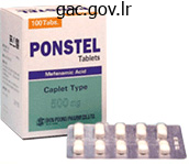
Purchase mefenamic 500 mg free shipping
Note that the lung has totally reexpanded and the mediastinum has shifted again to the midline. Spontaneous (Closed) Pneumothorax Spontaneous pneumothorax is attributable to the rupture of a subpleural lung bleb with little or no trauma and may be categorized as both major or secondary based on the presence of underlying lung illness. The typical patient with spontaneous pneumothorax is a tall, thin, 20- to 40-year-old male smoker. The morbidity, mortality, and long-term complications related to pneumothorax improve in sufferers with underlying lung disease. Whereas a major pneumothorax may be managed by selective remark or might simply be aspirated, a secondary pneumothorax typically requires a more aggressive administration strategy. The sudden onset of pleuritic chest pain and dyspnea with exertion or at relaxation is the commonest finding. More subtle manifestations may also happen with little or no ache and only delicate dyspnea on excursion that the affected person could ignore for days. A individual with a small spontaneous pneumothorax may by no means search medical consideration, and the method will resolve without therapy. Tube thoracostomy is the most typical remedy, however new trials suggest that conservative administration or aspiration of first-time primary pneumothoraces leads to comparable outcomes as conventional tube thoracostomy, although with fewer complications, shorter hospital stay, and decrease value. However, to date no high-quality scientific trials have positively demonstrated that aspiration is a superior treatment methodology. In addition, in uncommon instances, a spontaneous pneumothorax might progress to tension pneumothorax as described previously, which can result in a doubtlessly lifethreatening condition. Traumatic Closed Pneumothorax Closed pneumothorax often occurs from a rib fracture that penetrates the lung, but can even happen when an alveolus or bleb ruptures after blunt trauma. The air leak from a closed pneumothorax is mostly self-limited but can, in rare instances, progress to a pressure pneumothorax. Traumatic Open Pneumothorax An open pneumothorax happens when the chest wall is penetrated and the adverse intrapleural pressure is misplaced. Each breath can increase intrapleural pressure, especially if the diameter of the chest wound is larger than the diameter of the trachea. With every respiratory try, air moves preferentially via the chest wall opening quite than down the trachea and thus prevents significant air flow of the involved lung. Tension Pneumothorax An open pneumothorax can occasionally manifest as a pressure pneumothorax, which is a life-threatening condition that requires instant intervention. A tension pneumothorax occurs when an injury creates a one-way "flap valve" mechanism that allows air into the pleural area with inspiration but then closes with expiration and traps the air. The progressive accumulation of air within the pleural area results in ipsilateral complete lung collapse after which impingement on the mediastinum with a shift of the center toward the uninvolved facet. On expiration, the injury/valve closes and traps growing amounts of air in the pleural area. Eventually, the mediastinum shifts and cardiac filling and in the end cardiac output are compromised. C, this elderly patient sustained a pressure hemopneumothorax after slipping and falling on ice. The left hemithorax is very darkish (radiolucent) because of total collapse of the left lung (large white arrow). Multiple posterior rib fractures are present however are difficult to respect on this movie (small white arrows). The air-fluid stage (black arrow) indicates the presence of air in the pleural cavity along with fluid (blood). D, A computed tomography scan of the identical affected person again demonstrates the findings seen on the standard radiograph. Consequently, any patient with a penetrating thoracic damage (even with out immediate proof of a hemothorax or pneumothorax) ought to be considered for a "prophylactic" chest tube before mechanical air flow. A pneumothorax may develop in sufferers with asthma or emphysema from the high pressure required for ventilation, which may additionally lead to a tension pneumothorax.
Diseases
- Cerebroretinal vasculopathy
- Opioid dependence
- Rosai Dorfman disease
- Knobloch Layer syndrome
- Basal cell carcinoma
- Rheumatoid arthritis
- Amelia X linked
- Acrorenoocular syndrome
- Primerose syndrome
- Frydman Cohen Ashenazi syndrome

Mefenamic 250mg with amex
Turn the device on, apply the patient electrodes within the appropriate positions, analyze the rhythm, and deliver a shock if a shockable rhythm is present. The pediatric resuscitation tips included findings from a complete evaluation of the information. To carry out pediatric defibrillation, a defibrillator monitor capable of changes in vitality appropriate for children is needed. To acquire the electrical rhythm and subsequently administer an efficient defibrillatory shock, place the appropriate-sized pads and paddles accurately on the chest. Furthermore, appropriate pad or paddle size-the largest surface area possible with out direct electrode-to-electrode contact-will lower transthoracic impedance and enhance defibrillation. If contact is made between the paddles, an electrical arc or short circuit might happen. Never use dry paddles because the resistance to flow of current will be very large. However, chorus from using saline-soaked pads in children as a result of they could cause arcing on account of the proximity of the pads on the chest. In addition, the utilization of ultrasound gel and alcohol pads is discouraged because of poor electrical conductivity and probably high impedance. An option when using self-adhesive pads is to place one pad simply to the left of the sternum and the other over the again so that they approximate the position of the guts. Procedure and Technique the process for pediatric defibrillation is just like the algorithm for grownup defibrillation. Shock vitality for defibrillation First shock 2 J/kg, second shock 4 J/kg, subsequent shocks four J/kg, most 10 J/kg or adult dose. Maintenance: 20-50 mcg/kg per minute infusion (repeat bolus dose if infusion initiated >15 minutes after intial bolus therapy). Note: the use of epinephrine and its safety and helpful effects for an improved cardiac arrest end result have lately been questioned. If the sufferer is unresponsive to verbal and tactile stimuli, start chest compressions immediately (30: 2) at a price greater than 100/min. If a spinal wire damage is suspected, use the jaw-thrust maneuver with out head tilt. Do not use extreme pressure when ventilating as a result of this might cause regurgitation or aspiration, impede venous return to the center, and reduce coronary blood move because of increased intrathoracic strain. After interposing the breaths, proceed to assess the circulation by checking for a pulse in both the carotid or femoral artery (<10 seconds). If a single rescuer is performing the compressions and ventilations, the compressionto-ventilation ratio should be 30: 2. If two rescuers can be found, the compression-to-ventilation ratio must be 15: 2. If an enough pulse is current, interpose 12 to 20 breaths/ min (1 breath each three to 5 seconds). The positions of the electrodes on a toddler correspond to the positions used in an grownup. As talked about earlier in the adult section of the chapter, two kinds of defibrillators can be found: biphasic and monophasic. Based on a evaluate of grownup and pediatric animal data, when a handbook defibrillator is used for the first shock try, an power degree of two J/kg should be used with both a biphasic or a monophasic defibrillator. If a second or subsequent defibrillation is indicated, four J/kg must be used with both type. Before defibrillation, examine to make sure that the defibrillator is set to the unsynchronized mode. Once the defibrillator has been charged and the affected person cleared, apply firm strain to the defibrillation paddles (25 lb) to increase contact and deflate the lungs to the end-expiration state. This will usually be followed by a perceptible whole-body muscle twitch by the affected person. Once the shock has been delivered, resume resuscitation with quick chest compressions. If two rescuers can be found, use a 15: 2 ratio and change compressors when the primary compressor fatigues. If further monitoring gadgets are in place in the hospital setting, modify this step accordingly as determined by the resuscitation team chief.

Mefenamic 250 mg with amex
If the set off is ready too high (not sensitive enough), the work of respiratory incurred by the patient can be substantial. Some suppliers have been recognized to set the sensitivity at a high level if the affected person is markedly overbreathing the set fee. Many ventilators are set to a pressure set off with a sensitivity of 1 to 3 cm H2O. A longer time is spent in inhalation to allow more time for oxygenation and recruitment. Most ventilators may be set to obtain spontaneous respiratory, volume-targeted ventilation, pressure-targeted air flow, or some combination. In volume-targeted air flow, the ventilator is about to attain a determined volume whatever the strain required to achieve this. Pressure-targeted modes are set to attain a determined strain regardless of the quantity generated. In this mode, the ventilator offers a supplemental inspiratory strain to every of the patient-generated breaths. The utilized strain is turned off once the circulate decreases to a predetermined share. It should be set in order that the patient can set off the ventilator with out nice effort. Note that because the circulate rate adjustments, there are corresponding alterations in the efficient instances for inspiration and exhalation. Deflections above the x-axis (time) indicate inspiration, and those beneath indicate exhalation. Uncertainties corresponding to these have led many clinicians to favor volume-targeted strategies or dual-controlled methods in the acute care setting. The ventilator will generate the required driving stress to attain this "target. It ought to be famous that different essential features of the mechanical ventilator could be managed on this setting, corresponding to waveform (decelerating or square), I: E ratio, move rate, set off, and sensitivity. The time of gas move is set by the set quantity, circulate price, and waveform of gasoline delivery. When the set quantity is reached, gas circulate is terminated and expiration passively begins. Both are acceptable, and no data have demonstrated a better outcome with either mode. Every breath is absolutely supported by the ventilator, regardless of whether the breath is initiated by the affected person or the ventilator. The clinician units the base ventilation fee, but if the affected person tries to breathe faster than the set rate, extra breaths may be initiated by the patient, known as spontaneous breaths. Both help breaths and management breaths will attain the set goal, be it a set quantity (in a volume-targeted mode) or a set strain (in a pressure-targeted mode). The patient can initiate an additional spontaneous breath between the mandated or preset number of ventilator-supported breaths. The ventilator will generate an inspiratory strain that has been set by the clinician. With this target, the ventilator alters gas flow to achieve and maintain a preset airway stress during a preset inspiratory time (Ti). In the setting of hypoxemia, Ti may be increased quite precisely to enhance mean airway stress and oxygenation. Pressure-targeted modes, which are growing in popularity, might need better stress distribution, improved dissemination of airway stress, and greater distribution of ventilation. Subsequent information have shown that this technique of liberation actually will increase the variety of ventilator days. This could result in elevated imply airway strain, alveolar overdistention, and biotrauma.
Buy generic mefenamic 500 mg online
It is essential to be proficient in numerous totally different methods and to tailor their use to the wants of the person patient. Rescuers ought to apply potential scenarios earlier than dealing with sufferers with a compromised airway. Failure to achieve this might lead to unnecessarily aggressive administration in some conditions or to irreversible hypoxic harm as a end result of hesitation in others. Deciding who requires a definitive airway and who wants solely supportive measures is a formidable task for even essentially the most expert clinician. The following parameters ought to be assessed before the decision is made to establish a definitive airway: � Adequacy of current air flow � Potential for hypoxia � Airway patency � Need for neuromuscular blockade (uncooperative, full stomach, teeth clenching) Cervical spine stability � � Safety of the approach and talent of the operator Consideration of those elements should guide the clinician in deciding if tracheal intubation is important, and in selecting the optimum method. Time turns into important as the chance for irreversible hypoxic damage and cardiac arrest rises. All clinicians who carry out emergency intubation ought to be prepared to perform a surgical airway when intubation strategies and backup ventilation methods fail. Flexible endoscopic intubation is the go-to procedure for many anesthesiologists and is described later in the chapter. If excellent topical anesthesia is achieved, some patients may be intubated with none sedation. Ideal Versus Emergency Technique the intubation plan that might be finest within the ideal/elective scenario is often not one of the best plan within the emergency setting. Consider the affected person with quickly growing higher airway swelling as a result of angioedema or anaphylaxis, inflicting impending full airway obstruction. Other intubation methods might need a better probability of success if time was not a factor. The patient will likely develop full airway obstruction, important hypoxia, and dying within the time required to set up and carry out endoscopic nasal intubation. In this situation the emergency provider is "forced to act"sixty five to complete well timed tracheal intubation. In these patients, intubation often needs to be expedited so that different life threats could be addressed in a well timed method. When giving a paralytic agent, the supplier takes full duty for airway maintenance, air flow, and oxygenation of the affected person. If a patient has a failed airway, and oxygenation may be maintained, the clinician ought to attempt intubation by another methodology. This might be an anesthesiologist, emergency physician, paramedic, or another clinician with important airway expertise. The algorithm introduced here summarizes the final strategy used in the Department of Emergency Medicine at Hennepin County Medical Center. Individual providers and establishments ought to decide their own algorithms based on the supply of abilities and resources. There are many similarities between this algorithm and those put forth by the American Society of Anesthesiologists and the Difficult Airway Society; nevertheless, our algorithm is simpler and more applicable to emergency airway administration. Many algorithms resemble wish lists of equipment and expertise which may be simply not out there to many emergency airway providers. Our algorithm is based on the idea that oxygenation, not intubation, is the vital thing. The tip of the straight blade goes under the epiglottis and lifts it instantly, whereas the curved blade matches into the vallecula and not directly lifts the epiglottis by way of engagement of the hyoepiglottic ligament to expose the larynx. The straight blade is commonly a better choice in pediatric sufferers, in sufferers with an anterior larynx or a protracted floppy epiglottis, and in people whose larynx is fixed by scar tissue. Use of the straight blade can be extra often associated with laryngospasm because it stimulates the superior laryngeal nerve, which innervates the undersurface of the epiglottis. Individuals and institutions ought to formulate their very own algorithms primarily based on technical skills and the provision of assets. The illumination provided by the laryngoscope can make a giant distinction in the ability to visualize the laryngeal inlet. It was found that solely 24% of all blades offered the brightness necessary for fantastic inspection. Larger tubes are theoretically desirable as a result of airway resistance will increase as tube dimension decreases, however in apply, a 7.
Cha-de-Negro-Mina (Cha De Bugre). Mefenamic.
- How does Cha De Bugre work?
- Are there any interactions with medications?
- What is Cha De Bugre?
- Are there safety concerns?
- Weight loss and obesity, reducing cellulite, cough, edema, gout, cancer, herpes, viral infections, fever, heart disease, and wound healing.
- Dosing considerations for Cha De Bugre.
Source: http://www.rxlist.com/script/main/art.asp?articlekey=97068
Generic mefenamic 250 mg visa
Adequate humidification, cleansing, and systemic hydration will assist cut back the incidence of mucous blockage. Early complications (developing within three weeks after the procedure) occur in approximately 30% of sufferers and include bleeding, an infection, pneumothorax, costochondritis, and dislodgement (which could be caused by coughing). Life-threatening airway obstruction resulting from the formation of a large mucous ball has been reported. Obstruction inside the catheter tubing may cause a whistling sound from the oxygen tank humidifier. Always examine the affected person for indicators of subcutaneous air and catheter dislodgement. If routine broad-spectrum antibiotics are used by the affected person, Candida infections can develop at the stoma. Such infections are more frequent in sufferers receiving systemic antibiotics or long-term corticosteroids or these with diabetes. Many patients with chondritis have a deep, indurated, nonfluctuant lump around the tract that might be tender to palpation. If important bleeding is recognized or suspected, consult a specialist on an emergency basis and handle the airway definitively as clinically indicated (see the part on Major Bleeding). Manage pores and skin and pulmonary infections with the methods discussed for tracheostomy care. Stents Tracheal stenosis and tracheomalacia are recognized complications of synthetic airways. Management options include surgery and placement of silicone stents within the trachea. Morbidly obese patients are significantly in danger for lifethreatening issues related to tracheostomy. Dislodgement is specifically related to increased charges of morbidity and mortality. In addition to standard monitoring, use continuous capnography, if out there, to stop delay within the recognition of tube dislodgement. The patient ought to be positioned with the neck in slight extension, and the pinnacle of the mattress must be elevated to approximately 30 degrees. If the stoma tract has not healed or appears to be infected, change the catheter through a modified Seldinger method. Ask the affected person to sit upright, hold a pillow to the stomach, and cough forcefully after three deep inspirations. If the tube is obstructed, deflation of the tracheostomy cuff is most likely not adequate to enable enough ventilation as a end result of external compression brought on by abnormal neck anatomy might occur. Laryngectomy Patients It is essential to distinguish the affected person with a routine tracheostomy from the affected person with a laryngectomy as this could potentially limit the ability to present ventilation by means of the higher airway. A affected person with a partial laryngectomy could have a contiguous upper and lower airway, and interventions by means of the higher airway may be limited. Partial laryngectomy patients must also be ventilated via their stoma, and their mouth and nose must be saved closed to keep away from air escape. First, it is important to examine the laryngectomy stoma for any mechanical obstruction. External ventilation with a pediatric face masks or laryngeal masks airway could be performed. The approximate distance between the skin of the stoma and carina is simply approximately 6 cm. Pediatric tracheostomy tubes rarely have inflatable cuffs, except for those used for sure special indications. Continuous capnography might detect sure tracheostomy problems and is beneficial. Age guidelines may be helpful, but they will not be reliable in pediatric patients due to complicated medical problems.
Buy discount mefenamic 250 mg on-line
The capitate (round) carpal is in the distal row of carpals and articulates with the base of the center (third) metacarpal. Dislocation of the pinnacle of the humerus typically occurs in an anterior and slightly inferior direction, with the pinnacle coming to lie just beneath the coracoid course of (a subcoracoid dislocation). The supraspinatus muscle lies superior to the backbone and initiates abduction of the arm on the shoulder. The clavicle is a bit uncommon as a end result of it ossifies by intramembranous ossification, is considered one of the first bones to ossify, and is probably certainly one of the last bones to fuse. All of the other bones of the appendicular skeleton ossify by endochondral bone formation. Nasal cavities and paranasal sinuses: type the uppermost part of the respiratory system. Neurovascular: two anterolateral compartments that include the widespread carotid artery, inside jugular vein, and vagus nerve; all are contained within a fascial sleeve referred to as the carotid sheath. Prevertebral: posterocentral compartment that incorporates the cervical vertebrae and the associated paravertebral cervical muscles. Zygomatic bone: the cheekbone, which protrudes beneath the orbit and is weak to fractures from facial trauma. Ear (auricle or pinna): skin-covered elastic cartilage with several constant ridges, including the helix, antihelix, tragus, antitragus, and lobule. Jugular (suprasternal) notch: midline despair between the two sternal heads of the sternocleidomastoid muscle. Eight of these bones type the cranium (neurocranium, which contains the brain and 437 438 Chapter eight Head and Neck Supraorbital notch Superciliary arch Infraorbital margin Zygomatic bone Helix Nasal bone Tragus Ala of nostril Antihelix Antitragus Lobule Philtrum Commissure of lips Angle of mandible Submandibular gland Tubercle of superior upper lip External jugular v. Clavicle Glabella Anterior nares (nostrils) Nasolabial sulcus Thyroid cartilage Clavicular head of sternocleidomastoid m. Using your atlas and dry bone specimens, notice the complexity of the maxillary, temporal, and sphenoid bones. Pterion: point at which frontal, sphenoid, temporal, and parietal bones meet; the middle meningeal artery lies beneath this region. Comminuted: presents with multiple fragments (depressed if pushed inward; can compress or tear the underlying dura mater). Any fracture that communicates with a lacerated scalp, a paranasal sinus, or the center ear is termed a compound fracture. Note hair impacted into wound Clinical Focus 8-2 Zygomatic Fractures Trauma to the zygomatic bone (cheekbone) can disrupt the zygomatic complicated and its articulations with the frontal, maxillary, temporal, sphenoid, and palatine bones. Often, fractures contain suture strains with the frontal and maxillary bones, leading to displacement inferiorly, medially, and posteriorly. Ipsilateral ocular and visual adjustments could embrace diplopia (an upper outer gaze) and hyphema (blood in the anterior chamber of the eye), which requires instant medical attention. Lowered lateral portion of palpebral fissure Subconjunctival hemorrhage Flattened cheekbone Lateral canthal lig. Each fossa has quite a few foramina (openings) for structures to move in or out of the neurocranium. Arachnoid mater: nice, weblike avascular membrane directly beneath the dural floor; the house between the arachnoid mater and the underlying pia mater is known as the subarachnoid Chapter eight Head and Neck Foramen cecum Anterior ethmoidal foramen Foramina of cribriform plate Posterior ethmoidal foramen Optic canal Emissary v. Foramen rotundum Foramen ovale Foramen spinosum Foramen lacerum Carotid canal for Hiatus for Hiatus for Internal carotid a. Pia mater: delicate membrane of connective tissue that intimately envelops the mind and spinal wire. An outer periosteal layer is hooked up to the inner aspect of the skull and is supplied by the meningeal arteries, which lie on its surface between it and the bony cranium. Imprints of those meningeal artery branches may be seen as depressions on the internal desk of bone. Falx cerebelli: sickle-shaped layer of meningeal dura mater that projects between the 2 cerebellar hemispheres. Tentorium cerebelli: fold of meningeal dura mater that covers the cerebellum and helps the occipital lobes of the cerebral hemispheres.
Order generic mefenamic online
This space is close to the lingula and the left pleural house, thus making pneumothorax a frequent complication. Withdraw it several millimeters to forestall laceration of the myocardium or coronary vessels. The parasternal approach is an alternative approach to the beforehand described strategies. First, establish the biggest area of the parasternal effusion on ultrasound if attainable. Advance the needle over the cephalad border of the rib to avoid the neurovascular bundle on the caudal facet of the rib. Avoid going too far laterally from the sternal border due to potential damage to the interior mammary artery. Tsang and co-workers161 described this method for ultrasound-guided pericardiocentesis in 1998. The perfect web site for skin puncture is where the most important space of fluid accumulation is closest to the pores and skin surface. On ultrasound, this is indicated by a large anechoic (black) area at the high of the display, normally similar to the left anterior chest wall (rather than the subcostal region). This approach also avoids harm to the liver (common with the subcostal approach). Avoid selecting a site that would puncture the internal mammary artery, which lies three to 5 cm from either parasternal border, or the neurovascular bundle, which is located on the inferior border of the rib. Ultrasound-Guided Pericardiocentesis Pericardiocentesis has historically been performed blindly. This approach was related to a low success fee and a excessive fee of issues, corresponding to inadvertent puncture of the lung, ventricle, or epicardial vessels. Subxiphoid/Subcostal Approach As talked about earlier, the standard blind subxiphoid strategy can nonetheless be used for pericardiocentesis in the Ed. The method is carried out as follows: introduce the needle 1 cm inferior to the left xiphocostal angle at a 30-degree angle to the skin. Because the guts is an anterior structure, angles greater than 45 degrees could lacerate the liver or stomach. Aim towards the left shoulder and advance the needle slowly while continuously sustaining adverse stress on the syringe to aspirate any fluid. Aspirate with an "in-andout" vector only, not "side-to-side," which may lacerate tissue. If no fluid is aspirated, withdraw the needle completely and redirect it in a deeper posterior trajectory. Recommendations regarding needle trajectory range widely, including toward the right shoulder, sternal notch, and left shoulder. Be conscious that repositioning the patient alters the place of the heart and pericardial sac throughout the chest, so reassessment might be needed. Prepare the pores and skin antiseptically and place a sterile cowl over the ultrasound probe. If time permits, anesthetize the selected space with 1% lidocaine, with the superior border of the adjoining rib being used as a landmark. Ideally, the needle should have a sheath that enables it to be withdrawn after the pericardial area is entered. Attach a saline-filled syringe to the needle, and gently aspirate while slowly advancing the needle. Keep the ultrasound probe on the chest wall, instantly adjacent to the aspiration web site. If the contrast material clears immediately after administration (as occurs with agitated saline) or persists temporarily within the cardiac chambers, an intracardiac location is recommended. Fluid Aspiration and Evaluation A Removal of even a small quantity of pericardial fluid. After any approach used for pericardiocentesis, place a brief drain not solely to ensure rapid entry into the pericardial sac but in addition to permit more fluid to be removed rapidly if hemodynamic collapse recurs. After needle placement is confirmed, a brief drain may be positioned by the Seldinger technique, described in Chapter 22. Remove the syringe from the needle, advance a guidewire via the needle, after which take away the needle. Remove the dilator and slide an introducer sheath dilator (6 to 8 Fr Cordis) over the wire.
Trusted mefenamic 500mg
If utilizing landmarks quite than ultrasound to locate the vein, palpate the femoral artery with one finger, and puncture the skin just medial to the artery. Enter the skin at a 30- to 45-degree angle approximately 1 cm beneath the inguinal ligament. If using a finder needle with a syringe, apply steady, light suction whereas inserting the needle. Gently move the wire by way of the needle into the proximal end of the vein when blood return is famous. Make certain that the proximal finish is always visibly protruding from the hub of the needle. If resistance to elimination of the wire is encountered, withdraw the needle and the wire together to stop shearing off the tip of the wire. Once the catheter is placed, remove the needle and advance the wire through the catheter. Once the wire is in place and the catheter or needle removed, make a small incision (1 to 2 mm) with a No. Withdraw blood from the catheter ports after which flush them with a sterile saline solution. Three approaches to inside jugular catheterization are attainable (including the anterior, median or central, and posterior approaches, as mentioned in Chapter 22). Use of ultrasound to guide cannulation of the internal jugular vein seems to improve success charges (see Chapters 22 and 66). For the medial or central approach, use the apex of the angle fashioned by the sternal and clavicular heads of the sternocleidomastoid muscle because the puncture web site. If a line have been drawn from the mastoid course of to the sternal notch, the apex of the angle formed by the 2 muscular heads would fall approximately along the middle third of that line. Introduce a needle hooked up to a syringe on the apex of the triangle at an angle of 30 levels downward and towards the ipsilateral nipple. Keep the syringe related to the needle always to maintain constant negative strain and avoid air embolism. After blood flow is obtained, take away the syringe and place a finger over the hub of the needle. Pass the catheter far enough to attain the junction of the superior vena cava and proper atrium. Check the catheter for blood return, secure the road with sutures, and apply a sterile occlusive dressing. Confirm proper location of the catheter and rule out pneumothorax with a chest radiograph. Subclavian Venous Catheterization the subclavian vein is used much less frequently for central venous access in children than in adults. The technique is tougher in pediatric sufferers due to the smaller measurement of the vessel, in addition to a more cephalad location under the clavicles. The proper side is most popular because the dome of the lung is extra cephalad on the left aspect. The needle insertion website is at the distal third of the clavicle within the despair created between the deltoid and the pectoralis main muscular tissues. Introduce the finder needle bevel up and advance it slowly whereas making use of negative strain with the connected syringe. Direct the needle medially and slightly cephalad, beneath the clavicle toward the posterior facet of the sternal finish of the clavicle. Turn the syringe in order that the bevel of the needle factors caudad to direct the guidewire to the superior vena cava. Remove the syringe from the needle and insert the wire throughout a constructive pressure breath or natural exhalation. Remove the needle and introduce the catheter over the wire via the Seldinger approach, as beforehand described for femoral catheterization.
Order 250 mg mefenamic otc
Brachytherapy is the transperineal implantation of radioactive seeds instantly into the prostate. Epidemiology: � 1% of all male cancer diagnoses � Peak incidence in males aged 20�40 years � More frequent in white males Table 6. In the course of incurable illness, palliative care extends from prognosis to transition from curative to palliative intent, to ongoing deterioration, terminal stage and bereavement. The palliative care function is influenced by components similar to cultural perceptions of dying and dying, want for treatment, age of the affected person and their familial duties. Opioids are analgesia of selection, spasmolytics such as hyoscine (Buscopan) could additionally be beneficial in colicky ache. Guidelines for opioid prescribing Palliative care goals neither to hasten nor extend the dying process and is unambiguously distinct from euthanasia. The precept of double effect focuses on the intent of relieving struggling, however acknowledges that, in terminal phases, abatement of futile life-sustaining treatment might foreseeably however unintentionally alter the time of demise. Non-pharmacological antinausea methods embrace avoiding the smell of cooking food, frequent and smaller meal parts, anxiety management strategies and acupuncture. Strategies embody upright posture, free clothes, controlled expiration, leisure, chest physiotherapy, airy surroundings. Anticholinergics (such as hyoscine, atropine and glycopyrrolate) scale back the secretions and gurgling. Fatigue Fatigue is usually multidimensional and could also be due to correctable causes (anaemia, infection, respiratory sickness, endocrine cause) or subjectively related to terminal sickness and deconditioning. Strategies similar to train, activity planning, sleep hygiene, vitamin and biochemical correction, glucocorticoids could also be trialled. Terminal restlessness that is multifactorial (secondary to pain, discomfort, opiate toxicity, psychological distress). Practise good sleep hygiene; medication of alternative are diazepam, levomepromazine and midazolam. Apart from supportive psychotherapy, there should be a low threshold for initiating pharmacologic measures. Correctable causes ought to be recognized and � handled, other than implementing numerous adjunct strategies. Legal points could embody discussions surrounding resuscitation standing, appointing a power of lawyer and guardianship. Prognostication is difficult however models are used to estimate disease trajectories of cancer, organ failure and dementia. This chapter describes these conditions as properly as providing a scientific method to electrolyte abnormalities. The resultant filtrate is then funnelled down into the proximal convoluted tubule. Na, K, Ca, Mg and glucose) are reabsorbed here, facilitated by microvilli on the lumen. The glomerulus � Around 25�30% of cardiac output goes on to the kidneys by way of the renal arteries. The renal arteries divide into interlobular arteries and ultimately kind afferent arterioles. Podocyte Macula densa Juxtaglomerular cells Renal nerve A erent arteriole Proximal convoluted tubule. This permits water to transfer by way of osmosis into the interstitial areas, leading to extra concentrated urine. This distinctive system allows a phenomenon (known as the counter-current multiplier system) to happen. This creates a hyperosmolar space and supplies an osmotic gradient for the reabsorption of water through the descending loop. They regulate the release of renin by the juxtaglomerular cells, situated adjoining to the Bowman capsule and afferent arterioles (the results of renin are mentioned in the part below).
Real Experiences: Customer Reviews on Mefenamic
Yugul, 30 years: Physicians who perform endoscopic intubations every day have a hit price of practically one hundred pc when utilizing this method for tough intubations.
Yorik, 42 years: Left lung: Superior lobe drains to pulmonary and bronchopulmonary (hilar) nodes and inferior tracheobronchial (carinal) nodes.
Jerek, 28 years: Mercury column versus Dinamap readings confirmed increased disparity when systolic blood pressure was larger than one hundred forty mm hg, the vary at which accuracy ought to be most rigorously sought to correctly identify hypertension.
Giacomo, 27 years: Sampablo I, Escarrabill J, Rosell A, et al: Transtracheal catheter acceptance and antagonistic events in long-term home oxygen remedy.
9 of 10 - Review by K. Sivert
Votes: 70 votes
Total customer reviews: 70

