
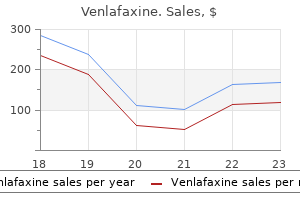
Venlafaxine dosages: 150 mg, 75 mg, 37.5 mg
Venlafaxine packs: 30 pills, 60 pills, 90 pills, 120 pills, 180 pills, 270 pills, 360 pills

Cheap venlafaxine 150mg free shipping
As the correlation between P2N2 changes and was remarkably consistent across medication from multiple lessons and at a quantity of doses, it means that whereas substantial variation exists in the potency of anesthetic medication in causing consolidation failure, a standard mechanistic pathway may be concerned. Furthermore, the observation supports the assertion that the underlying mechanism includes induction events within the consolidation cascade; consolidation failure evolving over several hours might be predicted by electrophysiologic events occurring inside several hundred milliseconds of publicity to the stimulus. At a presentation interval of 27 seconds, no degradation in memory efficiency was evident, but significant attenuation of the old/new impact was observed with propofol and midazolam. Again, the analysis distinguished the dominantly amnestic medicine from the dominantly sedative medicine and constrained the mechanism to early in the consolidation cascade. An intriguing risk raised by the connection between modulation of P2N2 and consolidation failure lies in the origin of the complex. P2N2 is understood to derive largely from synchronous oscillations,229 which as discussed earlier are important to non-Hebbian plasticity and strongly correlated with memory behavior. As such, one interpretation is that a common mechanism underlying the selective impact of anesthetic medication on the induction of consolidation entails adjustments in oscillations throughout a distributed cortico-hippocampal network. Unfortunately, no direct measures of synchrony in an Chapter 13: Consciousness, Memory, and Anesthesia 297 applicable reminiscence paradigm have yet been performed, and the placement of the hippocampus confers monumental methodologic barriers. Another intriguing discovering from the study was a pristine correlation between response time, P2N2, and the consolidation coefficient, which was preserved throughout all medication and dosages. In nonpharmacologic research, response time has lengthy been interpreted as an index of sedation, however in the anesthetic research, no relationship with sedation was recognized; this suggests caution in deciphering it as a behavioral marker of sedation in future drug studies. In a research utilizing tetanic stimulation of the Schaffer collateral-commissural pathway,232 isoflurane (0. The 5 �/� mutants are immune to the amnestic results of etomidate, but not to its basic anesthetic results. Memory deficits have been noticed in wild type mice, but not in 5 �/� mutants or in wild varieties receiving L-655,708. Perouansky and colleagues252 used a fear conditioning paradigm to study isoflurane zero. In some contrast, the nonimmobilizer F6, which causes amnesia with out sedation or a loss of motor activity, caused a lack of oscillatory power without slowing. The amygdala is recognized as critical to concern studying and memory, and the systematic examine of amygdala-dependent classical (Pavlovian) fear conditioning has produced a lot data relating to the mechanisms of associative learning. Thus, the amygdala is a key mechanistic site for anesthetic amnesia in certain experimental contexts. However, inhibitory avoidance, like many associative learning experimental paradigms carried out in animals, is selectively amygdala dependent. The experimental paradigms all involve the presentation of emotionally adverse and arousing pictures. Such gadgets are established to activate the amygdalo-hippocampal modulatory axis267,268 and should be better remembered than emotionally impartial items. An early behavioral research demonstrated that negative-arousing objects have been more proof against the amnestic effect of thiopental than have been neutral objects, suggesting that amygdala-dependent modulatory processes remained intact. However, the small numbers of topics in the dexmedetomidine cohort restrict interpretation of the unfavorable finding. Pryor and colleagues219 reported that activation of the amygdala in response to negative-arousing objects remains robust within the presence of propofol (0. This discovering means that the afferent processes serving higher-order interpretative capabilities and amygdala activation are unaffected by amnestic ranges of propofol, whereas efferent processes underlying amygdala-dependent modulation of hippocampal plasticity are disrupted. However, in contrast to propofol, a mnemonic advantage for these items is seen, and subsequent memory is correlated with activation within the amygdala and hippocampus on the left aspect. Taken together, these research suggest that propofol is more effective at attenuating the amygdalo-hippocampal modulatory axis. Amygdala hyperreactivity is of significance in a variety of fear-based psychopathologies, together with nervousness, phobia, panic dysfunction, and posttraumatic stress disorder. This is particularly relevant to the frequent incidence of posttraumatic stress disorder following intraoperative awareness272 and intensive care remedy,273,274 however further investigation is required earlier than any medical methods could be provided. Six coronal slices through the hippocampus (three leftmost slices, marked above by y coordinates �36, �33, �30) and amygdala (coordinates 0, three, 6) are chosen, with the coronal planes proven on the sagittal slice on proper. In the placebo group, important activations (negative-arousing > neutral) are seen in each right and left amygdala, and in each right and left hippocampus. In the propofol group, similar significant activations are seen in the proper and left amygdala, but no important activations are observed within the hippocampi.
Order generic venlafaxine on line
It is totally moral and legal to administer high doses of pain medication and sedatives for the intended impact of relieving struggling, even if the therapy has the aspect effect of hastening death. However, to administer any medication with the express intention of hastening demise is euthanasia and never medical therapy. When withdrawal of ventilator assist is anticipated in a affected person already receiving such medicine, they need to be withheld in all however extraordinary instances. Even worse, it can mask signs and indicators of distress and might thus prevent reduction of struggling through the dying process69 (see additionally Chapter 101). Pacemakers, implantable cardioverter-defibrillators, and ventricular help devices are increasingly utilized in each long-term medical remedy and intensive care (see also Chapters 48 and 101). Common examples embrace artificial joints, intraocular lenses, treatment delivery units, and orthopedic hardware. In fact, the ethical distinction between disabling a pacemaker in a pacer-dependent affected person and turning off a ventilator for a ventilator-dependent patient is minimal, if the request comes from a reliable affected person or the surrogate decision maker. Both actions contain discontinuance of synthetic therapies that the affected person no longer desires, and each could also be adopted by speedy death. As with different end-of-life therapies, discontinuance of such gadgets ought to embrace due consideration of whether the choice is made by a competent patient and with full informed consent. Management of discontinuance of gadget remedy ought to always embrace planning for remedy of distressing signs and administration of acceptable comfort measures. Euthanasia entails the administration of medication by somebody aside from the patient for the express purpose of inflicting death, within the perception that this may be finest for the patient (but not essentially on the specific request of the patient). Both practices differ ethically from withdrawing or withholding life-supporting medical remedies. In withdrawal of withholding of life-supporting remedy, the first intention is to discontinue remedies which are inflicting suffering with an understanding that death may or in all probability will result. The public persistently charges lack of autonomy and control, lack of ability to pursue beforehand valued actions, and lack of dignity as the most important considerations on the finish of life. Although many ethicists acknowledge that individual circumstances might make assisted suicide an ethically permissible action, most express concerns about potential abuse. Vulnerable members of society, such as the poor, old, and handicapped, could presumably be pressed by monetary and social factors into a suicide option rather than palliative care. More than 93% of patients enrolled in the Oregon program cited loss of autonomy as a main reason for pursuing the option. Advances in cardiopulmonary resuscitation and mechanical ventilation then made it potential to postpone demise, seemingly indefinitely. In 1968, the Ad Hoc Committee of the Harvard Medical School proposed redefining dying as the purpose at which all cardiorespiratory operate had irreversibly ceased, or all perform of the whole mind had irreversibly stopped (brain death). The first was to enable patients to be declared dead and not maintained on machines-thus limiting expense; reallocating medical sources to different, salvageable sufferers; and allowing the social rituals surrounding demise to happen. The public has been gradual to accept mind demise, in part because it requires complete trust in physicians and ignores indicators of death that the public already understands. In the United States, it requires demonstration that, in the absence of drugs, paralytic brokers, hypothermia, and other reversible circumstances that mimic loss of brain operate, no cortical or brainstem operate is current. The analysis is usually made either clinically, by demonstrating that cortical activity and brainstem reflexes are absent, or by radiographic research demonstrating the whole absence of cerebral blood flow. Although brain demise is a social and never a biologic definition of demise, medical, moral, theologic, and legal consultants typically agree that brain death adequately defines a situation in which a person with moral and authorized rights and ethical standing ceases to exist and will now not be treated as though alive. Expensive medical interventions could be discontinued without legal ramifications, and very important organs could be donated for transplantation if the affected person or the surrogate determination maker agrees. Before assuming care of a brain-dead organ donor, the anesthesiologist is obligated to review the chart for documentation of the declaration of mind demise and the factors on which it was based mostly. If any questions exist about the prognosis, organ donation ought to be postponed until the anesthesiologist is happy that these issues are addressed. Yet many protocols call for organ retrieval to begin only 2 minutes after circulation has stopped, and in at least one establishment, organ donation is allowed to start within seconds of cardiac arrest.
Diseases
- Vitamin A embryopathy
- Long QT Syndrome
- Quinsy
- Chromosome 12 ring
- Epidermolysis bullosa dystrophica, dominant type
- Myotubular myopathy
- Sanfilippo syndrome
- Bone neoplasms
- Muscular dystrophy, Duchenne and Becker type
- Occupational asthma - wood
Discount venlafaxine 37.5 mg amex
Primary atlanto-axial osteoarthritis is quite painful and responds poorly to nonoperative means. C1�2 instability because of rheumatoid arthritis could also be neither symptomatic nor a neurologic risk. Painful C1�2 rheumatoid involvement in the face of adequate medical remedy additionally signifies the necessity for fusion. Progressive C1�2 subluxation, especially with cranial settling, additionally has an unfavorable pure historical past. The complaints provided will differ with the presentation (eg, trauma, inflammatory arthritis, developmental, congenital). Patients with a traumatic damage may complain of isolated ache but additionally could present with neurologic deficits. A low threshold of suspicion should be maintained for patients with blunt trauma to the head or face, or with known noncontiguous fractures of the spine. An anterior atlantodental interval greater than 5 mm indicates probably damage to the transverse ligament and, in the setting of trauma, necessitates operative stabilization. An avulsion (arrow) of the transverse ligament from the ring of C1 indicates instability and may require arthrodesis of C1�2. Displaced odontoid fractures (type 2) have a better probability of a nonunion and will require a primary C1�2 fusion. Joint house narrowing is a sign of C1�2 osteoarthritis and responds poorly to nonoperative administration. Pseudo-pannus formation behind the dens in patients with rheumatoid arthritis can lead to cervical stenosis and myelopathy. Flexion (D) and extension (E) lateral radiographs show C1�2 instability in a patient with rheumatoid arthritis. Patients with major atlantoaxial arthritis will complain of extreme neck and head ache, most often unilateral, with varying degrees of refusal to rotate their head, particularly ipsilaterally toward the pain. Physical examination ought to embrace the following: Active self-limited rotation of the head, particularly toward the facet of the ache. Palpation of the suboccipital area near the interval between the posterior arches of C1 and C2 will elicit pain. The patient is examined supine together with his or her head resting comfortably on a pillow. In instances of C1�2 arthritis, this maneuver ought to present extra motion and fewer ache than comparable motion with an axial vertex load. With slight manual traction, head rotation is elevated, whereas an axial vertex load will trigger ache and lead to decreased rotation. When discovered, this should be treated with immediate anticoagulation to forestall thromboembolic problems. If a surgical process is important, anticoagulation is stopped earlier than and restarted after surgical procedure. Fractures of either C1 or C2 indicate a significant chance of additional cervical backbone fractures. A unilateral vertebral artery harm not often is symptomatic due to adequate collateral flow through the contralateral vertebral artery as nicely as the circle of Willis. A affected person with a vertebral artery harm who presents with neurologic deficits due to a concomitant spinal wire injury could additionally be particularly difficult to diagnose clinically. For sure fractures, use of a halo-vest may be applicable, and the patient is treated in the orthosis for three months. It is a time-tested "nonoperative" choice with well-defined success/failure charges. Some sufferers could require a halo for postoperative immobilization, relying on the fixation high quality, the anticipated stage of patient compliance with a tough collar, and other uncommon circumstances. Malreduction of C1�2, anomalous position or dimension of the vertebral arteries, and collapse of the lateral lots of C2 are relative contraindications to using the transarticular screw technique due to the danger for inadvertent penetration of the vertebral artery.
Discount venlafaxine 150 mg visa
Carefully shut the tibialis anterior sheath, superior extensor retinaculum, and subcutaneous tissue before skin closure. Immobilization in a minimum of 5 degrees of dorsiflexion is essential to avoid tension on the wound edges. The authors concluded that surgical restore of a ruptured tibialis anterior tendon could be useful no matter age, sex, medical comorbidities, or delay in diagnosis. Ouzounian et al4 reported on seven sufferers with tibialis anterior rupture treated with a variety of surgical reconstructive methods. The lack of statistical significance was probably because of the bimodal age distribution within the examine, with older, extra sedentary patients receiving nonoperative remedy. The literature is scarce regarding the results and complications of surgical reconstruction of the tibialis anterior tendon due to the rarity of this injury. Tendon has numerous insertions on bones of plantar midfoot, spring ligament, and medial aspect of navicular. Common peroneal nerve palsy may lead to progressively worsening equinocavovarus foot deformity because of overpull of plantarflexors and inverters powered by intact tibial nerve and lack of dorsiflexors and everters powered by compromised frequent peroneal nerve. Flaccid paralysis remains relatively stable since each sets of antagonists are compromised. Inability to dorsiflex ankle May verify by asking patient to walk on heels Manual muscle testing with patient seated on examining desk with knee flexed Lack of eversion Varus hindfoot Over time, might turn into a fixed inversion contracture In some disease processes (eg, Charcot-Marie-Tooth disease) toe dorsiflexion is spared, creating claw toe deformities. Patient makes an attempt to compensate for lack of ankle dorsiflexion with toe extensors, worsening claw toe deformities. Even when toe extensors are involved in the palsy, flexor tendons might become contracted. With equinocavovarus foot contracture, calluses might type under metatarsal heads, significantly the fifth. Absence of restoration at 1 year and notably at 18 months is extremely suggestive of no recovery. We advocate consultation with a neurologist to affirm interpretation of electrodiagnostic research. Flexible versus mounted deformities Flexible deformity sometimes corrects with tendon switch alone. Fixed deformity May require capsular release and even arthrodesis Toe contractures Although claw toe deformity is in all probability not evident with the ankle plantarflexed, once the deformity is corrected, toe contractures might become apparent. Dorsiflexing the ankle will put the contracted flexor hallucis and digitorum on stretch, thereby revealing the toe contractures. The surgeon must be ready to tackle toe contractures as a part of the process. Tendon switch anchoring We routinely use interference screws to anchor tendon transfers to bone. Need to have an anchoring system obtainable Alternatively, anchoring to present distal tendon or existing soft tissues in the foot could also be potential. In our experience, anesthesia should keep full muscle rest and paralysis through the procedure; otherwise, the success of the tendon switch could also be compromised. Approach Multiple comparatively small incisions are wanted; extensile exposures are pointless. Occasionally patients preserve an lively stretching program, thereby avoiding an Achilles contracture. Weakening of the gastrocnemius�soleus advanced may be helpful since a transfer of a wholesome muscle�tendon unit is subject to an automated onegrade loss of power (5/5 manual muscle testing drops to 4/5 with transfer). Equinus with knee in flexion and extension suggests tight gastrocnemius and soleus. Second Achilles hemisection (opposite path from first), to be adopted by third and last hemisection in identical path as first. Accidentally transecting the nerve results in loss of sensation within the plantar medial forefoot. Violating the veins could make it tough to obtain passable hemostasis as these veins may then retract underneath the foot. The medial incision could need to be prolonged proximally to enable access to the posterior medial malleolus, a common location the place the tendon may bind. Preparation of the Dorsal Foot Anchor Site Fluoroscopically determine the middle of the lateral cuneiform.
Order venlafaxine 150mg without a prescription
In distinction, as a result of resection of more than 50% of the aspect joint can lead to facet instability, resection of the superior side lateral to the pedicle is unnecessary. A Positioning Proper affected person positioning is crucial when performing posterior cervical foraminotomy to scale back blood loss and enhance visualization of the operative area. This desk is quite versatile and permits for intraoperative alterations in patient positioning all through the operation. Two separate ropes are used so the neck is maintained in correct alignment, depending on the process being carried out: one of many ropes is placed in-line and horizontal to the desk via a pulley system, and the other is placed over a cross-bar on the Jackson frame to facilitate placement of the top into extension. Although not essential, a horseshoe could also be used ventral to the face to catch the head if the tongs slip. This could weaken the lateral mass and result in a fracture, or more commonly it makes placement of the lateral mass screw harder if a fusion is being carried out along with a foraminotomy. Approach A posterior cervical foraminotomy could be performed using open, endoscopic, or microscopically assisted approaches. With either method, the lamina, the junction between the lamina and the aspect joint, and the side joint itself have to be exposed while preserving the side capsule. Dissection of the posterior cervical spine along the midline within the avascular aircraft. The posterior cervical backbone after meticulous dissection of the posterior elements with lateral extension over the facet capsules. Model of the cervical backbone showing the C5-6 interspace with the intralaminar V (yellow lines). This is the important thing anatomic landmark that should be acknowledged to perform an sufficient foraminotomy. An intraoperative picture exhibiting the C5-6 interspace with the intralaminar V (yellow lines). Model of the cervical spine exhibiting the C5-6 interspace with resection of the inferior aspect, which must be resected to the lateral margin of the pedicles to expose the underlying superior articular facet. To determine whether or not sufficient of the inferior aspect has been resected, a small angled microcurette can be utilized to palpate the pedicle. An intraoperative image exhibiting the C5-6 interspace with resection of the inferior side. During the decompression, copious irrigation (20-mL syringe with a 2-inch-long 18-gauge angiocath) should be used to forestall thermal harm to the surrounding tissues. Typically we suggest the utilization of a burr over Kerrison rongeurs as a result of inserting instruments (such as Kerrison rongeur, which may have a relatively thick footplate) into the already stenotic canal and foramen may cause neurologic harm. An intraoperative image exhibiting that when the inferior articular facet is resected, the superior articular aspect underneath may be identified. An intraoperative picture exhibiting the finished resection of the superior articular aspect. The remaining small ledge of bone could be eliminated utilizing a small angled microcurette or 1-mm Kerrison rongeur. Model of the cervical backbone displaying the C5-6 interspace showing C- or sickle-shaped decompression, which may lead to iatrogenic impingement on the nerve root. Model showing completion of the foraminotomy with full decompression of the foramen. An intraoperative image displaying palpation of the medial pedicle border after completion of the foraminotomy. If meticulous midline publicity was performed, the preserved interspinous ligaments with the muscular attachments are used as the first layer of closure. The amount of muscle included into the suture is minimized, since all such muscle will necrose. With a well-exposed spine, one can find a thin fascial layer enveloping the muscle that can be utilized to shut the layers. The closure progresses from deep to superficial with the placement of deep, center, and superficial drains.
Syndromes
- Use a needle to draw fluid out of a cyst, which will be examined under a microscope to look for cancer cells
- Emphysema
- If you have a child who is allergic to certain foods, introduce one new food at a time in small amounts so you can recognize an allergic reaction.
- Frequent visits to the doctor are needed to adjust the lengthening device. How long the lengthening device is used depends on the amount of lengthening needed. Physical therapy is needed to maintain normal range of motion.
- CRPS 2 is caused by an injury to the nerve.
- Improve weight distribution
Buy 37.5 mg venlafaxine visa
Once the Morse taper is secured, remove the wrench on the stem and the composite base plate and stem combination is in a position to be absolutely seated. A narrow handle attaches to the anterior aspect of the bottom plate to control rotation because the tibial element is impacted. Talar part In our opinion, that is essentially the most challenging step of the procedure, significantly if the joint was distracted to decrease bone resection or to right deformity. In this case, the joint space is quite tight by design, to achieve optimum soft tissue balance and ligament pressure. We routinely assemble a 10-mm stem to the talar dome element on the back desk for the dimensions 2 and three prosthesis, using the devoted assembly device to safe the Morse taper. Typically, a 14-mm stem is simply too lengthy to be linked to the talar dome part earlier than implantation. Since the Morse taper has not been secured, the rib wrench must remain under the 14-mm talar stem. The joint must then be gently distracted with a lamina spreader, adopted by insertion of the talar dome component. The toothless lamina spreader might have to go under the talar dome element to get hold of the distraction, while the talar element is rigorously compelled posteriorly into position. A handle attached to the talar dome element facilitates driving the talar dome posteriorly. A protecting plastic sleeve inserted onto the tibial base plate protects the talar dome from being scratched. Once the talar dome element seats on the stem, use the talar dome impactor to secure the Morse taper, with the rib wrench nonetheless between the talar dome component and the ready talar surface. Remove the rib wrench and examine the interface between talar dome and stem to make certain that the two talar components are securely connected. While impacting the talar part, use the handle that inserts into the talar dome to management refined modifications in rotation of the talar element. With the ankle in neutral place, there ought to be a stability with varus and valgus stress. If not, the polyethylene thickness may be inappropriate or, more probably, stability needs to be established. The ankle should dorsiflex to no much less than 5 degrees, preferably 10 degrees beyond neutral. Once the polyethylene has cleared the superior dome of the talar element, ease off on the plantarflexion of the insertion device and have the assistant or co-surgeon compress the joint, thereby forcing the polyethylene into the locking mechanism. Remove the insertion system and absolutely seat the polyethylene with the devoted impactor. Polyethylene insertion device that screws down and pushes polyethylene onto tibial component tracks. Apply sterile dressings on the wounds, adequate padding, and a short-leg cast with the ankle in neutral position. Therefore, perform a tendo Achilles lengthening to get the talus in a impartial position earlier than securing the leg within the leg holder. The foot and leg may be properly positioned in the leg holder and fluoroscopy could recommend proper alignment, however the ankle should still be malrotated, leading to symmetric but malrotated tibial and talar preparation. Place a skinny osteotome in the medial gutter of the tibiotalar joint to determine optimal rotation; the osteotome ought to be parallel to the aspect of the leg holder. For varus perform the medial launch; for valgus, the ankle is often free and simply wants the lamina spreader to realign the talus inside the ankle mortise. Therefore, first place the ankle within the heart of the fluoroscopic beam, and then make changes. Note additionally that as adjustments are made to the operating desk to optimize alignment, the ankle could "drift" from the center of the monitor and might need to be recentered in the fluoroscopic beam while alignment is being set. Assessing the place of any instrument fluoroscopically demands that correct alignment has been confirmed first. For instance, when positioning the cutting block relative to the reference drill, first verify that alignment is perfect, and then assess the slicing block place. The tibial base plate and the talar dome parts attach to their respective stems with Morse tapers; ensure these are fully secured before seating either composite (combination main element and stem) fully. Rotation Varus ankle and valgus malalignment Place the ankle at the heart of the fluoroscopic monitor. However, considered use of lamina spreaders is again attainable to facilitate insertion of the talar part.
Order venlafaxine overnight delivery
No tenderness is found in the Achilles tendon proximal to its insertion on the calcaneus. Lateral foot radiograph demonstrating the posterior calcaneal prominence and calcification inside the Achilles tendon insertion. Modalities: ultrasound, iontophoresis Extracorporal shockwave therapy may have some benefit but is essentially unproven. Corticosteroid injection could result in Achilles rupture and is contraindicated until the process is isolated to retrocalcaneal bursitis, by which case a even handed injection of only the retrocalcaneal bursa could be carried out. Insertional Achilles teninopathy with central calcific tendinosis could additionally be less amenable to nonoperative administration. The restoration following surgical administration for insertional Achilles tendinopathy is extended and may take a full yr earlier than the patient returns to full activity. We routinely inflate the thigh tourniquet with the patient supine on the stretcher, then flip the affected person to the prone place on the working room table. Preoperative Planning Preoperative medical clearance Even in wholesome sufferers, the skinny pores and skin on the posterior heel is in danger. Carefully inspect pores and skin to be certain that the patient is an inexpensive candidate for a posterior approach to the Achilles tendon insertion. The scalpel is moved by way of pores and skin and into central portion of distal Achilles tendon. The aim is to keep away from pointless delamination of the soft tissues and to elevate full-thickness flaps. More than half of the Achilles tendon insertion could be elevated with out compromising the integrity of the insertion. We elevate the Achilles tendon till all the diseased portion of tendon may be excised. Reattachment is facilitated by a proximal Achilles tendon lengthening that additionally serves to unload the Achilles tendon. After a full-thickness incision is made through the diseased portion of the tendon, lateral (B) and medial (C) tendon slips are developed. If needed, a single fluoroscopy spot image may be used to outline the trajectory of the noticed blade. This helps slender the heel and cut back the majority of the residual calcaneus, medial, and lateral prominences that may result in persistent pressure and impingement experienced by the affected person. While these chamfers are close to the medial and lateral insertion factors of the Achilles tendon, sometimes they can be excised with out compromising the residual tendon attachment. Touch-up to ensure an acceptable quantity of bone was removed and an enough "healing" cancellous floor is uncovered. Chamfer preparation to decompress the lateral (F) and medial (G) dimensions of the prominent calcaneus. While one study suggested that up to 75% of the tendon attachment may be launched with out compromising the integrity of the insertion, we routinely reattach the elevated portion of tendon to the exposed cancellous calcaneal floor. In our opinion, reattachment not only strengthens the repair but additionally facilitates direct tendon therapeutic to the calcaneus. If they need to fail, we would prefer for them to fail now so we can rectify the issue. The medial suture anchor (D) is positioned symmetrically relative to the lateral anchor and secured to bone (E). The sutures must not only be tensioned appropriately within the longitudinal airplane however must even be balanced nicely within the medial-to-lateral plane, in order that the two tendon slips can also be reapproximated aspect to aspect and reconfigure the physiologic Achilles attachment. Have the assistant hold the ankle in plantarflexion in order that the tendon slips absolutely contact the calcaneus. Once the Achilles tendon insertion is again wholesome and asymptomatic, it has been our experience that the gastrocnemius and soleus muscles accommodate. Sterile dressings, plentiful padding, and a posterior splint with the ankle in its resting pressure complete the closure. The two Achilles tendon slips must be reattached in a balanced manner on the uncovered cancellous floor of the calcaneus. Before tying the sutures of the suture anchors, verify that the tension appears nearly equal for the two tendon slips. Transfer the tendon as far posteriorly on the uncovered cancellous floor of the calcaneus as potential for the greatest mechanical benefit.
Buy venlafaxine 150mg
Apply a compression dressing and splint the foot into slight equinus with the posterior splint and sugar-tong "trilaminar splint. Develop entire operative area from posteromedial to posterolateral corner so panoramic view of Achilles tuberosity attachment. Experience permits elimination of paratenon and further elimination of small ruptures and/or ossification in chosen cases and conditions. Postoperative routine is (a) non�weight-bearing for two to 3 weeks, (b) partial weight-bearing for 2 to three weeks, and (c) to maximize strength of posterior tibial and peroneals as quickly as mobilization permits. Patients wear footwear with a heel counter and return to normal day by day perform in 8 weeks. All athletes returned to their previous level of activity in a median of 12 weeks. Patients might have an extended period of cast immobilization after d�bridement of the Achilles tendon or important Achilles tendinopathy. These outcomes compared with those revealed by van Dijk et al13: their 20 sufferers resumed collaborating in sports at a median of 12 weeks. The time of surgery after diagnosis of retrocalcaneal bursitis averaged 20 months. Indications for operative intervention included failed nonoperative measures, history and bodily examinations in preserving with retrocalcaneal bursitis, and Haglund deformity causing mechanical impingement or Achilles tendinopathy. Patients were prospectively adopted from 1997 to 2003, with a imply follow-up of 35 months (range 3 to sixty two months). An wonderful result was defined as pain-free activity with complete return to activity, and a poor outcome was defined as having persistent signs and incapability to return to exercise. The cohort was stratified into "every day athletic activity" and "athletic" teams and the groups had been in contrast. All sufferers reported satisfaction with the cosmetic look of their portal websites. The results of local steroid injections on tendons: a biomechanical and microscopic correlative examine. The clinical diagnosis is acute and chronic pathology of the Achilles tendon insertion and its surrounding tissues. Between the distal Achilles tendon and the dorsal-posterior calcaneal prominence, immediately proximal to the Achilles insertion, is the retrocalcaneal bursa. Most probably some preliminary harm happens, adopted by a number of minor reinjuries that lead to persistent signs. In the acute section, the method may have some inflammatory traits; nonetheless, the persistent process is degenerative, with a relative paucity of inflammatory tissue. In addition, the patient notes a progressively enlarging prominence on the posterior heel. Putting the Achilles tendon on stretch aggravates the signs, corresponding to when the affected person walks uphill. High-energy extracorporeal shock wave remedy as a treatment for insertional Achilles tendinopathy. Surgical administration of insertional calcific Achilles tendinosis with a central tendon splitting approach. Achilles tendon and paratendon microcirculation in midportion and insertional tendinopathy in athletes. Insertional Achilles tendinosis: surgical treatment through a central tendon splitting strategy. Eccentric loading compared with shock wave treatment for continual insertional Achilles tendinopathy: a randomized, controlled trial. Change in plantarflexion power after full detachment and reconstruction of the Achilles tendon. Technique and results of Achilles tendon detachment and reconstruction for insertional Achilles tendinosis. McGarvey et al9 famous an 82% satisfaction fee in 22 patients at mean follow-up of 33 months. Thirteen of 22 patients had been pain-free and and an equal quantity might return to full acitivities. Intratendinous degeneration leads to ectopic calcification and ossification at the Achilles tendon insertion to the calcaneus. Inflammatory enthesopathies corresponding to psoriasis, Reiter syndrome, and inflammatory bowel disease may be current.
Order genuine venlafaxine on-line
We warning towards using solely a single screw since this fixation might show rotationally unstable. Perform the dorsiflexion osteotomy with an oscillating saw, eradicating a dorsal wedge of bone within the proximal third of the metatarsal and leaving the plantar cortex intact. Secure it with Kirschner wires, a small dorsal plate, or a screw and tension band method. Place two Hohmann retractors to defend the gentle tissues and drill a gap centrally in the first metatarsal bone with sequentially larger-diameter drill bits: first 2. However, when the deformity is isolated to a set, plantarflexed first ray, a dorsiflexion first metatarsal osteotomy may be enough. Likewise, world cavus of the complete forefoot could additionally be effectively treated with a dorsiflexion midfoot osteotomy (Cole procedure). In select circumstances of versatile hindfoot varus, a Dwyer lateral closing wedge calcaneal osteotomy (see below) could additionally be carried out in lieu of hindfoot arthrodesis. The lateral method is carried out with an S-shaped skin incision, starting 2 cm distally and dorsally to the lateral malleolus, proceeding in an arch shape to the navicular, distally to the palpable talar head. Expose the sural nerve within the proximal wound edge with its accompanying vessels and retract it. Using a concave chisel, detach its origin from the anterior processes of the calcaneus bone. The hindfoot arthrodesis could additionally be performed with preservation of the subchondral bone structure or as a corrective wedge resection. If cavus was not corrected by the Steindler process, a dorsally based wedge should be taken from the Chopart joint. With excessive forefoot and midfoot adduction, the dorsal wedge resection might have to include a further lateral-based wedge resection. The extra conservative arthrodesis that maintains subchondral bone architecture of the joints is reserved for gentle to average deformity. Remove the cartilage and penetrate the subchondral bone with a chisel or drill to promote fusion. If a wedge resection is required to correct the deformity, we favor to use an oscillating saw. After the entire launch of the Chopart joint, the cavus foot can be manually corrected and the navicular centered on the talar head. We routinely stabilize the lowered joints with Kirschner wires (two via the talonavicular joint, two via the calcaneocuboid joint). In extreme deformity, a laterally based mostly wedge can be removed from the subtalar joint. Dorsal impingement of the talus on the tibia, in instances with limited ankle dorsiflexion or extreme hindfoot equinus, could warrant a modified Lambrinudi process. For each the triple arthrodesis and modified Lambrinudi process the sinus tarsi is free of all soft tissue constructions (interosseous ligaments and fat). The most important structure to be dissected is the interosseous ligament between the talus and calcaneus. To expose the subtalar joint, use a lamina spreader in the subtalar joint and place a Vierstein retractor under the apex of the lateral malleolus. Prepare the surfaces at the arthrodesis site with a concave chisel or with the oscillating noticed, depending on the amount of correction needed. If a Lambrinudi fusion is required, a dorsally based wedge is taken out of the subtalar joint. The willpower of the osteotomy strains is necessary for the size of the remaining bone. The first osteotomy runs parallel to the ankle joint line and thru the talar head. Both osteotomies unite within the posterior edge of the subtalar joint, forming a dorsally primarily based wedge with its apex within the posterior aspect of the subtalar joint. After resecting the cartilage or the bony wedge, assess the effect of correction by the reposition of the talocalcaneal and the Chopart joint. In addition to the correction of the cavus hindfoot varus elements, it is extremely important that the foot may be repositioned in a plantigrade place. A dorsally based wedge is faraway from the navicular�cuneiform joints and the cuboid. The distal osteotomy must be pushed exactly by way of the cuneiforms and the cuboid; the proximal osteotomy runs via the cuboid and navicular.
Order venlafaxine 37.5 mg overnight delivery
Excision of posterolateral talar dome lesions by way of a medial transmalleolar strategy. Osteochondral lesion of the talus in a sports drugs clinic: a new radiographic technique and surgical approach. Rates as excessive as 7 per a thousand person-years have been reported in the basic population. Ligament ruptures are mostly midsubstance tears or avulsions off of the talus. Patients are at elevated threat for recurrent lateral ankle sprains after sustaining the preliminary damage and failing to rehabilitate utterly. Chronic lateral instability could lead to progressive lack of perform and osteoarthritic modifications of the ankle. It is usually sick outlined and, in the chronically sprained ankle, may be manifest as a capsular growth. The anterior margin of the talus is wider than the posterior margin, which makes the ankle more vulnerable to inversion injuries whereas in plantarflexion. Duration of symptoms, the kind of incidents that cause sprains, the necessity for practical bracing, and previous therapies are necessary for figuring out treatment suggestions. If pain is present between episodes of instability, other lesions in regards to the ankle must also be thought of. Anterior translation 5 mm larger than the contralateral ankle, or an absolute worth of larger than 9 mm is suggestive of instability. Stress radiographs could additionally be useful, but physical examination remains the gold standard for evaluation of instability. Proprioceptive coaching and peroneal tendon strengthening are an important options. This is most helpful in the acute setting to decide which structures are injured. The ankle is held in plantarflexion, and the talus is translated ahead relative to the tibia. A talar tilt angle larger than 10 levels, or 5 levels higher than the contralateral ankle, is taken into account pathologic laxity. With the nonoperated leg protected, a platform could additionally be used to facilitate positioning of the operated leg. Alternatively: positioning within the lateral decubitus position, utilizing a stack of folded sheets to serve as a rest for the operated leg. The duration of therapy varies based mostly on energy deficiencies and the depth of the program. Orthotic units and shoe wear modification may also be used when foot or ankle malalignment contributes to the instability. A relative contraindication for this anatomic restore is generalized ligamentous laxity as could be encountered in Ehlers-Danlos syndrome. If an osteochondral lesion is present, the ligamentous reconstruction ought to be accomplished at the aspect of arthroscopic or open therapy of the osteochondral defect. This strategy facilitates entry to the peroneal tendons ought to there be related peroneal tendon pathology. The patient is positioned as described, a thigh tourniquet is positioned, and a regular orthopaedic prep and drape is carried out. With the bump positioned proximal to the ankle, a dissection is carried out to isolate the inferior extensor retinaculum. The joint capsule is then incised according to the pores and skin incision and simply distal to the leading edge of the fibula. This inspection, along with the preoperative evaluation, is used to determine whether or not or not a repair of this ligament is needed. A subperiosteal dissection is carried out at the anterior and lateral facet of the fibula, raising a flap 3 to 6 mm extensive. Using curettes and rongeurs, a trough is made in the anterior and lateral facet of the fibula at its forefront, about three mm deep and 3 mm extensive. If further shortening is required, the capsule may be trimmed from the distal reduce edge. A second reinforcing layer of restore is created by suturing the inferior extensor retinaculum to the periosteal flap with absorbable 2-0 figure eight sutures.
Real Experiences: Customer Reviews on Venlafaxine
Flint, 53 years: Further manual compression and impaction can be carried out throughout the arthrodesis sites before the proximal interlocking screws are inserted. Previous exposure of the anterior lumbar spine, notably if it involved mobilization of the nice veins, makes revision approaches much more dangerous due to the larger probability of vascular harm. We use a suture for this system when the Gallie graft is employed along side Magerl transarticular fixation, as a outcome of the Gallie configuration is relied on for maintenance of graft position, not for mechanical stability. It could additionally be necessary both comparatively early after the index process, or delayed because of late mechanical failure.
Roland, 28 years: There is usually enough bone to insert one or two screws for fixation, or a tension band wire method could possibly be used. If soft tissue issues are avoided, wonderful practical outcomes and full return to earlier exercise can be anticipated. Eventually the deformity will improve and turn into rigid, with the complaints ranging from a drained, weak foot with medial arch ache and lateral-sided "ankle" ache to growing ankle deformity and joint pain and doubtlessly ipsilateral knee and hip pain. After the lateral and medial column guidewires (fourth, first, and second metatarsals) are inserted to maintain the corrected foot position, the frame is removed and the foot is reprepped.
Onatas, 25 years: Patients with a traumatic injury might complain of isolated ache but also could present with neurologic deficits. Unlike ankle arthrodesis and total ankle arthroplasty, should defend ankle cartilage. To prevent kickout, the caudal endplate should be ready parallel to the ground, such that the shear vector is minimized. Caution should be exercised in such circumstances and interbody fusion deserted if not felt to be secure.
Rakus, 57 years: Osteotomy being fastidiously opened with an osteotome while preserving the lateral cortical hinge. These films should be evaluated for fractures of the fifth metatarsal, lateral talar process and anterior strategy of the calcaneus, as properly as fractures to the malleoli. Ankle and subtalar joint stability, stability of the peroneal tendons, and Achilles tendon tightness must be determined by examination beneath anesthesia. At this level, bone graft could be added to fill voids between the tibia and calcaneus and the anterior tibia and neck of the talus.
Emet, 64 years: Lateral column lengthening-as minimal as possible- is completed to place the talonavicular joint in neutral alignment. The appropriately sized talar trial is positioned on the prepared talus and impacted. The deep neurovascular bundle (deep peroneal nerve and anterior tibial and dorsalis pedis artery) is retracted laterally and dissection is carried through the joint capsule to expose the ankle. Once the reduction is confirmed fluoroscopically, provisional fixation can be positioned.
Bengerd, 32 years: Remember also to clearly see the complete separation of the plantar fascia with the flexor digitorum brevis muscle plainly visible. General Points Narrowest in mediolateral dimension Horizontal angulation Angulation is medial at all ranges except T12. A 2-mm drill bit is used to make a channel in the sagittal plane through the midportion of the inferior articular process, exiting through the articular floor into the joint. Properly sized tibial trial in place, with trial polyethylene for assist (we routinely obtain fluoroscopic affirmation within the lateral airplane that the tibial trial is flush on the ready tibial surface).
9 of 10 - Review by X. Kerth
Votes: 320 votes
Total customer reviews: 320