Combivent dosages: 100 mcg
Combivent packs: 1 inhalers, 3 inhalers, 6 inhalers, 9 inhalers, 12 inhalers
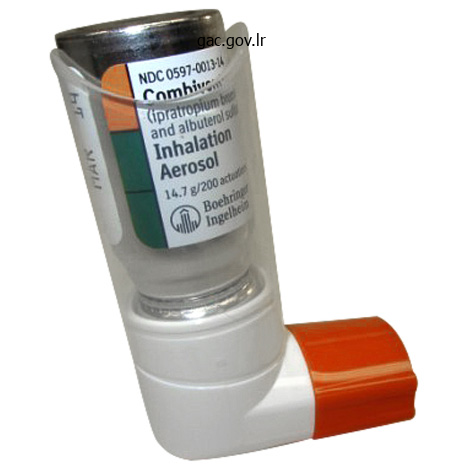
Trusted 100mcg combivent
Other causes of cor neal opacity include persistent uveitis, interstitial keratitis, corneal edema, lattice corneal dystrophy (amyloid depo sition), and long-standing glaucoma. Polysaccharides are deposited in the corneas in some of the mucopolysac charidoses (see Chap. The corneas are also diffusely clouded in sure lysosomal storage diseases (see Chap. Arcus senilis occurring at an early age (because of hyperlipidemia), typically combined with yellow lipid deposits in the eyelids and periorbital skin (xanthelasma), serves as a marker of atheromatous vascular disease. In the anterior chamber of the attention, a common problem is obstacle to the outflow of aqueous fluid, associ ated with excavation of the optic disc and visible loss, i. In more than ninety percent of circumstances (of the open angle type), the cause for this syndrome is unknown and a genetic issue is suspected. In roughly 5 % of cases, the angle between iris and the peripheral cornea is slender and blocked when the pupil is dilated (angle closure glaucoma). In the remaining instances, the situation is a results of some disease course of that blocks outflow channels-inflamm atory particles of uveitis, purple blood cells from hemorrhage in the anterior chamber (hyphema), new formation of vessels and connective tissue on the surface of the iris (rubeosis iridis), a relatively rare complication of ocular ischemia secondary to diabetes mellitus, retinal vein occlusion, or carotid artery occlu sion. The visual loss is gradual in open-angle glaucoma and the attention looks regular, in distinction to the red, painful eye of angle-closure glaucoma that was described above in ref erence to pharmacologic dilation of the pupil to facilitate fundoscopy. Intraocular pressures which are persistently above 20 mm Hg may damage the optic nerve over time. This could also be manifest first as an arcuate defect in the upper or lower nasal area or as a paracentral subject defect, which, if untreated, may proceed to blindness. Other characteristic glaucomatous subject patterns are winged extensions from the blind spot (Seidel scotoma) and a narrowing of the superior nasal quadrant which will progress to a horizontal edge, cor responding to the horizontal raphe of the retina (nasal step). It is now appreciated that elevated intraocular pressure is just a concurrent discovering and a danger factor for glaucoma and that optic harm could also be seen in sufferers with close to normal pressure. This represents a major revision of the previous view that stress was the basic trigger of harm in glaucoma. The "sugar cataract" of diabetes mellitus is the end result of sustained high ranges of blood glucose, which is modified within the lens to sorbi tol, the accumulation of which outcomes in a excessive osmotic gradient with swelling and disruption of the lens fibers. Galactosemia is a a lot rarer cause, however the mechanism of cataract formation is analogous, i. In hypoparathyroidism, decreasing of the concentration of calcium in the aqueous humor is in some way liable for the opacification of newly forming superficial lens fibers. Prolonged excessive doses of corticosteroids, in addition to radiation therapy, induce lenticular opacities in some patients. Subluxation of the lens, the results of weakening of its zonular ligaments, happens in syphilis, Marfan syndrome (upward displacement), and homocystinuria (downward displacement). In the vitreous humor, hemorrhage might happen from rupture of a ciliary or retinal vessel. On ophthalmo scopic examination, the hemorrhage appears as a diffuse haziness of half or all the vitreous or, if the blood is between the retina and the vitreous and displaces the lat ter quite than mixing with it, takes the form of a sharply outlined clot. The most common vitreous opacities are benign "floaters" caused by the condensation of vitreous collagen fibers, which seem as darting gray flecks or threads with changes within the place of the eyes; they might be annoying or even alarming until the person stops looking for them. A sudden burst of flashing lights associated with a rise in floaters could mark the onset of retinal detach ment. Patients complaining of bright flashes and spots in imaginative and prescient ought to be examined with the indirect ophthal moscope to rule out tears, holes, or detachments of the vitreous or retina. Another widespread prevalence with advancing age is shrinkage of the vitreous humor and retraction from the retina, causing persistent streaks of sunshine, usually within the periphery of the visible area. These phosphenes, also known as Moore lightning streaks, had been thought to be fairly benign, however they may at instances, indicate incipient retinal or vitreous tears or detachment, and their first look requires prompt analysis by an ophthalmologist. They are most prominent on transfer ment of the globe, on closure of the eyelids, at the moment of lodging, with saccadic eye movements, and with sudden exposure to darkish. The time period uveitis refers to an infective or noninfec tive inflammatory illness that impacts any of the uveal buildings (iris, ciliary physique, and choroid). According to Bienfang and colleagues, uveitis accounts for 10 per cent of all instances of authorized blindness within the United States. The irritation may be within the anterior part of the eye or in the posterior half, behind the iris and extending to the retina and choroid. Visual stimuli entering the eye traverse the inner layers of the retina to reach its outer (posterior) layer, which contains two classes of photoreceptor cells: the flask-shaped cones and the slender rods. The photore ceptors relaxation on a single layer of pigmented epithelial cells, which kind the outermost floor of the retina.
Comb Flower (Echinacea). Combivent.
- Dosing considerations for Echinacea.
- How does Echinacea work?
- Are there safety concerns?
- Is Echinacea effective?
- Are there any interactions with medications?
- Urinary tract infections (UTIs), migraine headaches, chronic fatigue syndrome (CFS), eczema, hayfever, allergies, bee stings, attention deficit-hyperactivity disorder (ADHD), influenza (flu), and other conditions.
- Preventing vaginal yeast infections when used with a medicated cream called econazole (Spectazole).
- Preventing recurrent genital herpes.
- What other names is Echinacea known by?
- What is Echinacea?
Source: http://www.rxlist.com/script/main/art.asp?articlekey=96942
Buy combivent 100mcg low price
The broadly adopted Glasgow Coma Scale, constructed initially as a fast and easy means of quantitating the responsiveness of sufferers with cerebral trauma, can be utilized in the grading of other acute coma-producing diseases as talked about earlier on this chapter (see additionally Chap. It is usually possible to determine whether or not coma is related to meningeal irritation. In all but the deep est stages of coma, meningeal irritation from either bacte rial meningitis or subarachnoid hemorrhage will trigger resistance to the initial excursion of passive flexion of the neck however not to extension, turning, or tilting of the head. Meningismus is a fairly specific however somewhat insensitive signal of meningeal irritation as commented in Chap. In the toddler, bulging of the anterior fontanel is at instances a more reliable signal of meningitis than is a stiff neck. A temporal lobe or cerebellar herniation or decere brate rigidity can also create resistance to passive flexion of the neck and be confused with meningeal irritation. A coma-causing lesion in a cerebral hemisphere can be detected by cautious observation of spontaneous move ments, responses to stimulation, prevailing postures, and by examination of the cranial nerves. Hemiplegia is revealed by an absence of stressed actions of the limbs on one facet and by insufficient protective actions in response to painful stimuli. The weakened limbs are normally slack and, if lifted from the mattress, they "fall flail. A lesion in a single cerebral hemisphere causes the eyes to be turned away from the paralyzed facet (toward the lesion, as described below); the alternative occurs with brainstem lesions. In most circumstances, a hemiplegia and an accompanying Babinski sign are indicative of a contralateral hemispheral lesion; however with lateral mass impact and compression of the other cerebral peduncle in opposition to the tentorium, extensor posturing, a Babinski sign, and weakness of arm and leg may seem ipsilateral to the lesion (the earlier-mentioned Kernahan-Woltman sign). A moan or grimace may be provoked by painful stimuli applied to one side but to not the other, reflecting hemianesthesia. Of the various indicators of brainstem function, essentially the most useful are pupillary size and reactivity, ocular move ments, oculovestibular reflexes and, to a lesser extent, the pattern of respiration. These functions, like consciousness itself, are depending on the integrity of constructions within the midbrain and rostral pons. As a transitional phenomenon, the pupil could turn into oval or pear-shaped or appear to be off middle (corec topia) because of a differential lack of innervation of a portion of the pupillary sphincter. The light-unreactive pupil continues to enlarge to a measurement of 6 to 9 mm diameter and is quickly joined by a slight outward deviation of the attention. In uncommon situations, the pupil contralateral to the mass might enlarge first; this has reportedly been the case in 10 p.c of subdural hematomas but has been far much less frequent in our experience. As midbrain displacement continues, each pupils dilate and become unreactive to gentle, in all probability as a end result of compression of the oculomo tor nuclei in the rostral midbrain (Rapper, 1990). The final step within the evolution of brainstem compression tends to be a slight discount in pupillary measurement on both sides, to 5 mm or smaller. Normal pupillary dimension, form, and light-weight reflexes indicate integrity of midbrain buildings and direct consideration to a reason for coma apart from a mass. Pontine tegmental lesions trigger extraordinarily miotic pupils (<1 mm in diameter) with barely perceptible response to sturdy gentle; this is attribute of the early part of pon tine hemorrhage. The ipsilateral pupillary dilatation from pinching the side of the neck (the ciliospinal reflex) is usu ally lost in brainstem lesions. The Homer syndrome (miosis, ptosis, and lowered facial sweating) could additionally be noticed ipsi lateral to a lesion of the brainstem or hypothalamus or as an indication of dissection of the internal carotid artery. With coma attributable to drug intoxications and intrin sic metabolic disorders, pupillary reactions are normally spared, however there are notable exceptions. High-dose barbiturates may act equally, however the pupillary diameter tends to be 1 mm or more. Systemic poisoning with atropine or with medication that have atropinic qualities, particularly the tricyclic anti depressants, is characterized by extensive dilatation and repair ity of the pupils. Hippus, or fluctuating pupillary dimension, is sometimes attribute of metabolic encephalopathy. A unilaterally enlarged pupil is an early indicator these are altered in a wide selection of ways in coma. In gentle coma of metabolic origin, the eyes rove conjugately from side to aspect in seemingly random trend, sometimes resting briefly in an eccentric position. These actions disappear as coma deepens, and the eyes then remain immobile and slightly exotropic. A lateral and slight downward deviation of 1 eye suggests the presence of a third-nerve palsy, and a medial deviation, a sixth-nerve palsy.
Buy 100 mcg combivent overnight delivery
Transient international amnesia could additionally be ischemic or per haps migrainous in nature, although not atherosclerotic thrombotic, however not often (if ever) do the assaults progress to stroke. From oblique evidence of retrograde blood circulate in the inside jugular arteries in the course of the Valsalva maneuver (occasion ally reported to precipitate an attack), Sander and col leagues and Chung and coworkers have advised that venous congestion of the temporal lobes was operative. The precipitation of similar attacks by, vertebrobasilar and coronary angiography is also sugges tive of an ischemic or migrainous causation. This pro vides a possible explanation for the association of extremely emotional events prior to an episode. In addition, the mode of answering and fixing problems provides invaluable information about the psychological operations of the topic and should be incor porated into any evaluation of cognition. A perplexed or slowed individual may in the end perform adequately but nonetheless have critically flawed cortical or subcor tical operate. Each of the checks under is necessarily an abstraction however ones that separate particular features of the mind. As already emphasized, the patient should have regular, or almost so, attentiveness to perform these tasks and a deficiency in any certainly one of them might disrupt the per formance of others. Verbal trail making (reciting alternating letters of the alphabet and their ordinal place, i. The capacity to repro duce them at intervals after committing them to memory is a test of memory span. Visual facility: Show the patient an image of a quantity of objects; then ask him to name the objects. Subtraction of serial 3s and 7s from a hundred is an efficient check of calculation in addition to of focus. Constructions: Ask the patient to draw a clock and place the palms at 7:forty five, a map of the United States, a flooring plan of her house; ask the patient to copy a dice and other figures. General habits: Attitudes, common bearing, evidence of hallucinosis, stream of coherent thought and atten tiveness (ability to keep a sequence of psychological operations), mood, manner of costume, etc. Special exams of localized cerebral features: Grasping, sucking, aphasia battery, praxis with each palms, and corticosensory operate. To enlist the complete cooperation of the patient, the physician should put together him for questions of this type. If the patient is agitated, suspicious, or belligerent, intellectual functions should be inferred from his remarks and from data equipped by the household. We additionally find it helpful to quiz the affected person about cultural icons of the past which are appropriate to his age. Recent past: Tell me about your latest sickness (com pare with earlier statements). Immediate recall (attention, short-term working mem ory): Repeat these numbers after me (give series of 3, 4, 5, 6, 7, eight digits at a speed of 1 per second). In our experience, a excessive level of perfor mance on all checks eliminates the potential of dementia in nearly all circumstances. It might fail to identify a dementing illness in an uncooperative affected person and in a highly intel ligent individual in the earliest stages of illness. The question of whether to resort to formal psycho logic checks is certain to arise. A score of 24 on the widely used "mini-mental" is taken into account regular and scores under 21 usually indicate cognitive impairment. Patients with lower levels of education and older age have lower nor mative scores, however even people of their eighties with a highschool education rating 23 or above if not demented (see Crurn et al for age and education adjusted normal score). In this test, an index of decay is supplied by the discrepancy between the vocabulary, picture-completion, and object-assembly tests as a gaggle (these correlate properly with premorbid intelligence and are comparatively insensitive to dementing brain disease) and different measures of general perfor mance, specifically arithmetic, block-design, digit-span, and digit-symbol checks. Questions that measure spatial and temporal orientation and reminiscence are the important thing objects in most of these abbreviated scales of dementia. All of the aforementioned scientific and psychologic tests, and several other others as nicely, measure the same elements of behavior and intellectual operate. X-J Ask the affected person to copy a pair of intersecting pentagons onto a piece of paper. The primary responsibility of the doctor is to diagnose the treatable types of dementia and to insti tute applicable therapy. To this question we normally reply that they may, but that extra time is required to make sure. Reassurance that the physician will be obtainable to help the patient and family handle the state of affairs is of utmost worth.
Order combivent 100 mcg without prescription
A affected person with lumbosacral pressure or disc illness (except within the acute phase or if the disc fragment has migrated laterally) can usually prolong the backbone with little or no aggravation of pain. Maneuvers within the lateral decubitus place yield less info but are useful in eliciting joint disease. In cases of sacroiliac joint illness, abduction of the upside leg against resistance reproduces ache in the sacroiliac area, generally with radiation of the ache to the buttock, posterior thigh, and symphysis pubis. Hyperextension of the upside leg with the draw back leg flexed is another test for sacroiliac illness. Rotation and abduction of the leg evoke pain in a diseased hip joint and with trochanteric bursitis. A helpful indicator of hip illness is the Patrick test: with the patient supine, the heel of the offending leg is placed on the opposite knee, and pain is evoked by depressing the flexed leg and exter nally rotating the hip. It is preferable to first pal pate the regions which are the least more likely to evoke ache. Localized tenderness is seldom pronounced in disease of the backbone as a outcome of the involved structures are so deep. Nevertheless, tenderness over a spinous process or jarring by mild percussion may indi cate the presence of deeper, local spinal inflammation (as in disc space infection), pathologic fracture, metastasis, epidural abscess, or a disc lesion. Tenderness over the interspinous ligaments or over the area of the articular sides between the fifth lumbar and first sacral vertebrae is according to lumbosacral disc disease. Tenderness in this region and in the sacroiliac joints is also a frequent mani festation of ankylosing spondylitis. Tenderness over the costovertebral angle usually signifies genitourinary disease, adrenal disease, or an injury to the transverse means of the first or second lumbar vertebra. Tenderness on palpation of the paraspinal muscle tissue may signify a strain of muscle attachments or injury to the underlying transverse processes of the lumbar vertebrae. Focal ache in the same parasagittal line alongside the tho racic spine factors to inflammation of the costotransverse articulation between spine and rib (costotransversitis). Other sites of tenderness and the structures implicated by illness are proven in the figure. In palpating the spinous processes, it is important to note any deviation in the lateral aircraft (this could also be indicative of fracture or arthritis) or within the anteroposterior aircraft. A "step-off" ahead displacement of the spinous process and exaggerated lordosis are important clues to the presence of spondylolisthesis (see further on). Many of the processes discussed above can coexist, particularly in the older particular person, who could have hip and lumbar spine osteoarthropathy. This makes the interpre tation of assorted signs tough except the symptoms are first analyzed correctly. On completion of the examination of the again and legs, one turns to a search for motor, reflex, and sensory adjustments within the decrease extremities (see "Herniation of Lumbar Intervertebral Discs," further on on this chapter). Region of sacrosciatic notch (tenderness = fourth or fifth lumbar rlisc rupture and sacroiliac sprain). Radiographs of the lumbar spine may be helpful in the routine analysis of low again pain and sciatica and could be carried out with the patient in flexed and extended positions in the anteroposterior, lateral, and oblique planes. Readily demonstrable in plain movies are narrowing of the intervertebral disc spaces, bony facetal or vertebral overgrowth, displacement of vertebral bodies (spondy lolisthesis), and an unsuspected infiltration of bone by most cancers. This results in an anterior displacement of 1 vertebral physique in relation to the adjacent one, spondylolisthesis. The main reason for spondylolisthesis in older adults is degenerative arthritic disease of the backbone as discussed additional on. Patients with progressive vertebral displace ment and neurologic deficits require surgical procedure. Reduction of displaced vertebral bodies earlier than fusion and direct repair of pars defects are possible in particular circumstances. A common anomaly is fusion of the fifth lumbar vertebral body to the sacrum ("sacralization") or, con versely, separation of the first sacral section, giving rise to 6, rather than the standard 5 lumbar vertebrae ("lum barization"). However, neither of those is constantly associated with any kind of back derangement. Another one or several of the lumbar vertebrae or of the sacrum less-common finding is an absence of fusion of the laminae of (spina bifida). The anomaly could additionally be accompanied by malformation of vertebral joints and often induces ache only when aggravated by harm. The neurologic features of defective fusion of the backbone (dysraphism) are discussed in Chap.
Generic combivent 100 mcg on-line
As a rule, lesions of the cerebral cortex and white matter are asso ciated not with ache but with hypalgesia. Schott For instance, Dyck and colleagues, in a examine of painful versus nonpainful axonal neuropathies, concluded that fiber degeneration. Also, the incidence of ectopic impulse there was no distinction between them when it comes to the kind of technology all along the surface of injured axons and the possibility of ephaptic activation of unsheathed axons appear relevant notably to some causalgic states. Stimulation of the nervi nervorum of larger nerves by an expanding intraneural lesion or a vascular change was postulated by Asbury and Fields because the mechanism of nerve trunk ache. The sprouting of adrenergic sympathetic axons in response ostensible explanation for the abolition of causalgic pain by sympathetic blockade. This has given rise to the term to nerve damage has already been talked about and is an sympathetically sustained pain as discussed below. Particular illnesses giving rise to neuropathic ache are considered of their appropriate chapters but the fol lowing remarks are of a general nature, relevant to all the painful states that compose this group. The sensations that characterize neuropathic pain differ and are often multiple-burning, gnawing, aching, and taking pictures or lancinating qualities are described. There is an almost invariable affiliation with a quantity of of the signs of hyperesthesia, hyperalgesia, allodynia, and hyperpathia (see above). The irregular sensations coexist in plenty of cases with a sensory deficit and native autonomic dysfunction. Furthermore, the pain usually responds poorly to remedy, together with the administra tion of opioid medications. Regenerating axonal sprouts, as in a neuroma, are additionally hypersensitive to mechanical stimuli. On a molecular level, it has been proven that voltage-gated sodium chan nels accumulate at the site of a neuroma and all alongside the axon after nerve injury, and that this provides rise to ectopic and spontaneous activity of the sensory nerve cell and its axon. This mechanism is concordant with the aid of neurogenic pain by sodium channel-blocking anti-epileptic medication. Spontaneous exercise in nociceptive C fibers is thought to give rise to burning pain; firing of enormous myelinated A fibers is believed to produce dys esthetic ache induced by tactile stimuli. The abnormal response to stimulation can be influenced by sensitization of central ache pathways, probably in the dorsal horns of the spinal cord, as outlined within the evaluation by Woolf and Mannion. Hyperalgesia and allodynia are thought to result from such a spinal wire mechanism. Several obser vations have been made concerning the neurochemical mechanisms that might underlie these modifications however none provides a constant explanation. Possibly multiple of those mechanisms is operative in a given periph eral nerve disease. Evidence that the sodium channel can generate neu ral ache is given by the extraordinary disease "paroxys mal excessive pain dysfunction" also referred to as "familial rectal ache syndrome. Pain states of peripheral nerve origin far outnumber those attributable to spinal cord, brainstem, thalamic, and cerebral illness. Although the pain is local ized to a sensory territory equipped by a nerve, plexus or nerve root, it typically radiates to adjacent areas. Sometimes the onset of pain is quick on receipt of damage; extra often it appears at some point through the evolution or recession of the disorder. The illness of the nerve may be obvious, expressed by the standard sensory, motor, reflex, and autonomic adjustments, or these changes could also be unde tectable by standard checks. Similar but extra diffuse painful states such as erythromelalgia and paroxysmal excessive ache disorder are being uncovered which are predi cated on similar voltage-gated sodium channel mutations and extra impressively, by the congenital absence of the flexibility to expertise pain due to a lack of operate muta tion in a sodium channel gene and a mutation within the tyro sine kinase receptor gene. He noted that when a gaggle of neurons is deprived of its pure innervation, they become hyperactive. Others point to a lowered density of certain types of fibers in nerves supplying a causalgic wne as the basis of the burning pain but the comparability of the density of nerves from painful and nonpainful neuropathies has not proved to be constantly totally different. The term causalgia is, in our view, best reserved for the syndrome described above-i. Some neurologists use "causalgia" to describe only the burning feature of ache because of partial nerve injury. Others have applied the time period to a variety of conditions which might be characterized by persistent burning pain but have only an inconstant association with sudomotor, vasomotor, and trophic adjustments and an unpredictable response to sympathetic blockade. The medical features of each the causalgic and dystonic elements of the syndrome have been somewhat unusual in the cases reported.
Buy combivent with amex
The baby awakens abruptly 1 in 5 sleepwalkers has a household history of this disorder. Motor efficiency and responsiveness in the course of the sleep walking incident differ significantly. The most typical behavioral abnormality is for a affected person to sit up in bed or on the edge of the bed with out actually strolling. When walking about the home, he might turn on a lightweight or per type some other familiar act. There could also be no outward present of emotion, or the patient could additionally be frightened (night terror), however the frenzied, aggressive behavior of some grownup sleepwalkers, described below, is uncommon in the youngster. Usually the eyes are open, and such sleepwalkers are guided by vision, thus avoiding familiar objects; the sight of an unfamiliar object may awaken them. If spoken to , they make no response; if advised to return to mattress, they might achieve this, but extra typically they should be led again. Sometimes they repeatedly mutter strange phrases or perform sure repetitive acts, such as push ing against a wall or turning a doorknob backwards and forwards. Children with night terrors are sometimes sleepwalkers as nicely, and each sorts of attack may happen simultaneously. The entire episode lasts only a minute or two, and in the morning the child recalls nothing of it or only a vague unpleasant dream. The persistence of such issues into grownup life, however, has, in a small number of cases, been asso ciated with psychopathology (Kales et al). It has been found that diazepam, which reduces the length of the deep stages of sleep, will stop evening terrors. Selective serotonin reuptake inhibitors have additionally been used suc cessfully, especially when night terrors are related to sleepwalking. Frequent evening terrors have report edly been eradicated by having dad and mom awaken the child for several successive nights, just previous to the standard time of the assault or at the first sign of restlessness and auto nomic arousal (Lask). Frightening desires or nightmares are way more frequent than night terrors and affect kids and adults alike. Sleepwalking should be distin guished from fugue states and ambulatory automatisms of advanced partial seizures discussed in Chap. Children often out develop this disorder; mother and father should be reassured on this rating and disabused of the notion that somnambulism is an indication of psychiatric or some other disease. Almost all the time, the grownup sleepwalker has a historical past of sleepwalking as a baby, although there may have been a interval of freedom between the child hood episodes and their reemergence within the third and fourth a long time. If one extends the class of somnambulism to all forms of nocturnal wandering, it seems to be remarkably widespread, with a lifetime prevalence of 29% of U. Somnambulism in the grownup, as in the baby, can be a purely passive event unaccompanied by fear or different indicators of emotion. More incessantly, however, the attack is characterised by frenzied or violent behavior associ ated with concern and tachycardia, like that of a night terror and sometimes with self-injury. Very rarely, crimes have reportedly been committed during sleepwalking, but the authors are skeptical that organized and planned sequen tial exercise is feasible. The finding of regular sleep pat terns on polysomnography distinguishes these assaults from complicated partial seizures. Some patients reply higher to a combination of clonazepam and phenytoin or to fluraz epam (Kavey et al). Also, within the provocatively named "sexomnia," the individual, male or female, engages in sexual exercise, generally forcefully, and has no recollection of the events. It is characterised by attacks of vigorous, agitated, and infrequently dangerous motor activity accompanied by vivid goals (Mahowald and Schenck). The characteristic options are angry speech with shout ing, violent exercise with damage to self and mattress mate, a really high arousal threshold, and the variable however some times detailed recall of a nightmare of being attacked and combating again or attempting to flee. The episodes range in frequency in affected people, occurring as soon as every week or two or several instances nightly. Polysomnographic recordings dur ing these episodes have disclosed augmented muscle tone but no seizure exercise. The rare appearance of this disorder with pontine infarctions has been talked about earlier within the chapter. These observations have led to the sug gestion that this dysfunction is an early manifestation of a degenerative mind disease characterized by the deposi tion of alpha-synuclein in certain neuronal methods, as summarized by Boeve and associates. The episodes could be suppressed by the administra tion of clonazepam in doses of zero.
Syndromes
- Black, tarry stools
- Methods to cause vomiting
- Malabsorption
- Rapid breathing
- Dust
- Chronic sinusitis is when swelling and inflammation of the sinuses are present for longer than 3 months. It may be caused by bacteria or a fungus.
Generic combivent 100 mcg visa
Large and heavily myelinated fibers enter the twine just medial to the dorsal hom and divide into ascending and descending branches. The descending fibers and some of the ascending ones enter the grey matter of the dorsal hom inside a couple of segments of their entrance and synapse with nerve cells in the pos terior horns as properly as with large ventral hom cells that subserve segmental reflexes. Some of the ascending fibers run uninterruptedly in the dorsal columns of the identical side of the spinal cord, terminating within the gracile and cune ate nuclei within the higher cervical spinal wire and medulla. The central axons of the first sensory neu rons are joined within the posterior columns by other second ary neurons whose cell our bodies lie within the posterior horns of the spinal wire (see below). The fibers in the posterior columns assume a medial place as new fibers from every successively greater root are added laterally, thereby creating somatotopic laminations. Of the long ascending posterior column fibers, that are activated by mechanical stimuli of pores and skin and subcuta neous tissues and by movement of joints, solely about 25 percent (from the lumbar region) reach the gracile nuclei at the upper cervical twine. The remaining fibers ship collater als to , or terminate in, the dorsal horns of the spinal wire, a minimum of in the cat (Davidoff). There are also descending fibers in the posterior columns, including fibers from cells within the dorsal column nuclei. The posterior columns comprise a portion of the fibers for the sense of contact as properly as the fibers mediating the senses of touch-pressure, vibration, course of movement and place of joints, and stereoesthesia-recognition of surface texture, form, numbers, and figures written on the pores and skin and two-point discrimination-all of which rely upon patterns of touch-pressure. The nerve cells of the nuclei gracilis and cuneatus and accessory cuneate nuclei give rise to a secondary afferent path, which crosses the midline in the medulla and ascends because the medial lem niscus to the posterior thalamus. In addition to the well-defined posterior column pathways, there are cells in the extra loosely structured "reticular" part of the dorsal column that obtain second ary ascending fibers from the dorsal horns of the spinal wire and from ascending fibers in the posterolateral col umns. These dorsal col umn fibers project to brainstem nuclei, cerebellum, and thalamic nuclei. Many other cells of the dorsal hom nuclei are intemeurons, with each excitatory and inhibitory results on local reflexes or on the first ascending neurons. The major somatosensory pathways emphasizing the posterior column-lemniscal system (thicker tract lines). The dorsal horn cells, in flip, give rise to secondary sensory fibers, some of which may ascend ipsilaterally however most of which decussate and ascend within the spino thalamic tracts, as described in Chap. Observations primarily based on surgical interruption of the anterolateral funiculus indicate that fibers mediating contact and deep strain occupy the ventromedial part (anterior spinothalamic tract). The lemniscal system is located in a medial to the descending corticospinal system. After coming into the pons, the pain and tempera ture fibers turn caudally and run via the ipsilateral medulla as the descending spinal trigeminal tract, termi nating within the lengthy, vertically oriented nucleus caudalis, or spinal trigeminal nucleus, that lies beside it and extends to the second or third cervical phase of the wire, the place it becomes continuous with the posterior horn of the spi nal grey matter. Axons from the neurons of this nucleus cross the midline and ascend as the trigeminal quintotha lamic tract (also termed, somewhat imprecisely, trigemi nal lemniscus) alongside the medial aspect of the spinothalamic tract. The first area (Sl) corresponds to the publish central cortex or Brodmann areas three, 1, and a pair of. Electrical stimulation of this space yields sensations of tingling, numbness, and heat in particular regions on the alternative facet of the body. The data transmitted to Sl is tactile and proprioceptive, derived mainly from the dorsal column-medial lemniscus system and concerned mainly with sensory discrimination. The second somatosensory area (S2) lies on the higher bank of the sylvian fissure, adjoining to the insula. Localization of operate is much less discrete in S2 than in Sl, but S2 is also organized somatotopically, with the face rostrally and the leg caudally. The sensations evoked by electrical stimulation of S2 are a lot the same as these of Sl but, in distinction to the latter, could additionally be felt bilaterally. Undoubtedly, the notion of sensory stimuli involves extra of the cerebral cortex than the 2 discrete areas described above. Some sensory fibers probably proj ect to the precentral gyrus and others to the superior pari etal lobule. This reciprocal association most likely influences motion and the transmission and interpretation of pain, as discussed in Chap. The "sensory homunculus," or cortical illustration of sensation in the postcentral gyrus; compare this to the distribution of physique areas within the motor cortex as shown in.
Purchase combivent 100mcg with mastercard
A extra extensive syndrome was originally described by Bartleson, Swanson, and Whisnant under the title "A migrainous syndrome with cerebrospinal fluid pleocytosis". One-quarter of this group had a history of previous migraine and a similar number had a viral-like illne ss within three weeks of the neu rologic downside. The transient neurologic deficits have been primarily sensorimotor and aphasic; solely 6 patients had visible symptoms. The patients had been asymptomatic between attacks and in none did the entire illness persist beyond 7 weeks. The causation and pathophysiology of this syndrome and its relation to migraine are obscure. The distinction between this syndrome and the recurrent aseptic meningitis of Mollaret and different chronic meningitic syndromes as well as cere bral vasospasm or vasculitis is difficult (see "Chronic Persistent and Recurrent Meningitis" in Chap. Tension and other emotional states, which are claimed by some migraineurs to precede their attacks, are so incon sistent as to be no extra than potential aggravating fac tors. The puzzle is how this genetic predisposition is translated periodically right into a regional neurologic deficit, unilateral headache, or each. For many years, our excited about the pathogenesis of migraine was dominated by the views of Harold Wolff and others-that the headache was brought on by the distention and excessive pulsation of branches of the exterior carotid artery. Certainly, the throbbing, pulsating high quality of the headache and its reduction by compression of the common carotid artery supported this view, as did the early remark of Graham and Wolff that the headache and amplitude of pulsation of the extracranial arteries diminished after the intravenous administration of ergotamine. The importance of vascular elements continues to be emphasised by more recent findings however not in the finest way envisaged by Wolff. For instance, in a group of 11 sufferers with traditional migraine, Olsen and colleagues, utilizing the xenon inhalation methodology, famous a regional reduction in cerebral circulation spreading forward from the occipital area during the period when neurologic signs appear. They concluded that the discount in blood flow was in keeping with the cortical spreading depression syndrome described beneath. In reference to the extracranial vessels, Iversen and associates, by means of ultrasonography, documented a dilatation of the superior temporal artery on the side of the migraine during the headache period. The same dilatation within the center cerebral arteries has been inferred from observations with transcranial Doppler insonation. The complication of cerebral infarction can additionally be consistent with a vascular hypothesis, nevertheless it includes solely a tiny proportion of migraineurs. The original opinion expressed by Wolff that a vascular component is answerable for the cranial pain of migraine can be unconfirmed. The relationship between the vascular modifications and evolving neurologic signs of migraine are notewor thy. Lashley, who plotted his personal visible aura, calculated that the cortical impairment progressed at a price of two to 3 mm/min over the floor of the brain. Both of these occasions are intrigu ingly similar to the above-mentioned phenomenon of "spreading cortical despair," first noticed by Leao in experimental animals. He demonstrated that a noxious stimulus utilized to the rat cortex was adopted by vaso constriction and slowly spreading waves of inhibition of the electrical exercise of cortical neurons, moving at a rate of approximately three mm/min. Lauritzen and Olesen attri bute each the aura and spreading oligemia to the unfold ing cortical melancholy, and appreciable work since then has corroborated this idea. An various, but not necessarily unique hypoth esis hyperlinks the aura and the painful section of migraine through a neural mechanism originating in the trigemi nal nerve as proposed by Moskowitz. This relies on the innervation of extracranial and intracranial vessels by small unmyelinated fibers of the trigeminal nerve that subserve both pain and autonomic features (the "trigeminovascular" complex). The small molecules released from nerve end ings adjacent to the cortex would then incite spreading melancholy on this mannequin. Against this hypothesis is the incidence of headache as usually as not on the aspect reverse the side of technology of the aura and the lack of clinical impact of medicine that work on this experimental mannequin. These authors level to their findings that occlusion of blood circulate via the scalp or widespread carotid circulation fails to alleviate the pain of migraine in one-third to one half the sufferers. Lance (1998) has instructed that the trigeminal pathways are in a state of persistent hyperex citability in the migraine patient and that they discharge periodically, perhaps in response to hypothalamic stimu lus appearing on the endogenous pain control pathways.
Purchase 100 mcg combivent amex
Lesions of the premotor cor tex (area 6) end in weakness, spasticity, and increased stretch reflexes (Fulton). Resection of cortical areas 4 and 6 and subcortical white matter in monkeys causes complete and everlasting paralysis and spasticity (Laplane et al). The one place the place corticospinal fibers are totally isolated is the pyramidal tract within the medulla. In people, there are a quantity of documented cases of a lesion roughly confined to this location. Similarly in monkeys-as was shown by Tower in 1940 and subse quently by Lawrence and Kuypers and by Gilman and Marco-interruption of each pyramidal tracts leads to a hypotonic paralysis; ultimately; these animals regain a variety of actions, although slowness of all movements and lack of particular person finger actions remain as everlasting deficits. Also, the cerebral peduncle had prior to now been sectioned in patients in an effort to abolish involuntary movements (Bucy et al). In some of these patients, a slight degree of weak point or solely a Babinski sign was produced however no spasticity devel oped. Furthermore, to reiterate a earlier comment, management over a variety of voluntary actions depends at least partially on nonpyramidal motor pathways. Animal experiments recommend that the corticoreticulospinal pathways are par ticularly important in this respect, because their fibers are arranged somatotopically and affect stretch reflexes. Further research of human disease, presumably utilizing diffu sion tensor imaging strategies, are necessary to settle issues related to volitional movement and spasticity. The distribution of the paralysis attributable to upper motor neuron (supranuclear) lesions varies with the locale of the lesion, but certain options are attribute of all of them. A group of muscles is always concerned, never individual muscle tissue, and if any movement is pos sible, the proper relationships between agonists, antago nists, synergists, and fixators are preserved. On careful inspection, the paralysis never entails all of the muscles on one aspect of the body, even in the severest types of hemi plegia. Movements that are invariably bilateral-such as these of the eyes, jaw, pharynx, higher face, larynx, neck, thorax, diaphragm, and abdomen-are affected little or by no means. Upper motor neuron lesions are characterised fur ther by sure peculiarities of retained movement. There is decreased voluntary drive on spinal motor neurons (fewer motor models are recruitable and their firing charges are slower), resulting in a slowness of movement. There can be an increased diploma of co-contraction of antagonistic muscles, mirrored in a decreased fee of fast alternating movements. These abnormalities in all probability account for the greater sense of effort and the manifest fatigability in effecting voluntary motion of the weakened muscular tissues. Another phenomenon is the activation of paralyzed muscular tissues as parts of certain automatisms (synkinesias). Attempts by the patient to move the hemiplegic limbs may lead to quite so much of associated movements. Thus, flexion of the arm could end in involuntary pronation and flexion of the leg or in dorsiflexion and eversion of the foot. Also, volitional actions of the paretic limb often evoke imitative (mirror) movements in the regular one or vice versa. In some sufferers, as they get well from hemiplegia, a variety of movement abnormalities emerge, such as tremor, atheto sis, and chorea on the affected aspect. These are expressions of damage to basal ganglionic and thalamic buildings and are mentioned in Chap. If the higher motor neurons are interrupted above the level of the facial nucleus in the pons, hand and arm muscles are affected most severely and the leg muscles to a lesser extent; of the cranial musculature, solely muscles of the tongue and lower part of the face are involved to any vital diploma. At lower levels, such because the cervical wire, full, acute, and bilateral lesions of the upper motor neurons not solely cause a paralysis of voluntary movement but also quickly abolish the spinal reflexes of segments beneath the lesion. With some acute cerebral lesions, spasticity and paralysis develop together; in others, especially with parietal lesions, the limbs stay flaccid but reflexes are retained. The antigrav ity muscles-the flexors of the arms and the extensors of the legs-are predominantly affected.
Generic combivent 100mcg on-line
With unilateral sciatica, the patient lists to one facet and strongly resists bending to the other side, and the popular posture in standing is with the leg barely flexed on the hip and knee. When the herniated disc lies lateral to the nerve root and displaces it medially, tension on the basis is decreased and pain is relieved by bending the trunk to the facet opposite the lesion; with herniation medial to the foundation, tension is lowered by inclining the trunk to the aspect of the lesion. In the sitting position, flexion of the hips could be per shaped more simply, even to the point of bringing the knees in touch with the chest. The reason for that is that knee flexion relaxes tightened hamstring muscles and relieves the stretch on the sciatic nerve. This characteristic may be evident in instances of lumbar disc disease, mak ing the maneuver less sensitive than others. Examination with the patient in the reclining position yields much the identical info as within the standing and sitting positions. With vertebral illness, passive flexion of the hips is free, whereas flexion of the lumbar spine could also be impeded and painful. Among the most useful indicators in detecting nerve root compression is passive straight-leg raising (possible as a lot as nearly ninety levels in normal individuals) with the patient supine. This places the sciatic nerve and its roots under rigidity, thereby producing radicular, radiating pain from the buttock through the posterior thigh. Straight elevating of the alternative leg ("crossed straight-leg raising," Fajersztajn sign) may trigger sciatica on the other facet and is a extra particular signal of prolapsed disc than is the Lasegue signal. Several of the various derivatives of the straight-leg elevating signal are mentioned within the section on lumbar disc illness. Asking the seated patient to lengthen the leg so that the only real of the foot could be inspected is a means of checking for a feigned Lasegue sign. Many different congenital variants have an result on the decrease lumbar vertebrae: asymmetrical side joints, abnormali ties of the transverse processes, are seen occasionally in patients with low back symptoms, but apparently with no greater frequency than in asymptomatic individuals. Spondylolysis consists of a congenital and probably genetic bony defect within the pars interarticularis (the phase on the junction of pedicle and lamina) of the decrease lumbar ver tebrae. In extreme acute injuries from direct impact the examiner should be cautious to avoid additional damage and movements should be kept to a minimum until an approximate analysis has been made. Furthermore, what was formerly referred to as "sacroiliac pressure" or "sprain" is now identified to be caused by, in some cases, disc disease. The term acute low back pressure may be preferable for minor, self-limiting accidents which might be usu ally related to lifting heavy masses when the back is in a mechanically disadvantaged position, or there could have been a fall, prolonged uncomfortable postures such as in air travel or automotive rides, or sudden sudden motion, as may occur in an auto accident. Nonetheless, the discomfort of acute low back pressure may be extreme, and the affected person could assume unusual pos tures associated to spasm of the lower lumbar and sacrospi nalis muscle tissue. The ache is normally confined to the lower a part of the again, in the midline, across the posterior waist, or simply to one side of the backbone. The prognosis of lumbo sacral strain relies on the biomechanics of the damage or activity that precipitated the pain. The injured structures are identified by the localization of the pain, the finding of localized tenderness, augmentation of pain by postural changes-e. In greater than eighty p.c of cases of acute low again strain of this type, the ache resolves in a matter of several days or every week, even with no specific therapy. The defect assumes importance in that it predis poses to refined fracture of the pars articularis, generally precipitated by slight trauma however typically in the absence of an appreciated harm. The ache of muscular and ligamentous strains is normally self-limiting, responding to simple measures in a relatively quick period of time. The primary precept of therapy in both issues is to keep away from reinjury and reduce the discomfort of painful components. Despite this method, the authors can affirm from personal expertise that some accidents pro duce such discomfort that arising from a mattress or chair is simply not possible within the early days after injury (see Vroomen et al). Lying on the aspect with knees and hips flexed, or supine with a pillow beneath the knees favor aid of ache. With strains of the sacrospinalis muscles and sacroiliac ligaments, the optimum position is hyper extension, which is effected by having the patient lie with a small pillow under the lumbar portion of the backbone or by mendacity susceptible. Local physical measures-such as application of ice within the acute section and, later, warmth diathermy and massage-often relieve ache temporarily. When weight bearing is resumed, discomfort may be diminished by a lightweight lumbosacral assist, however many orthopedists chorus from prescribing this aid. Spinal manipulation-practiced by chiropractors, osteopaths, and others-has all the time been a contentious matter partly because of unrealistic therapeutic claims made in treating illnesses other than low back derange ments.
Real Experiences: Customer Reviews on Combivent
Varek, 52 years: They trigger high-energy collisions with atomic nuclei, principally hydrogen in the tissues. Several of the numerous derivatives of the straight-leg raising sign are mentioned in the part on lumbar disc illness.
Aschnu, 62 years: At the end of part 1 of the motion potential, greater than 99% of Na+ channels transit from an open (activated) state to an inactivated state. There is much less danger of adhesions if organs and tissues are dealt with gently and trauma to the visceral peritoneum averted.
Thorek, 33 years: Some patients who die of epilepsy do so because of uncontrolled seizures of this kind, complicated by the consequences of the underlying sickness or an harm sustained as a end result of a convulsion. Communicating, normal-pressure, or obstructive hydrocephalus (usually with ataxia of gait) 6.
8 of 10 - Review by R. Hurit
Votes: 140 votes
Total customer reviews: 140

