Cefdinir dosages: 300 mg
Cefdinir packs: 30 pills, 60 pills, 90 pills, 120 pills, 180 pills
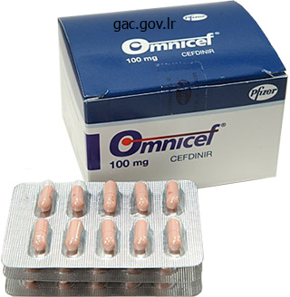
Order cefdinir 300mg with visa
An endoanal sizer is placed within the rectum, whereas two fingers are positioned in to the vagina to palpate the ischial spines. Nonabsorbable figure-of-eight suture are then placed through the uterosacral ligaments at the degree of the ischial spines, approximately 2 cm medial to the ischial backbone and away from the rectum, and introduced through the ipsilateral vaginal apex, avoiding entry in to the vaginal canal. During suture placement, the rectum is pushed to the contralateral facet to keep away from harm. This study is consistent with other smaller collection with a 75� one hundred pc remedy fee at less than 1 year and a complication price of 2�9. The procedure is as described above, nevertheless, the uterosacral ligaments are sutured collectively using one purse-string permanent suture via the uterosacral ligaments near the sacrum and then by way of the posterior cervical ring. A second permanent suture plicating the uterosacral ligaments together in the midline, 2 cm posterior to the primary suture offers extra reinforcement [4]. Several research counsel that ladies with cervical elongation higher than 5 cm are at elevated danger for failure, citing sensation of symptomatic vaginal bulge from the elongated cervix extending down the vaginal canal [4]. While the literature on laparoscopic uterosacral ligament suspension accommodates no large-scale, prospective, comparative trials, the results from the above case collection combined with the proven historical past of uterosacral vault suspension through the vaginal approach counsel that this might be a promising addition to the array of procedures used to tackle pelvic organ prolapse. It may have a particular role in patients who wish to preserve the uterus and in sufferers wishing to avoid the use of synthetic mesh. A fifth port (5 mm) could also be placed in the left decrease quadrant, or a suture could additionally be used to retract the sigmoid colon. In the unique report, a single piece of Gortex mesh was attached to the posterior vaginal apex and sutured or stapled to the anterior longitudinal ligaments of the sacrum. Since its inception, modifications to the process have been made, together with the utilization of anterior and posterior pieces of polypropylene mesh. The procedure can be modified for sufferers desirous of preserving the uterus by using a single piece of posterior mesh or an anterior Y-shaped piece of mesh and a posterior mesh. Most methods described for laparoscopic sacrocolpopexy involve the same initial steps. After acquiring access utilizing either the Hasson open method or the Verress needle, a 10�12-mm intra- or infra-umbilical port is placed. One or two further 5-mm ports are positioned 2�3 cm cephalad and 2�3 cm medial to the anterior superior iliac spines, avoiding ilioinguinal and iliohypogatric nerve damage or entrapment. After the sigmoid is retracted, the sacral promontory, proper common iliac artery, and right ureter are recognized. The posterior peritoneum over the sacral promontory is incised longitudinally to the extent of the vaginal apex. The bladder may be stuffed to help demarcate this aircraft, or by introducing a cystoscope gentle in to the bladder. This aircraft is dissected no much less than three cm distal to the vaginal apex to allow space for placement of the anchoring sutures. The lack of direct tactile suggestions makes this dissection challenging; resulting in a cystotomy or sutures in the bladder in 10. Similar dissection is carried out on the posterior vaginal wall to deperitonealize this area and separate the vagina from the rectum posteriorly. The mesh, either in two separate strips (3�5 cm x 12�15 cm) or prefashioned in a Y-configuration, is passed in to the field and sutured with nonabsorbable suture to the posterior and then the anterior vaginal wall. Cystoscopy ought to be carried out at the finish of the procedure to guarantee ureteral patency and that none of the sutures have passed in to the bladder. The vagina is suspended to the sacral promontory, recreating normal vaginal anatomy. Depending on surgeon desire, a single piece of posterior mesh (3�5 cm x 12� 15 cm) or an anterior Y-shaped piece of mesh and a posterior mesh are used. In cases using a single piece of mesh, the mesh is sutured to the posterior vaginal cuff and posterior cervix using 0-nonabsorbale sutures. When using two pieces of mesh, the posterior mesh is placed as described, whereas each arm of the anterior Y-shaped mesh is handed via the broad ligament. Laparoscopic sacrocolpopexy seems to efficiently recapitulate the open approach that has demonstrated durable outcomes for a number of many years. Several research have demonstrated the laparoscopic method to be successful, with a 90�96% treatment rate and a low mesh erosion rate, ranging from 1% to 8%.

Order cefdinir 300mg online
Correlation of pathologic findings with progression after radical retropubic prostatectomy. Estimation of prostate cancer quantity by a quantity of core biopsies before radical prostatectomy. Estimation of prostate cancer volume by endorectal coil magnetic resonance imaging vs. The influence of the inclusion of endorectal coil magnetic resonance imaging in a multivariate evaluation to predict clinically unsuspected extraprostatic most cancers. Correlation of margin standing and extraprostatic extension with development of prostate carcinoma. The role of typical and useful endorectal magnetic resonance imaging within the decision of whether or not to preserve or resect the neurovascular bundles during radical retropubic prostatectomy. Blood loss during radical retropubic prostatectomy: relationship to morphologic options on preoperative endorectal magnetic resonance imaging. Detection of domestically recurrent prostate cancer after cryosurgery: analysis by transrectal ultrasound, magnetic resonance imaging, and three-dimensional proton magnetic resonance spectroscopy. Diffusion-weighted imaging with obvious diffusion coefficient mapping and spectroscopy in prostate most cancers. In vivo measurement of the apparent diffusion coefficient in regular and malignant prostatic tissues utilizing echo-planar imaging. Differentiation of noncancerous tissue and most cancers lesions by obvious diffusion coefficient values in transition and peripheral zones of the prostate. Lesion localization in sufferers with a previous negative transrectal ultrasound biopsy and persistently elevated prostate particular antigen stage utilizing diffusion-weighted imaging at three Tesla earlier than rebiopsy. Citrate as an in vivo marker to discriminate prostate most cancers from benign prostatic hyperplasia and regular prostate peripheral zone: detection via localized proton spectroscopy. Diffusion-weighted imaging of the prostate at three T for differentiation of malignant and benign tissue in transition and peripheral zones: preliminary outcomes. The objective of this chapter is to present an introduction to the usage of angioembolization in trendy urology. Renal angiography and embolization the indications for renal angiography and embolization have changed since 1973, when Almgard et al. Indications for renal embolization diversified from refractory bleeding and recurring infections to nonoperative candidates for definitive remedy. The pediatric literature additionally contains reviews of renal embolization as an different to simple nephrectomy for extreme hypertension or to pretransplant nephrectomy, and for nephritic syndrome and ablation of irreversible rejected renal allograft [4]. In one sequence, 10 of 12 sufferers developed extreme flank ache, which was the most significant symptom of postinfarction syndrome. Note the enlarged capsular department partially supplying the tumor, which is typical of renal cell carcinoma. Some of the perceived advantages of preoperative embolization include a reactive edema of tissue planes facilitating dissection and permitting early ligation of the renal vein, a lower in intraoperative blood loss, and cytoreduction of the tumor thrombus quantity. No specific recommendations can be gleaned from these heterogeneous patient information; subsequently, the course of action is still left to the clinician to decide. Renal surgical procedure is a type of managed trauma; the use of conservative management in addition to embolization in the remedy of renal trauma has been supported by knowledge from the University of California, San Francisco General Hospital [13]. The most typical complication is postinfarction syndrome, which occurs 24�48 h after the procedure. It may be related to nausea, vomiting, fever, flank ache, and mild leukocytosis. Fourteen % of sufferers required repeat embolization in the meta-analysis [9]; nonetheless, this carried little added morbidity for the affected person. The incidence in the open partial nephrectomy collection from the Cleveland Clinic was 3 of 698 (0. This area is essential as a end result of the femoral head provides the posterior floor on which the physician can apply direct stress after access has been discontinued, so as to obtain hemostasis. The Angioseal gadget makes use of a vascular plug to preserve hemostasis, in distinction to Starclose, which makes use of a vascular clip. Identification of acute bleeding website Renal arterial anatomy is delineated, and any source(s) of hemorrhage is recognized. If no apparent supply of bleeding is elicited on initial angiography, then we catheterize every of the segmental arteries, particularly arteries near the area of surgical procedure (percutaneous stone extraction tract or partial nephrectomy renorrhaphy sites).
Syndromes
- The pituitary gland is injured (secondary adrenal insufficiency) and it cannot release ACTH
- Medicine (antidote) to reverse the effect of the poison
- Chest discomfort or tightness
- Difficulty breathing (dyspnea) that develops slowly and gets worse
- Convulsions
- Brain damage
- Wheezing
- Triaminic DM
- Poor feeding or irritability in children
- Gentamicin: 5 to 10 mcg/mL
Purchase genuine cefdinir
Understanding that surgeon choice is in all probability going the most vital factor in figuring out which approach is employed, we choose a transperitoneal, anterior approach with antegrade dissection. Using the precept of triangulation, dissection in the caudal direction provides for higher visualization of the buildings being dissected, as compared to making an attempt to see across the cumbersome prostate and dissecting back in the path of the bladder. It also permits the surgeon to perform the usually tough apical dissection after mobility of the prostate has been established. As for the right arm, whereas some prefer to use a monopolar hook or monopolar flat spatula, we choose a pair of monopolar scissors. Anterior bladder mobilization and retraction suture After profitable placement of all trocars and confirmation that all instruments are in working order, using a 0� laparoscope, an inverted "U"-shaped incision is made in the anterior peritoneum from one inguinal ring to the other, with the bottom of the "U" positioned near the umbilicus across both medial umbilical ligaments. The arms of the "U"-shaped incision must be carried right down to the inguinal ring until the crossing vas deferens is recognized, thus permitting for sufficient mobilization of this bladder-containing "flap. Care must be taken to not traumatize the pelvic bone as this can result in meddlesome bleeding that can typically be troublesome to control. Once the bladder is absolutely mobilized, we suggest a full-length 3-0 monocryl retraction suture be positioned on the prime of the urachus and be brought out via the 5-mm assistant port present in the proper upper quadrant of the stomach. It also serves to protect each the massive and small bowel from inadvertent upstream thermal damage because the bladder "flap" separates all instruments from the bowel (see Video 91. The fatty tissue overlying the prostate is removed to have the ability to improve visualization of the puboprostatic ligaments, dorsal vein, and anterior bladder neck. Although not routinely performed in any respect establishments, we believe that this maneuver facilitates the apical dissection and reduces the danger of anterior apical constructive margins. Furthermore, the lateral limits of this fatty tissue are in direct anatomic continuation with area three of an extended pelvic lymph node dissection. This ensures a more secure staple line by compressing all edema and vascularity out of the tissues. However, we prefer the constant and reproducible effect of stapling because it reliably gives a visual landmark to transect the urethra, therefore lowering positive apical surgical margins (see Video 91. Bladder neck transection this is the commonest a half of the process with which inexperienced surgeons struggle. There are numerous causes for this, however most distinguished we imagine is the absence of obvious visual landmarks, the innate pure anatomic variability of the junction, and the want to "really feel" the difference between the prostate and the muscle of the bladder. After the digital camera is switched to the 30� down scope, we first determine the posterolateral contour of the prostate. Next it helps to see or "really feel" the prostatovesical junction by compressing or pinching the bladder at its junction with the prostate. The prostate will end in minimal excursion of tissue, whereas compression of the bladder wall might be evidenced by an obvious mobility within the tissue. In and out traction on the Foley balloon also aids in figuring out the anterior bladder neck. Using the principles of traction and counter-traction to aid in defining the detrusor muscle fibers, electrocautery is used to divide the abundance of arteries and veins that traverse this area. Once the anterior bladder is opened all the means down to the urethra within the midline, the Foley catheter is recognized and deflated. The catheter tip is delivered in to the surgical field and the robotic fourth arm grasps the Foley by way of its eyelet and pulls it anteriorly towards the belly wall. Counter-traction is supplied by clamping the outer portion of the Foley catheter to the surgical drapes. The bladder neck is then dismembered from the prostate utilizing a bladder necksparing approach; nonetheless, it is important to open the bladder neck adequately to visualize the trigone and assist in defining the angle of transection of the posterior bladder neck. Sometimes the bladder attaches with a Control of dorsal venous advanced and apical dissection After the endopelvic fascia is incised with chilly scissors, the prostate is mobilized laterally down to the membranous urethra, sweeping the levator muscle fibers off the prostate with little to no cautery used. It is essential to note that constructive apical margins happen due to transection too close to the apical prostate. This dissection is carried out to the extent of the prostatic apex, staying near the posterior floor of the prostate. It is essential to carry this dissection far sufficient distally to utterly mobilize the rectum off the prostatic apex, and thereby cut back the chance of rectal harm through the apical dissection and transection of the urethra. Posterior counter-traction of the rectum provided by the surgical assistant facilitates this dissection.

Order cefdinir pills in toronto
Active surgical drains are closed system drains that exert a adverse low stress, which removes fluid gradually. Opening the plug and squeezing the air out of the bulb followed by plug substitute creates the self-suction system [14]. The other hand pinches the tubing to transfer blood clots and tissue via the tubing, thus facilitating improved suction. Two surgeons positioned drains in all instances and the kind of drain positioned was left to the discretion of each surgeon (37. Although there was variation in the incidence of issues by drain type, none was statistically vital [22]. Common complications with either kind of drain embrace pores and skin infection or irritation, perinephric wound infections, bleeding, pain, loss of the drain, and retrograde drain migration. Therefore, the risks and advantages of drain placement should be thought-about after each operation and a drain should be removed as soon as its function is not necessary. Conclusions Successful laparoscopic surgery depends on a secure and effective methodology of specimen entrapment and removing, and correct closure of the stomach wall. A systematic approach to the laparoscopic exit begins with a diligent visual inspection of the operative subject and trocar sites for extreme bleeding, organ damage, and organ entrapment. Proper closure of the stomach fascia and pores and skin can be performed with a selection of hand-suturing techniques in addition to laparoscopic closure gadgets, which permit for direct visualization both throughout or immediately following the closure. Basic information relating to using retroperitoneal drains (indications and complications) can be useful in attaining a successful end result after laparoscopy by limiting the number of postoperative complications. Veterinarian 121 scientific apply web site "wounds and wound care lesson" loudoun. Passive drains provide an exit port for fluid, blood, purulent materials, and particles, and act via gravity and capillary motion. Penrose drains are sometimes used when the fluid to be eliminated is too viscous for a closed self-suction drain. Following placement, the drain is sutured in to place on the pores and skin degree to stop retrograde migration in to the belly cavity. The distal finish of the drain is roofed with drain and dressing sponges to contain the drainage in the postoperative period. Closed-suction drains are thought to perpetuate a urinary fistula postoperatively and presumably result in delayed hemorrhage with removing of the drain. Incisional hernia after laparoscopic nephrectomy with intact specimen elimination: caveat emptor. Intact specimen extraction in laparoscopic nephrectomy procedures: Pfannenstiel versus expanded port web site incisions. Comparison of various extraction websites used during laparoscopic radical nephrectomy. A multi-institutional study on the safety and efficacy of specimen morcellation after laparoscopic radical nephrectomy for medical stage T1 or T2 renal cell carcinoma. Modified renal morcellation for renal cell carcinoma: laboratory experience and early scientific application. Feasibility of pathological evaluation of morcellated kidneys after radical nephrectomy. Safety and efficacy of laparoscopic radical nephrectomy with handbook specimen morcellation for stage cT1 renal cell carcinoma. Transperitoneal laparoscopic renal surgery utilizing blunt 12 mm trocar without fascial closure. Randomized potential examine comparing typical subcuticular pores and skin closure with dermabond skin glue after saphenous vein harvesting. Metaanalysis of skin adhesions versus sutures in closure of laparoscopic port-site wounds. For instance, laparoscopic access is often obtained through quite lots of initially blind punctures where viscera and blood vessels are vulnerable to unique accidents. Laparoscopy due to this fact requires a specialised data base, and demands a unique set of troubleshooting abilities.
Discount 300 mg cefdinir with amex
The incidence of elective conversion during laparoscopic easy nephrectomy was three. Dissection and securing of the hilar vessels is the step related to the best variety of "crash" conversions due to uncontrollable hemorrhage. Reported open conversion rates for attempted laparoscopic nephrectomy for renal tuberculosis are as high as 80% in essentially the most skilled hands [122], with spillage of caseating materials reported by a number of groups [126, 127]. The want for extended multidrug remedy following this kind of prevalence and the difficulty of those operations makes this another questionable disease for which laparoscopic nephrectomy ought to be utilized. The significant dimension of these specimens requires initial cyst decompression utilizing the Harmonic shears, with irrigation and aspiration of the launched fluid to permit adequate publicity of the hilum for dissection [129]. Mean laparoscopic occasions have been, nonetheless, significantly longer than in the open group (247 vs 205 min) with one case transformed to an open process. Complications the reported complications with establishment of transperitoneal and retroperitoneal access for any laparoscopic method also can happen during laparoscopic easy nephrectomy. Several reviews have particularly assessed the problems inherent to nephrectomy and stratified them Chapter 81 Renal Surgery for Benign Disease 967 from misplacement over previously placed clips [90]. They additionally noted a three:1 ratio for the incidence of visceral harm in the course of the transperitoneal versus the retroperitoneal method, respectively [131]. These include disproportionate single trocar website ache, stomach distention, diarrhea, and leukopenia followed rapidly by cardiopulmonary collapse [140]. The major indications for surgical intervention embody an ectopic ureter associated with a poorly functioning dysplastic upper pole moiety or an higher pole moiety with chronic obstruction and resultant poor operate. Recurrent infections, flank or abdominal ache, and incontinence may also be current [4]. Surgical intervention contains removing of the diseased moiety in addition to all or a portion of the related ureter. The laparoscopic approach to upper pole heminephrectomy was first described by Jordan et al. This has since turn out to be an increasingly popular technique for the therapy of patients with this situation. Both the transperitoneal and retroperitoneal approaches have been described [5, 141�144], though most authors appear to favor the transperitoneal exposure due to the extra familiar orientation of the hilum and improved working area [142]. Benzoin Open nephrectomy surgical pan 968 Section 6 Laparoscopy and Robotic Surgery: Laparoscopy and Robotics in Adults Step four: Exposure of the retroperitoneum this step is equivalent to that outlined for laparoscopic nephrectomy utilizing both the transperitoneal or retroperitoneal approach. Mobilization permitting unobstructed access to the hilar buildings is important as dissection and securing of the higher pole blood supply is important, as is the ability to move the transected ureter behind the renal artery and vein as described below for an higher pole heminephrectomy. Step 5: Identification and dissection of the upper or decrease pole and ureter the dilated higher or decrease pole ureter is usually readily identifiable as a medial tortuous tubular construction, which can be intimately connected to the ureter draining the conventional moiety. This latter construction can normally be differentiated by gently drawing an instrument across the surface of the suspected location of the ureter and feeling the resistive catch as it passes over the underlying stent. The two ureters could be intimately hooked up and the proximal facet of the dilated system is dissected out with care taken not to injure the wall or vasculature supplying the conventional system. Dissection and exposure of the hilar vessels is similar to that described for nephrectomy. Complete mobilization is facilitated by performing the dissection prior to transection of the ureter as distention of the diseased moiety usually helps to define the correct plane. This dissection ought to continue until a right-angled clamp could be handed freely behind the vessels for an higher pole heminephrectomy. The Harmonic scalpel is utilized to separate the adrenal gland from the higher pole system when performing an higher pole heminephrectomy. Complete mobilization of the affected moiety to the sting of the traditional pole parenchyma must be carried out to facilitate eventual removing of the diseased portion of the kidney. Step 6: Division of the ureter and vasculature of the concerned moiety the ureter is then transected on the point where it may be easily separated from the normal ureter to forestall Steps of the procedure Step 1: Placement of an ureteral catheter Some authors favor preliminary cystoscopic insertion of a ureteral catheter in to the ureter draining the nondiseased moiety previous to performing the laparoscopic a part of the process [141, 143]. The catheter does, however, assist within the identification of the normal ureter while dissecting out the dilated ureter draining the diseased moiety.
Effective 300 mg cefdinir
This culminated within the improvement of laparoscopic surgical procedure, the place an endoscope and devices passed through a quantity of small incisions are used to function on organs inside the abdominal cavity. Urologists have been on the forefront of minimally invasive surgery from the beginning. They have been the pioneers in using pure orifices to function on genitourinary organs. The pioneering efforts of Clayman and Gaur heralded the entry of urologists in to laparoscopy and retroperitoneoscopy [1, 2]. It is now over 20 years since Clayman carried out the first laparoscopic nephrectomy, and laparoscopy is now routinely carried out for many benign and malignant conditions of the genitourinary tract. It is now frequent to carry out even advanced reconstructive surgeries in a minimally invasive fashion. It was turning into clear that this was an emerging subject of minimally invasive surgery, and regulation of procedures performed and guidance to physicians meaning to carry out these procedures was important to the expansion of those surgeries. The bladder may be closed with endoscopic suturing strategies and a small rent can also heal with simply catheterization. It continues to set the standards for the development of the specialty, documents new techniques, and maintains a database of the procedures that have been accomplished. In these methods, which remain experimental, transgastric/ transcolonic routes have been used for cadaveric small bowel resection, and the transgastric/transvaginal strategy has been used for porcine nephrectomies [15�17]. The lack of effective clipping/stapling gadgets for use via flexible endoscopes make it a lot easier to perform these duties by way of the extra laparoscopic port. Also, use of an extra transumbilical entry, albeit a needlescopic port for visualization of entry in to the peritoneal cavity by way of the viscus, makes the procedure much safer. This has been the most typical sort of procedure accomplished in humans [18, 19] and has been used for quite lots of procedures, together with radical nephrectomy and cholecystectomy [11, 20]. It helps the surgeons to achieve expertise in the use of and the view with versatile endoscopes, whereas making certain security for the patient undergoing these new procedures. The most attention-grabbing growth on this space is using flexible robotics and mini-robots to carry out different procedures. Flexible robotics has been used clinically with success to carry out renoscopy to fragment renal calculi [23�25]. Gastrointestinal endoscopists are probably the most familiar with this route and so they have driven the innovation in this field. The main difficulties associated with this route appear to be the dearth of a tool for secure closure of the stomach [7], tough orientation after retroflexing the scope in the peritoneal cavity, significantly for a cholecystectomy or upper stomach procedures, in addition to inadequate illumination of the capacious peritoneal cavity. The transesophageal route also has been used to entry the mediastinum in an animal mannequin and varied procedures may be performed, together with pleural biopsy and cardiomyotomy [8]. It is the most commonly used route in urologic functions with a fair variety of reports of procedures carried out in animals by this route [4, 9]. Some gynecologists stay concerned about adhesions in the pelvis and subsequent infertility after these procedures. There are also concerns about spread of endometriosis and that this additionally might lead to dyspareunia within the postoperative period [12]. In vivo mini-robots have been described experimentally that may be launched in to the peritoneal cavity through a pure orifice to explore the peritoneum and carry out surgical procedures [27, 28]. The perforation of a "regular" viscus to get at an irregular organ seems to contravene the natural means of commonsense, apart from being hazardous, should the closure be insecure, or leak publish process. The passage of two instruments down the channel of the endoscope considerably limits their use [5]. It is difficult to hold and retract regular organs with the flimsy endoscopic devices, not to mention organs inflamed and enlarged by illness. Use of a versatile endoscope to move these devices has the drawback of excessive mobility, making these procedures extra cumbersome as the endoscope simply will get lost in the insufflated and capacious abdominal cavity. Development of newer supplies and devices will doubtless enhance the follow of these varieties of surgeries. Overcoming the remaining obstacles will require shut cooperation between engineers and surgeons. They reported this procedure in an animal mannequin in addition to in the first human scientific instances [32]. The umbilicus is a ubiquitous cicatrix from birth and can be used to conceal the access in to the belly cavity. Gynecologists have been performing tubal ligation with a single puncture laparoscope since it was introduced within the late Nineteen Seventies [34, 35].
Norway Spruce (Hemlock Spruce). Cefdinir.
- Are there safety concerns?
- What is Hemlock Spruce?
- Dosing considerations for Hemlock Spruce.
- How does Hemlock Spruce work?
- Coughs, the common cold, bronchitis, fevers, inflammation of the mouth and throat, muscular and nerve pain, arthritis, bacterial infection, arthritis pain, nerve pain, muscle pain, tuberculosis, and other conditions.
Source: http://www.rxlist.com/script/main/art.asp?articlekey=96451
Purchase cefdinir amex
Nephron sparing surgical procedure for appropriately chosen renal cell carcinoma between four and seven cm leads to end result much like radical nephrectomy. Comparison of outcomes in elective partial vs radical nephrectomy for clear cell renal cell carcinoma of 4�7 cm. Laparoscopic partial nephrectomy for hilar tumors: analysis of short-term oncologic end result. Laparoscopic partial nephrectomy for central tumors: evaluation of perioperative outcomes and problems. Minimally invasive nephron sparing administration for renal tumors in solitary kidneys. Laparoscopic partial nephrectomy for a number of ipsilateral renal tumors using a tailored surgical approach. Feasibility of laparoscopic partial nephrectomy after previous ipsilateral renal procedures. Laparoscopic partial nephrectomy: comparison of transperitoneal and retroperitoneal approaches. Retroperitoneoscopic radical and partial nephrectomy in the affected person with cirrhosis. Prospective randomized comparison of transperitoneal versus retroperitoneal laparoscopic radical nephrectomy. Laparoscopic partial nephrectomy using FloSeal for hemostasis: approach and experiences in 102 patients. Improved hemostasis during laparoscopic partial nephrectomy utilizing gelatin matrix thrombin sealant. A comparative research of a number of brokers alone and combined in protection of the rodent kidney from heat ischaemia: methylprednisolone, propranolol, furosemide, mannitol, and adenosine triphosphate-magnesium chloride. Laparoscopic ice slush renal hypothermia for partial nephrectomy: the initial expertise. Robotic-assisted laparoscopic partial nephrectomy: method and preliminary medical expertise with DaVinci robotic system. Robotic partial nephrectomy with sliding-clip renorrhaphy: method and outcomes. Robotic and laparoscopic partial nephrectomy: a matched-pair comparability from a high-volume centre. Robot assisted partial nephrectomy versus laparoscopic partial nephrectomy for renal tumors: a multi-institutional evaluation of perioperative outcomes. Effects of intermittent versus continuous renal arterial occlusion on hemodynamics and performance of the kidney. Ischemia with intermittent reperfusion reduces practical and morphologic damage following renal ischemia in the rat. Ischemic preconditioning and intermittent clamping enhance the tolerance of fatty liver to hepatic ischemia-reperfusion damage in the rat. Effects of ischemic liver preconditioning on hepatic ischemia/ reperfusion damage in the rat. Positive margins in laparoscopic partial nephrectomy in 855 cases: a multiinstitutional survey from the United States and Europe. Multiple studies have reported important advantages with the laparoscopic method in comparison with the standard open strategy. This minimally invasive method offers sufferers less postoperative pain, a shorter hospital keep, a sooner return to normal activities, and a better beauty result. In the past decade, robotic expertise has been launched to help laparoscopic procedures. With a threedimensional (3D) show to improve depth notion and instruments containing a "wrist" joint to improve dexterity, robot help may offer benefits over standard laparoscopy, doubtlessly reducing the technical complexity of the process and enabling less skilled surgeons to ship minimally invasive surgery to their sufferers. Occasionally, symptomatic adrenal cysts and myelolipomas could also be eliminated laparoscopically. Nonfunctioning adrenal lesions larger than 4�5 cm or tumors that have proven development in measurement on serial imaging may also be excised laparoscopically.
Cheap cefdinir 300 mg fast delivery
The position of computerized tomography in the evaluation of complication after laparoscopic urological surgical procedure. Randomized trial evaluating a radial expandable needle system with cutting trocars. Comparison of laparoscopic and open retropubic urethropexy for therapy of stress urinary incontinence. Laparoscopic orchiopexy for the palpable true undescended testicle: Preliminary experience. Access and trocar placement is of crucial importance as improper placement could lead to issue later. In this chapter we outline the necessary steps for correct affected person position, entry, and secondary trocar placement for each renal retroperitoneal surgical procedure and pelvic extraperitoneal surgical procedure. In addition, we define potential exclusion standards for retroperitoneal renal/adrenal surgical procedure and extraperitoneal pelvic surgical procedure. Finally, we briefly summarize critical issues related to laparoscopic exit on the finish of the case. No definitive exclusion criteria exist for retroperitoneal entry, although a few instances might present more challenges for retroperitoneal surgery. Renal tumors larger than 10 cm in measurement are greatest approached via a transperitoneal strategy. Those positioned on the posterior hilar side might current challenges with retraction and lifting of the kidney and as such may be better suited to the transperitoneal method. Prior percutaneous entry for stone surgical procedure has not introduced a significant challenge in our expertise. Retroperitoneal renal/adrenal surgical access Direct entry to the retroperitoneum for renal or adrenal surgical procedure could offer a number of potential advantages over the transperitoneal method. Ligation of the renal artery and vein throughout radical nephrectomy could also be aided by circumventing the necessity for bowel dissection. In addition, in circumstances of posterior small renal plenty, direct access to the posterior facet of the kidney might obviate the need for full renal mobilization. Patients with a number of prior transabdominal surgical procedures or massive anteriorly located mesh can also profit from the retroperitoneal method for renal or adrenal surgery. Patient place and entry approach After induction of common anesthesia, the affected person is positioned in the full flank position. The patient is secured to the desk with 3-inch tape at the degree of the chest (without impeding mechanical ventilation) and hips. Securing the patient on this means permits the desk to be rotated from right to left. Pillows are placed between the legs and strain points (knees, hips) are correctly padded. The tip of the twelfth rib is marked with semi-permanent ink previous to sterile prep and drape, as palpation of the rib after preparation and gloving may be impaired, especially within the obese patient. At the intersection of this line and the decrease facet of the 12th rib, the left trocar is inserted. S-retractors are used to split the external oblique muscle and often the inner oblique muscular tissues all the method down to the lumbodorsal fascia. At this point a 30� lens, 10-mm laparoscope is inserted by way of the dissecting balloon port and the anatomy is appreciated. The psoas muscle can typically be assessed posteriorly and the peritoneum anteriorly. The balloon dissecting trocar is deflated and a balloon working trocar (Covidien) is positioned. Placement of secondary trocars Under direct imaginative and prescient the 2 remaining trocars are positioned. This trocar may be angled in a barely more cephalad course, which can lower the amount of torque on the trocar and surgeon fatigue during the case. The peritoneum may be visualized and, if essential, may be dissected extra medially using the surgical left trocar and a laparoscopic Kitner instrument. Access for extraperitoneal pelvic surgery the appliance of pelvic extraperitoneal surgical procedure is best for robot-assisted laparoscopic prostatectomy. Other urologic applications include distal ureter surgical procedure, partial cystectomy of anterior lesion, and restricted pelvic lymph node dissection.
Order cefdinir line
Uncorrected coagulopathy, pregnancy, extreme skeletal deformity, and morbid weight problems have been accepted contraindications of this modality. Treatment success for renal stones of 20 mm or smaller was reported to be above 90%, and 92. Recent research emphasize the significance of skin�stone distance in stone clearance, measured because the imply value of three distances from the renal stone to the skin at 0�, 45�, and 90� and utilizing a radiographic caliper or computational measuring system [23, 24]. Overall stone-free rates had been 91%, 97%, and 94% in regular weight, obese, and morbidly obese sufferers, respectively. They found no vital difference by method of stone location (renal/proximal, mid-, and distal ureteral stones) and measurement (cut-off dimension was 1 cm) in all teams. The absolute contraindications are active urinary tract infection and uncontrolled coagulopathy. The authors also reported that technical modifications and acceptable instrumentation had been necessary to improve the stone-free rate in this group of patients. The complication price and size of hospital stay have been much like those for the nonobese group. The anesthesiologist and surgeon usually struggle to use this posiion in morbidly overweight patients as a result of compromised cardiopulmonary status and body weight. The more recent literature has focused on alternatives in affected person positioning, widening affected person selection, or new techniques. In addition, urologists are extra snug adopting a sitting posture during stone management. Although the supine position seems to have some advantages for overweight and morbidly obese patients and sufferers with staghorn calculi, the susceptible place has higher outcomes over the supine position. Percutaneous access in to the pelvicalyceal system could be more difficult in overweight patients. Segura described a method to incise the skin and fats, with incision extending right down to the muscular fascia, reducing the size required to reach the stone [49]. In addition, selecting the shortest tract for percutaneous access and utilizing longer devices, similar to an Amplatz sheath longer than 17 cm and longer nephroscope, might facilitate the procedure. Use long Amplatz sheaths and nephroscopes Choose the shortest potential tract Stitch two sutures on the Amplatz sheath Use the supine place in selected sufferers (for air flow problems) inflating the balloon. Several strategies have been described to overcome these limitations in some patients. To handle the obese patients with urolithiasis safely and effectively, the urologist should have data and experience of the potential diagnostic and remedy problems, as well as modifications of the techniques. References Conclusions Epidemiologic research indicate that obesity increases the danger of urolithiasis. Several pathophysiologic mechanisms have been proposed for abnormalities in urine composition in obese patients. These embody carbohydrate-induced calciuria, increased protein consumption, results of insulin resistance on renal ammonia metabolism and urine pH, elevated prevalence of gout and hyperuricosuria, or some other unidentified abnormality in renal electrolyte transport. Dietary modifications have an important function as a result of metabolic abnormalities are extra common in overweight stone formers. Obesity poses a quantity of issues in the management of stone disease from prognosis and imaging through to anesthesia and surgery. Impact of obesity in sufferers with urolithiasis and its prognostic usefulness in stone recurrence. Extracorporeal shockwave lithotripsy in the remedy of renal pelvicaliceal stones in morbidly obese sufferers. Ureteroscopic remedy of renal calculi in morbidly overweight sufferers: a stone-matched comparability. Outcomes of contemporary percutaneous nephrostollithotomy in morbidly overweight sufferers. Clinical worth of minimally invasive percutaneous nephrolithotomy within the supine place underneath the guidance of real-time ultrasound: report of ninety two circumstances. Modified supine versus prone position in percutaneous nephrolithotomy for renal stones treatable with a single percutaneous access: a prospective randomized trial.
Cefdinir 300 mg online
Holman and Toth described 15 sufferers handled with laparoscopy-assisted percutaneous transperitoneal surgery in the Trendelenburg place to facilitate displacement of bowel away from the pelvic kidney [20]. These authors cautioned that a transperitoneal method to the pelvic kidney adds pointless threat and morbidity due to the potential for damage to the bowel and violation of the natural peritoneal barrier. This may lead to hemorrhage, peritoneal urine leak, peritonitis, or postoperative ileus. Extraperitoneal access to the pelvic kidney reduces the danger of intestinal harm and leaves the peritoneal cavity intact. Puncture of the specified calyx within the pelvic Chapter sixty two Associated Conditions and Treatment of the Pelvic Kidney kidney was carried out in an antegrade fashion, utilizing each fluoroscopic and laparoscopic control. The proposed advantage of transmesenteric access is a reduced threat of bowel harm related to bowel manipulation and mobilization. A posterior strategy is less advantageous in an overweight patient, in whom the transgluteal distance to the pelvic kidney is likely to be greater than with a standard flank incision. A case of incomplete femoral neuropathy has been reported, in all probability on account of direct traumatic harm to the dorsal columns of the lumbar plexus [25]. With more medial posterior approaches adjacent to the paraspinal muscles, insufficient size of the nephroscope might inhibit intrarenal manipulation. Additionally, a more medial posterior approach that creates a tract adjoining to the paraspinal muscles and alongside the quadratus lumborum or psoas major muscle tissue places several nerves in danger for injury. The iliohypogastric and ilioinguinal nerves lie anterior to the quadratus lumborum, the genitofemoral nerve lies anterior to the psoas major, and the femoral nerve lies lateral to the psoas major muscle. Consequently, with the anomalous location of an ectopic kidney, meticulous consideration must be paid to understanding the spe- 711 cific anatomy so as to scale back the chance of harm to structures that lie alongside the access tract. Ultrasound steering may help depict viscera, and the ultrasound probe itself can be used to deflect bowel away from the kidney. Laparoscopy as an adjunctive tool within the endoscopic therapy of difficult stones has yielded stone-free rates between 91% and one hundred pc within the few available case sequence. The upper calyx was accessed in all 4 cases, with three stones in the renal pelvis and one in a calyx. These sufferers have been left "tubeless," without external drainage nephrostomy tubes, and instead had inside double-J ureteral stent placement [26]. Laparoscopy was helpful in permitting direct viewing of aberrant vessels and peripelvic inflammation. Once the pelvic kidney was dissected and uncovered appropriately, a 2-cm pyelotomy was created, and the calculi have been extracted with grasping forceps. The depth and site of the pyelotomy made it troublesome to carry out endoscopic or suture closure, so the renal pelvis was left open. When the renal pelvis was not easily visible, the ureter was identified, traced proximally to the renal pelvis, and isolated with a vessel loop. Because the stones have been large and solitary, the vessel loop was used for traction and identification quite than prevention of stone migration [30]. Percutaneous access was gained via the transgluteal approach by way of the higher sciatic foramen. A suction drain is positioned within the perinephric space via a working port to forestall urine leak [31]. A laparoscopic transmesocolic pyelolithotomy, if feasible, is an option that avoids colon mobilization [35]. Intraoperative fluoroscopy permits identification of the renal pelvic and allows for periodic re-evaluation of pending stone burden. With enchancment in laparoscopic suturing strategies and laparoscopic suturing devices, pyelotomies are sutured closed in a operating trend. However, the number of published circumstances for laparoscopic pyelolithotomy is small, with the need for intracorporeal suturing making it a extra technically challenging procedure. Following interventional radiologic principles of deep pelvic abscess drainage, the strategy via the higher sciatic foramen rendered the patient stone free with an uncomplicated postoperative course. Recommendation is to place the tract as near the sacrum as potential to avoid vascular or neurologic structures. The patient had an uncomplicated hospital course and remained stone free on follow-up imaging (unpublished observations). The laparoscopic approach provided good surgical exposure, and operative occasions were corresponding to those of laparoscopic pyeloplasty in anatomically regular kidneys. The authors commented that laparoscopic pyelolithotomy may be carried out concomitant with pyeloplasty when kidney stones are present.
Real Experiences: Customer Reviews on Cefdinir
Arakos, 42 years: For robotic renal surgical procedure, the assistant is positioned on the aspect reverse the surgical web site. This prevents charring around the probe and permits extra vitality to be delivered in to the tumor. In the recrystallization process, smaller ice crystals fuse to kind larger buildings that inflict extra injury on cell buildings. Understanding that surgeon desire is likely the most significant consider figuring out which approach is employed, we choose a transperitoneal, anterior approach with antegrade dissection.
Tyler, 40 years: Preservation of ejaculation through a modified retroperitoneal lymph node dissection in low stage testis most cancers. The trumpet-like configuration was a constant finding through the follow-up interval. In a comparative research from the identical institution, five patients underwent laparoscopic ureterolysis and five robotassisted ureterolysis [89]. When attributable to a Veress needle puncture or trocar tip, laparoscopic restore of huge or small bowel with intracorporeal suturing is readily completed.
8 of 10 - Review by I. Murat
Votes: 341 votes
Total customer reviews: 341

