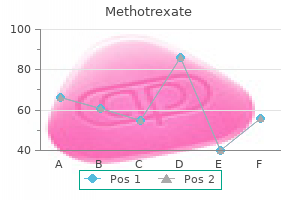
Methotrexate dosages: 10 mg, 5 mg, 2.5 mg
Methotrexate packs: 10 pills, 20 pills, 30 pills, 60 pills, 90 pills, 120 pills, 180 pills, 270 pills, 360 pills

Buy 2.5 mg methotrexate free shipping
A suspected net on the cervical level additionally can be seen in Video 5-1 on the Evolve website accompanying this text. Patients typically report that signs are intermittent and less doubtless if they choose their meals correctly and chew carefully (see the section on Differential Diagnosis). Conversely, signs are more probably if the patient eats away from house or carries on a dialog while eating; in these conditions the choice of meals is extra restricted and correct preparation of meals before swallowing is harder. Once the food is dislodged, the affected person typically can return to the meal without further issue. The extent to which consideration to the mechanics of slicing and chewing controls symptoms is restricted. When the lumen is severely compromised, the patient might discover it impossible to keep the level of consideration required to stay symptom free with out avoiding solids completely. The affected person might describe signs without any apparent development in frequency or severity that date again for many years. Radiographically, rings and webs seem as thin (2 to 4 mm) bands that type shelflike constrictions anyplace alongside the esophagus. Although radiologists occasionally check with thicker lesions as webs or rings, these are in all probability quick strictures or abnormal muscular contractions. Treatment of webs or rings entails dilatation or rupture of the ring by any one of a wide selection of esophageal dilator systems. Dilatation may provide everlasting relief, though a large proportion of patients want periodic redilatation at variable intervals. The majority of benign esophageal strictures are acquired in adulthood as a consequence of esophagitis. In a circular structure such because the esophagus, edema ensuing from ongoing inflammation and fibrosis as part of the healing course of happens on the expense of luminal diameter. However, dysphagia is progressive, with episodes becoming extra frequent and extreme over a period of months or years. As luminal narrowing increases, the patient reports trouble swallowing meals that beforehand caused no issue. Stenosis occasionally can become so extreme that even thick liquids cause dysphagia. Even then, however, dysphagia is nearly always higher for solids than liquids. Esophagitis could vary in severity from microscopic irritation to mucosal edema to erosion, ulcerations, and stricture. Gastroesophageal reflux Infections (Candida, viral) Trauma (prolonged nasogastric intubation) Acute chemical ingestion (lye, industrial acids) Drug-induced esophagitis (tetracycline, iron, potassium, quinidine, nonsteroidal antiinflammatory drugs) 6. Skin conditions (pemphigus, cicatricial pemphigoid, epidermolysis bullosa dystrophica, lichen planus, toxic epidermal necrolysis, Stevens-Johnson syndrome) eight. Because sudden onset of a swallowing disorder is rare in youthful individuals, the one potential cause that came to mind was pillinduced esophagitis. When I asked whether he was taking treatment, she reported that he had simply began taking tetracycline for his pimples and the day earlier than he had forgotten to take his medicine at residence using the conventional amount of water. The barium throughout the narrowed lumen has a somewhat irregular look because of the erosions. In some patients the esophagus seems to be comparatively insensitive to acid publicity. These individuals never expertise vital reflux signs regardless of extreme esophagitis and development to stricture formation. Although most benign esophageal strictures are a result of reflux esophagitis, any source of esophagitis may cause stricture formation (Box 5-1). Drug-induced or capsule esophagitis could be seen in younger or older grownup patients (see Practice Note 5-1). Typically, commonly administered medicines which would possibly be bigger in measurement (tetracycline, potassium, quinidine) turn out to be lodged at the level of the aortic arch and dissolve, inflicting inflammation and stricture.
Buy methotrexate 2.5mg on-line
Generous sampling is really helpful as higher invasion is designated as frank invasion. A urothelium with reactive atypia is at higher left, whereas urothelial carcinoma in situ is seen decrease right. The differential includes pseudosarcomatous stromal response and primary bladder sarcoma. The nuclear positivity for p63 is most helpful given its nearly absent expression in mesenchymal lesions. Spindle Cell Lesions: Rhabdomyosarcoma Spindle Cell Lesions: Rhabdomyosarcoma (Left) A spindle cell sarcoma with fibrosarcomatous morphology from the bladder dome and urachus of a pediatric affected person is proven. MyoD1 showed a similar pattern, whereas a smaller subset of cells also showed induction of desmin expression. Urothelial-Associated Markers Urothelial-Associated Markers (Left) S100p, or placental S100, is an emerging marker utilized in urothelial histogenesis, positive in urothelial and pancreaticobiliary primaries. Urothelial-Associated Markers Uroplakins: Highly Specific (Left) Uroplakin-3 is exquisitely particular for urothelial neoplasms when scored for plaque-like membranous positivity. Helpfully, keratins and p63 must be negative, whereas chromogranin and synaptophysin optimistic, with S100 constructive sustentacular cells. Enteric-Type Adenocarcinoma Enteric-Type Adenocarcinoma (Left) A moderately differentiated enteric-type adenocarcinoma is seen within the bladder dome. In a bladder/urachal main, membranous accentuation with relative nuclear sparing (pictured) is predicted. The remaining prostate is coronally sectioned at 3- to 5-mm intervals and submitted sequentially. Submission of prostate ranges from systematic partial sampling (at least 50%) to entire gland processing. Whole-mount sections present ease in determining location of extraprostatic extension and optimistic margin but are technically demanding and require particular submitting and storage. Egevad L et al: International society of urological pathology consensus convention on dealing with and staging of radical prostatectomy specimens. Seminal vesicles, vas deferens, & relatively flatter posterior floor are orientation landmarks. The prostatic urethra and ejaculatory duct can be used as landmarks to establish these different zones histologically. Prostate Microanatomy Prostate Base, Seminal Vesicles and Vas Deferens (Left) Anterior-superior view shows the urethra on the base. Paired bilateral seminal vesicles are connected to the posterior side of the base, lateral to the tubular vas deferens. Knowledge of prostate microanatomy and landmarks allows histologic orientation of submitted sections. Handling of Prostate Biopsy Handling of Prostate Biopsy (Left) the prostate cores are ideally submitted with solely 12 cores per cassette. Too many cores per block might trigger tissue loss since cores may be cut at completely different ranges. Inked cores are more seen and help in the optimal slicing of the tissue throughout the paraffin blocks. This strategy ensures illustration of the main target of curiosity that could be misplaced in subsequent deeper ranges. Hong H et al: Anatomic distribution of periprostatic adipose tissue: a mapping examine of 100 radical prostatectomy specimens. Note the hyperplasia of verumontanum mucosal glands, which frequently comprise corpora amylacea of their lumina. Basal cells are located internal to the glandular basement membrane define and with scant cytoplasm. The glands are variable in dimension, and the luminal outline is normally not sharp or rigid. Benign Prostatic Duct Corpora Amylacea (Left) Benign glands contain corpora amylacea with its distinctive magenta color and round lamellation. Corpora amylacea are frequent in benign prostate and verumontanum glands and will solely rarely be seen in prostate carcinoma.
Syndromes
- Difficulty swallowing
- Prothrombin time (PT, a different measure of blood clotting, often abnormal from liver disease)
- Radiation therapy or exposure
- Swollen clitoris (very rare)
- Heart defibrillation (purposeful shocking of the heart by medical personnel)
- Notice a lump in your neck
- CT scan of the head
- Delayed or slowed start of urinary stream
- A slow weight loss of 1 or 2 pounds a week, until the desirable body weight is reached, is best.
Discount 10 mg methotrexate with amex
Fever, Sepsis: Rule Out Biliary/Gallbladder Source this is a frequent indication in septic, postoperative, intubated sufferers with multisystem failure. Ultrasound is less delicate and particular for acute cholecystitis given the issue in detecting gallbladder tenderness and the reality that fasting, whole parenteral nutrition, hypoalbuminemia, sepsis, and coronary heart failure might contribute to gallbladder distension and wall thickening. The intrahepatic and extrahepatic bile ducts should be evaluated for dilatation or thickening secondary to cholangitis. Ascending cholangitis may be associated with biliary sludge or pus and obstructing stones. Cholangitis could also be difficult by hepatic abscesses, typically clustered across the irregular bile ducts. The liver, pancreas, and different organs should also be screened for causes of sepsis throughout ultrasound. Palpable Gallbladder A palpable gallbladder could presumably be secondary to gallbladder carcinoma or other tumors, mucocele, or benign obstruction. More superior tumors may obliterate the gallbladder lumen and extend into the close by liver, making it tough to determine the origin of the tumor. Bile-filled obstructed noninflamed gallbladders usually result from non-stone illness corresponding to pancreatic or distal bile duct carcinomas. Gallbladder mucoceles secondary to persistent stone obstruction are typically minimally tender with no wall thickening. Adjacent vessels are splenic vein, superior mesenteric artery, aorta, and inferior vena cava. This ought to be distinguished from emphysematous cholecystitis by demonstrating mobility and correlating with signs of wall irritation. The gallbladder is distended with sludge and wall thickening, findings indicating acute calculous cholecystitis within the correct medical context. Demographics � Epidemiology Biliary sludge � Similar epidemiology to cholelithiasis � M<F � More frequent in middle-aged, overweight ladies Natural History & Prognosis � Biliary sludge Approximately 50% of cases resolve spontaneously over 3-year interval 20% persist and remain asymptomatic 5-15% develop gallstones 10-15% turn into symptomatic 294 Echogenic Bile Diagnoses: Biliary System (Left) Transverse ultrasound reveals sludge filling the gallbladder. Demographics � Age More widespread in middle age � Gender M<F � Epidemiology 5% of inhabitants have polyps; 50% are ldl cholesterol polyps 6% of cholecystectomy specimens 298 Gallbladder Cholesterol Polyp Diagnoses: Biliary System (Left) Longitudinal left lateral decubitus ultrasound exhibits a ldl cholesterol polyp with a normal gallbladder wall. Demographics � Age Typically > 25 years � Gender M:F = 1:three � Epidemiology 302 Acute Calculous Cholecystitis Diagnoses: Biliary System (Left) Supine transverse ultrasound reveals a thickened edematous gallbladder wall with dependent sludge (stones not shown). There is a group with low-level echoes (abscess) medial to the thick-walled gallbladder. Atar E et al: Percutaneous cholecystostomy in critically sick patients with acute cholecystitis: issues and late consequence. Demographics � Age More common in middle-aged and elderly � Gender M:F = 3:1 � Epidemiology zero. Seretis C et al: Metaplastic modifications in continual cholecystitis: implications for early analysis and surgical intervention to forestall the gallbladder metaplasia-dysplasia-carcinoma sequence. Pathology of the Pancreas, Gallbladder, Extrahepatic Biliary tract, and Ampullary Region. Bennett G et al: Cholelithiasis, cholecystitis, choledocholithiasis, and hyperplastic cholecystoses. Schnelldorfer T: Porcelain gallbladder: a benign course of or concern for malignancy Demographics � Age > 35 years � Gender Cholesterolosis: F > M Adenomyomatosis: F > M � Epidemiology Cholesterolosis more frequent, 12% prevalence Adenomyomatosis comparatively much less common, 5% prevalence 316 14. Cariati A et al: Gallbladder cancers: related situations, histological types, prognosis, and prevention. Cairns V et al: Risk and Cost-effectiveness of Surveillance Followed by Cholecystectomy for Gallbladder Polyps. Kai K et al: Clinicopathologic options of superior gallbladder most cancers related to adenomyomatosis. Extrahepatic: Calculus, stricture, pancreatic head adenocarcinoma, cholangiocarcinoma, lymph node compression, etc. The reason for gentle biliary ductal dilatation was due to obstructing stone (not shown).
Cheap 2.5 mg methotrexate otc
On admission, the sufferers with probably the most extreme dysphagia had been those with traumatic mind harm, followed by stroke. For occasion, patients entering a rehabilitation setting could not have as many accompanying medical issues and dysphagia as these coming into a nursing home. Conversely, infants born prematurely may have many medical problems which will secondarily end in dysphagia. In a survey of the entire inhabitants of an acute basic hospital, fewer patients with dysphagia would be discovered in the general population in contrast with a survey of a particular part of that hospital, such as the stroke unit. Special Populations Some major medical diagnoses are more probably to precipitate dysphagic symptomatology, such as illnesses that have an effect on the central and peripheral nervous system and issues affecting the structures of the alimentary tract, similar to most cancers. An estimated 300,000 to 600,000 persons in the United States annually are affected by dysphagia from neurologic issues alone; most instances happen after a stroke. Stroke Prevalence reports of dysphagia after stroke rely upon when in the midst of restoration the detection of a swallowing impairment was made. For instance, in acute stroke (less than 5 days after onset) the prevalence of dysphagia may be as excessive as 50%, whereas 2 weeks after stroke solely 10% to 28% of sufferers could also be dysphagic. Immediately after stroke, 51% were believed to be at risk for Community Estimates of the prevalence of dysphagia amongst older persons living in the neighborhood range from 16% to 22%. Acute and Chronic Geriatric Care Of the 211 sufferers admitted to an acute geriatric unit in Singapore, the prevalence of dysphagia was 29% on admission and 28% at discharge. These results counsel that early detection is important in preventing dysphagic issues and that a significant number of patients will enhance without intervention particular to their dysphagia. Similarly, comparable prevalence figures for dysphagia on admission (43% to 51%) were discovered by Gordon et al. Of these, two thirds did so silently, suggesting that occasions of aspiration might be detected solely by videofluoroscopy, not the bedside examination. After analyzing prevalence reports from two large stroke databases, Gonzalez-Fernandez et al. Dysphagia may finish up from the removing of tissue, with subsequent sensory and motor loss, and the consequences of radiation remedy and chemotherapy. In a large multicenter therapy trial of sufferers with laryngectomies who have been treated with both surgical procedure and radiation or radiation and chemotherapy, roughly 33% had some sort of swallowing-related problem at 2-year follow-up. In a sequence of 46 patients treated by supraglottic laryngectomy, 60% had dysphagia after their hospital keep. Evidence means that sufferers with pharyngeal tumor resections and people with tumors involving the tongue base usually have a tendency to have dysphagia. Incidence information are available for patients who survive head harm and enter a rehabilitation setting; the incidence ranges from 27% to 30%34,36 to 42% (218 of 524). Among patients with head injuries getting into a rehabilitation setting, Winstein36 found that 27% have been dysphagic on admission to rehabilitation and that only 6% have been dysphagic after 5 months of rehabilitation. In basic, the more severe the initial injury, the upper the incidence of dysphagia. However, as quickly as patients enter the rehabilitation setting, their possibilities of returning to oral feeding are good. On admission he was nonresponsive and a nasogastric feeding tube was placed to present vitamin and hydration. As his responsiveness improved, the nasogastric feeding tube was removed and he began oral feeding. As he fed himself, it was noted that he choked on most makes an attempt and dysphagia was suspected. For occasion, some sufferers report dysphagia as the preliminary symptom of the illness, whereas others may never mention dysphagia. Complications from dysphagia, particularly people who threaten pulmonary operate, might lead to aspiration pneumonia and death (see Chapter 6). Interestingly, the esophageal abnormalities were current even before overt manifestations of dysphagia have been current. One third of the sufferers sampled complained of dysphagia, whereas more than 80% had goal demonstrations of its presence. Dysphagic complaints in sufferers receiving follow-up care in an outpatient clinic ranged between 30% and 40%. Their examine confirmed a optimistic relation between dysphagia and illness severity and between dysphagia and brainstem involvement.
Order methotrexate 5 mg
Cytologic Atypia Comedonecrosis (Left) Enteric-type adenocarcinoma with comedonecrosis may be morphologically indistinguishable from colon cancer. In contrast, urothelial carcinoma with glandular differentiation has a urothelial morphology in some areas. Exophytic and Invasive Histology 412 Invasive Adenocarcinoma Urinary Bladder Adenocarcinoma of Bladder Adenocarcinoma of Bladder (Left) Primary vesical adenocarcinoma (enteric and colloid type) is often high stage and reveals apparent damaging invasion of the muscularis propria. This feature is very useful in the distinction from cystitis glandularis with mucin extravasation. Adenocarcinoma of Bladder Adenocarcinoma of Bladder (Left) Signet ring cell adenocarcinoma of the urinary bladder is rare however is morphologically indistinguishable from metastatic gastric signet ring cell adenocarcinoma. There may be complete immunophenotypic overlap between these 2 lesions and adenocarcinomas arising in the urachus. The whole vary of main mucosal-based vesical adenocarcinoma histology may also be seen in urachal adenocarcinomas. Urachal Adenocarcinoma Urachal Adenocarcinoma (Left) Urachal adenocarcinomas are indistinguishable from main vesical adenocarcinoma. The presence of a muscularis propria-based tumor within the dome and absence of surface lesions favor a urachal main. Metastatic Colonic Adenocarcinoma to Bladder M�llerianosis of Bladder (Left) Lesions in the spectrum of m�llerianosis may enter the differential analysis of adenocarcinoma due to the presence of glands deep inside the bladder wall, together with the muscularis propria. M�llerianosis of Bladder 414 Invasive Adenocarcinoma Urinary Bladder Prostatic Adenocarcinoma Prostatic Adenocarcinoma (Left) Prostatic adenocarcinoma might involve the lumina of the prostatic urethra and may prolong superiorly to involve the bladder. The columnar cells of ductal carcinoma extra closely mimic enteric-type vesical adenocarcinoma. In comparison to vesical adenocarcinomas, prostatic carcinoma has extra rounded monomorphic nuclei. Urothelial Carcinoma With Tubular Features Cystitis Glandularis With Intestinal Metaplasia (Left) In this area, the blandlooking tubular constructions invade deeply into the bladder wall and characterize a tubular component of otherwise classic urothelial carcinoma elsewhere within the tumor, effectively ruling out the analysis of adenocarcinoma. Some glands have prominent luminal cytoplasm, and others present extensive goblet cell metaplasia. Architectural variety is the rule rather than an exception in clear cell adenocarcinoma. Clear Cell Adenocarcinoma of Bladder: Tubulocystic Architecture Clear Cell Adenocarcinoma of Bladder: Invasion and Myxoid Stromal Reaction (Left) A low-power photomicrograph reveals clear cell adenocarcinoma with a traditional tubulocystic pattern. The papillae are lined by a single layer of cuboidal cells, a feature distinct from typical papillary urothelial carcinoma. There is subtle stalk invasion that, with haphazard progress when current, is diagnostic of adenocarcinoma. Clear Cell Adenocarcinoma of Bladder: Papillary Tufting and Stromal Invasion Clear Cell Adenocarcinoma of Bladder: Bland Morphologic Features (Left) the degree of cytologic atypia could also be variable inside a given clear cell adenocarcinoma. This papillary focus is relatively bland, which may lead to confusion with nephrogenic adenoma. Areas with more typical cytologic features of carcinoma ought to be sought and are typically recognized. Clear Cell Adenocarcinoma of Bladder: Nuclear Atypia and Prominent Nucleoli Clear Cell Adenocarcinoma of Bladder: Heterogeneous Tumor Patterns (Left) Multiple admixed architectural development patterns are frequent in clear cell adenocarcinoma of the bladder. The neoplastic cells have a extra eosinophilic cytoplasm, which can be very hanging in some cases. Clear Cell Adenocarcinoma of Bladder: Marked Eosinophilic Cytoplasm and Myxoid Stroma 418 Clear Cell Adenocarcinoma Urinary Bladder Clear Cell Adenocarcinoma of Bladder: High-Power View, Solid Growth Clear Cell Adenocarcinoma of Bladder: Round Uniform Cuboidal Cells (Left) the foci of strong intratubular progress might mimic a poorly differentiated urothelial carcinoma, especially if the cytoplasm is more eosinophilic, as on this example. Identification of more attribute patterns of clear cell adenocarcinoma is helpful. Clear Cell Adenocarcinoma of Bladder: Short Papillae and Nuclear Hobnailing Clear Cell Adenocarcinoma of Bladder: Invasive With Minimal Stromal Reaction (Left) Clear cell adenocarcinoma with small papillary projections is shown. Tumor cells include clear to eosinophilic cytoplasm with an occasional hobnail appearance.
Quality 2.5mg methotrexate
There is a big cystic element with a clean wall interspersed with nodular and papillary excrescences. Examination of cytologic features with immunostains is important in the differential diagnosis from mesothelioma. This micropapillary pattern has additionally been described in carcinomas of lung, bladder, and other places such that metastases should be ruled out. Narang V et al: Paratesticular papillary serous cystadenocarcinoma: a rare entity in Indian population. The differential diagnosis contains carcinomas arising from the rete testis and epididymis as nicely as metastases, and therefore correlation with gross and scientific history is essential. Large, confluent cribriform glands are lined by stratified columnar cells, resembling its feminine counterpart: Endometrioid carcinoma of the uterus or ovary. Endometrioid Carcinoma Clear Cell Carcinoma (Left) Clear cell carcinoma reveals high-grade nuclear features with plentiful clear cytoplasm and distinguished nucleoli. Like its ovarian counterpart, a variety of growth patterns, including papillary, tubular, and stable architecture may be seen. Clear Cell Carcinoma Clear Cell Carcinoma (Left) Clear cell carcinoma of m�llerian sort exhibits highgrade nuclear features with plentiful clear cytoplasm and outstanding nucleoli. Papillary development, basement membrane-like materials deposition, and areas of necrosis are seen. Brenner Tumor 876 Papillary Serous Carcinoma, M�llerian Subtype Testis and Paratesticular Structures Papillary Serous Tumor: Borderline Papillary Serous Tumor: Borderline (Left) Papillary serous borderline tumor shows polypoid excrescences, micropapillae, stratified cells with budding, and psammoma bodies. The primary differential diagnosis is with a mesothelioma at the welldifferentiated finish of the spectrum. Mucinous Tumor: Borderline Mucinous Tumor: Borderline (Left) this image shows a borderline mucinous tumor arising from the tunica vaginalis. Paratesticular Mesothelioma Metastatic Carcinoma (Left) this is a picture of paratesticular mesothelioma, which is one of the major differential diagnoses of m�llerian-type papillary serous carcinoma. Immunohistochemistry with a panel of mesothelial- and adenocarcinoma-associated markers is commonly essential to make a definitive diagnosis. Before a diagnosis of primary m�llerian carcinoma is made, a metastatic tumor has to be ruled out. There is a major variation in measurement and form of adipocytes, increased fibrous tissue between adipocytes, and scattered atypical stromal cells. The tumor contrasts itself from a lipoma by massive measurement, variation in size of adipocytes, and by presence of atypical stromal cells. Sclerosing liposarcomas are extra frequent within the inguinal area in comparability with other sites. Dedifferentiation Dedifferentiation (Left) Dedifferentiated liposarcoma shows cellular spindle cell nonlipogenic tumor of excessive grade. Dedifferentiation 880 Liposarcoma Testis and Paratesticular Structures Dedifferentiation Dedifferentiation (Left) Differentiated liposarcoma is composed of fascicles of spindle cells resembling that of fibrosarcoma or leiomyosarcoma. Myxoid Liposarcoma Myxoid/Round Cell Liposarcoma (Left) Myxoid liposarcoma reveals myxoid stroma and attribute "hen wire" thin arborizing vasculature. At low energy, myxoid malignant fibrous histiocytoma, embryonal rhabdomyosarcoma, and aggressive angiomyxoma are in the differential prognosis. Sclerosing Lipogranuloma Adenomatoid Tumor (Left) Sclerosing lipogranuloma is proven. These tumors are rarely large-sized lesions and tend to be primarily paratesticular quite than centered in the spermatic twine or inguinal canal. Melanotic Neuroectodermal Tumor: Small Blue Cells Melanotic Neuroectodermal Tumor: Melanin Pigments (Left) Melanotic neuroectodermal tumor entails efferent ductules. The tumor is composed of the attribute mobile elements of small blue cells and large epithelioid cells with brown melanin pigment amidst a dense fibrous stroma. A reexamination of a histogenetic downside based mostly on immunohistochemical, circulate cytometric, and ultrastructural study of 10 cases. There are massive cells forming clusters and scattered small blue cells in a dense fibrous stroma. There are clusters of small blue cells and huge epithelioid cells with brown pigment. Melanotic Neuroectodermal Tumor: Melanin Pigments Melanotic Neuroectodermal Tumor: Epithelioid Cells and Melanin Pigments (Left) High-power view of melanotic neuroectodermal tumor shows nests of large epithelioid cells with vesicular nuclei, distinguished nucleoli, and coarse melanin pigment.
Codonopsis Pilosula (Codonopsis). Methotrexate.
- Dosing considerations for Codonopsis.
- How does Codonopsis work?
- Are there safety concerns?
- What is Codonopsis?
- HIV infection, protection against radiotherapy in cancer treatment, brain disorders, anorexia, diarrhea, asthma, cough, diabetes, and other conditions.
Source: http://www.rxlist.com/script/main/art.asp?articlekey=96622
Buy generic methotrexate from india
Available isletspecific biomarkers routinely used to beneath stand islet well being in general embrace parameters that check the functionality of islets relative to the synthesis of insulin and concurrent regulation of blood glucose concentrations. A prolonged eleva tion (>120 min) in both plasma glucose and insulin consti tutes impaired glucose tolerance and insulin resistance. In addition to measurement of insulin, byproducts of insulin synthesis, corresponding to Cpeptide, can be assessed. As such, each Cpeptide and insulin can be used to consider endocrine function (Wu et al. When evaluating potential islet pancreatic toxicants, con siderations for timing of glucose challenge should be primarily based on peak focus of the toxicant achieved after single or multiple doses. Concurrent measurement of pancreatic hormones similar to insulin, Cpeptide, and glucagon along with serum glucose may be helpful in the interpretation of the flexibility of the endocrine pancreas to preserve glucose homeostasis. In the absence of concurrent vascular harm in non pancreatic tissues, adjustments in vascular injury biomarkers in the blood may be interpreted as probably originating from the pancreas. This expertise is still in the explor atory phases and desires further assay validation and quali fication (linkage to the vascular damage) earlier than this assay is out there for prospective biomarker functions. Testing species speci ficity, when reagents can be found, in addition to growing new speciesspecific reagents for these assays is an approach that could quickly result in prioritization of assays utilizing wellannotated pancreatic harm samples. Practically talking, there are bottlenecks in the improvement of reli in a position immunoassay reagents across species. A broad protein biomarker discovery method could also be challenging due to reproducibility issues on account of important proteolysis throughout the pancreas in comparison with other organs, such as the liver or kidney. However, testing wellannotated samples with the serum peptide biomarkers already reported in the literature is fea sible and could be executed inside cheap time and prices. Investigators will have the option to utilize newer platforms out there for broad biomarker profiling and discovery. In addition, a small set of biomarkers is what is required to tackle acute pancreatic harm rather than a larger panel of exploratory biomarker candidates sometimes used to perceive novel toxicity of a drug candidate. The primary benefit of in vitro work is that it permits one to mannequin drug candidate publicity and cellular response and discover possible mechanisms of toxicity within a simplified, outlined, and con trolled setting. However, if the mecha nism of toxicity entails greater than a single tissue component or has a extra advanced pathogenesis, as it may in the pancreas, given its complicated anatomy and physiology, this approach could also be inadequate to mannequin the in vivo toxicity. However, there are restricted information and literature pertaining to the isolation of reside pancreatic cell subsets, mechanistic modeling, and dependable excessive throughput screening for in vitro assessments of toxicity primarily due to the complicated homeostatic regulatory methods concerned. Acini and islets can both be isolated by perfusion of the common bile duct or direct infiltration of the pancreatic lobules with collagenase and a trypsin inhibitor (to prevent autodigestion), adopted by guide dissociation and dimension separation into the smaller acinar cells and bigger complete islets that comprise a number of subtypes of hormonesecreting cells. Unlike the mechanical isolation of many other paren chymal cell populations, a 3day incubation interval is recom mended to allow for recovery of the cells from the isolation course of. After isolation, the entire islets may be additional sub divided into particular person cells by additional dissociation and move cytometric cell purification. Autophagy is a fundamental catabolic mechanism that entails the uptake into vacuoles and degradation of unnecessary or dysfunctional cellular parts, together with zymogen granules, through the actions of lysosomes. Autophagy might have a physiological position in that it promotes cell survival throughout hunger by maintaining cellular vitality ranges; nonetheless, autophagy is also a trademark of sublethal injury to acinar cells and may precede acinar cell necrosis in drug candidateinduced damage (Wallig and Sullivan, 2013). The cells can be lysed to probe through Western blot for these autophagic markers (Meyer et al. Acinar cell degranulation results from signal transduction within the cells throughout a stress response that releases beforehand inactive proenzymes as secretory enzymes (such as trypsin, chymotrypsin, amylase, and lipase) from zymogen granules (Ji et al. Release of amylase and lipase from acinar cells throughout a stress response can be monitored in vitro by measuring the amount of enzyme secretions launched into the cell tradition medium using a chemistry analyzer. Drug candidate administration was related to vacuolization of acinar cells in H&Estained sections of the rat pancreas (a), which correlated ultrastructurally with autopha gosomes crammed with degenerating organelles (b). Cerulein, an oligopeptide that can stimulate digestive enzyme secretions, is often used as a optimistic control com parator for activation of amylase and lipase secretion from acinar cells (Kim, 2008). The activation and conversion of trypsinogen to trypsin or other activated proteases could be monitored in vitro by using fluorogenic substrates. Upon enzymatic cleavage, the nonfluorescent substrate is con verted resulting in an increase in fluorescence. The substrate can be utilized to constantly measure enzyme exercise in acinar cell extracts and purified enzyme preparations using a fluorometer or fluorescence microplate reader or can be used for intracellular protease assays with analysis by circulate cy tometry or fluorescence microscopy (Halangka et al. To monitor insulin regulation, cul tured islets can be treated with the drug candidate of selection and uncovered to physiological levels of glucose.
Order 5mg methotrexate with mastercard
Approach to Scrotal Sonography Diagnoses: Scrotum (Left) Transverse grayscale ultrasound of the scrotum shows an abnormal axis of the left testis in comparability with the proper. It is necessary to look at each testes utilizing the identical colour scale and similar shade Doppler acquire settings. This affected person shows decreased circulate on the right aspect, surgically confirmed to be right-sided partial testicular torsion. Comparison of either side utilizing the same scale is important to detect asymmetry in dimension and vascularity. In a affected person with acute right-sided ache, findings counsel acute epididymoorchitis. Note the compressed and near-complete replacement of regular testicular parenchyma. A few scattered microliths are also seen within the noninvolved portion of the testicle. Pathology revealed a mixed germ cell tumor with 95% embryonal part and 5% teratoma. Testicular Germ Cell Tumors Diagnoses: Scrotum (Left) Longitudinal grayscale ultrasound of the testis in a patient with a cystic teratoma shows a complex heterogeneous mass with cystic areas of various sizes and small echogenic foci. Pathology revealed a mixed germ cell tumor with choriocarcinoma and mature teratoma. Pathology after orchiectomy confirmed it to be a benign sex cord stromal tumor (unclassified type). Miliaras D et al: Adult type granulosa cell tumor: a very uncommon case of sex-cord tumor of the testis with evaluate of the literature. Testicular Metastases, Lymphoma, Leukemia � Often a number of; otherwise indistinguishable Intratesticular Hematoma � Scrotal trauma, no internal shade move in hematoma 7. The gross discovering is completely different from Leydig cell tumor, which usually is tanbrown to yellow because of high lipid content. These findings are in maintaining with tubular ectasia of the rete testis with associated spermatocele. Annual screening ultrasound is indicated due to increased threat for cancer in an atrophic testicle with microlithiasis. This needs to be annually screened by ultrasound as a outcome of elevated danger for cancer. The patient had a history of proper orchiectomy for germ cell tumor and was being screened annually for increased danger of most cancers. The affected person had a history of proper orchiectomy for germ cell tumor and was being followed-up by ultrasound on an annual basis. This limits adequate evaluation of the testicular parenchyma for tumor, hence these must be referred to specialist centers for alternate strategies of future screening. Patients with any intratesticular calcification must be thought of to be at greater threat of a testicular malignancy. Demographics � Epidemiology Infant & adolescent boys most frequently affected 702 Testicular Torsion/Infarction Diagnoses: Scrotum (Left) Transverse colour Doppler ultrasound of the left testis in a male with an acute painful scrotum for 2 hours shows full absence of internal blood move. Pathology confirmed it to be a hemorrhagic infarction of the testis with torsion > 360 levels. An undescended testis could additionally be situated wherever from the kidney to the inguinal canal. Testicular microlithiasis in a cryptorchid testis is associated with elevated threat of development of testicular carcinoma. The heterogenous echogenicity and lack of color flow inside this testis is consistent with testicular torsion. Abaci A et al: Epidemiology, classification and management of undescended testes: does medication have worth in its remedy The findings are consistent with epididymitis complicated by an epididymal abscess. In instances of focal intratesticular hematomas, follow-up to decision is beneficial to exclude underlying neoplasm. Rafailidis V et al: Sonography of the scrotum: from appendages to scrotolithiasis. Note that the stomach probe quite than the high-frequency linear probe was wanted to capture this large lesion.
Generic 5mg methotrexate
A lately described multicellular mannequin which will provide promise is the formation of liver buds from pluripotent stem cells cultured with endothelial cells and mesenchymal stem cells (Takebe et al. This coculture system selforganizes into a 3D bud of cells with a fancy vasculature and reveals improved hepatic perform in comparability with 2D controls. Despite this, the authors report a scarcity of biliary cell formation suggesting that this model, whereas being a step ahead, requires impartial reproduction and further improvement before absolutely recapturing the complex structure of the liver. Perfusion bioreactors present a extremely complex and complex approach for modeling druginduced hepatotoxicity by providing alternatives to combine a multicellular and 3D hepatic cell system with structural and perfusion capabilities that better mimic the physiological circumstances of the liver (Zeilinger et al. Human liver cell cultures in 3D formats placed in hole fiber bioreactors have been 27. For a wonderful review on bioreactor technologies in relation to liver cell tradition (see reference Ebrahimkhani et al. In scope are complete histological evaluations of novel biomarkers (Kleiner et al. This effort will require the adoption of standardized liver security databases, standardized protocols for biospecimen assortment and storage, and the initiation of large potential scientific trials, involving various illness populations and remedy with many alternative drugs. Despite vital progress in preclinical renal injury biomarker qualification, to date, medical biomarker qualification studies are ongoing to achieve this goal. Defining the context of use for novel biomarkers in man represents an necessary area of collaborative analysis interest. Further areas of analysis focus also wants to be focused towards the era of sturdy cross species bioanalytical assays that are standardized or pointofcare exams in parallel with a complete understanding of cross species variations in biomarker expression, mechanisms of release, and clearance, distribution, and kinetics. It is also necessary to perceive the costeffectiveness of a brand new biomarker and the added worth when transferring from an experimental device to the scientific setting. It is essential that pharmaceutical firms begin now to archive samples and hyperlink these specimens to the relevant liver safety information. Formal biomarker validation and qualification will warrant vital time to get hold of regulatoryendorsed exploratory standing via Letters of Support. Over the past 5 years, a paradigm shift towards the thorough elevation of hepatic in vitro models has proven that presently out there in vitro fashions and, furthermore, the endpoints in use with these models lack sufficient sensitivity and specificity to enable significant a priori threat assessment of the hepatotoxic potential of candidate medication in human. Single endpoint cytotoxicity assays have poor concordance with in vivo preclinical and medical readouts, most likely reflecting the measurement of a late occasion within the pathologic means of liver injury (Xu et al. Day, Accuracy of hepatic adverse drug response reporting in one English well being area. Biomarkers Definitions Working Group, Biomarkers and surrogate endpoints: preferred definitions and conceptual framework. Oshima, Caspase cleavage of keratin 18 and reorganization of intermediate filaments throughout epithelial cell apoptosis. Antoine, Stratification of paracetamol overdose patients using new toxicity biomarkers: present candidates and future challenges. Kajiwara, An approach for formation of vascularized liver tissue by endothelial cell coated hepatocyte spheroid integration. McGill, Serum glutamate dehydrogenase- biomarker for liver cell death or mitochondrial dysfunction Schmidt, Glutamate dehydrogenase: biochemical and medical elements of an attention-grabbing enzyme. Chalasani, Etiology of newonset jaundice: how typically is it brought on by idiosyncratic drug induced liver injury within the United States These small urinary proteins have been extensively Drug Discovery Toxicology: From Target Assessment to Translational Biomarkers, First Edition. Further nonclinical and clinical research are warranted to demonstrate the translatability of kidney safety biomarkers. Indeed, given the limitations, sCr is in all probability not a delicate or particular marker for an acute or subtle kidney injury. Urine samples are sometimes taken at a single time level, throughout an in a single day or timed assortment period. Urinalysis parameters normally include the evaluation of physical parameters (urine quantity, shade, readability, specific gravity, and osmolality), semiquantitative chemical examination by reagent strips (pH, glucose, protein, heme, ketones, bilirubin, urobilinogen, and nitrite), and sediment microscopic examination (different cell sorts, casts, abnormal crystals, and bacteria). Potentially, these qualitative and quantitative assays can provide common data on the hydration status, urogenital tract adjustments, acid�base steadiness, and renal concentrating capability. Quantitative urinalysis is carried out to measure the absolute amount of drugs excreted by kidneys over a specified time period. The information are usually normalized against urinary creatinine (relevant only if the check compound has no impact on urinary creatinine (uCr) excretion) or urine volume (normalize the effect of diluted or concentrated urine).
Buy 2.5mg methotrexate otc
Resistive index is the ratio of peak systolic velocity minus finish diastolic velocity to peak systolic velocity. Granata A et al: Renal transplant vascular complications: the position of Doppler ultrasound. Ultrasonographic Findings � Renal transplant could additionally be edematous � May have elevated resistive indices or absence of diastolic circulate � Look for hemorrhage, vascular thrombosis, or hydronephrosis 5. Nuclear Medicine Findings � Normal perfusion with accumulation of exercise in renal parenchyma utilizing Tc-99m mertiatide � Minimal if any excretion eight. Note pores and skin staples in the abdominal wall from current liver transplantation as well as ascites. Note the surrounding periadrenal fat infiltration, hepatic contusion, and perihepatic fluid on this affected person submit trauma. Note focal preservation of normal adrenal enhancement alongside the medial peripheral margin. Note the well-defined peripheral rim with central low attenuation and layering hyperdense debris degree. Central gentle tissue exhibits central T1 hypointensity, whereas peripheral intratumoral fat exhibits T1 hyperintensity. Note the India ink etching artifact at the interface between the fat and gentle tissue elements of the myelolipoma. Microscopic Features � 70% show excessive intracytoplasmic lipid content material � 30% are atypical with lipid-poor options 2. However, the appearance is nonspecific and indistinguishable from other small adrenal lesions. The measurement of the lesion and its nonspecific appearance prompted further evaluation. Note the hyperechoic foci related to the septum and low-level echoes in the smaller cystic component. Note hypervascularity of the mass, which generally results in necrosis and cystic change. The look is nonspecific, but the affected person introduced with elevated catecholamines and hypertensive urgency, suggesting a prognosis of pheochromocytoma. Pheochromocytoma Diagnoses: Adrenal Gland (Left) Longitudinal transabdominal ultrasound demonstrates a heterogeneous adrenal mass with mixed echogenicity as a outcome of necrosis and hemorrhage in the giant pheochromocytoma. The mass is distinct from the left kidney, but notice abutment/narrowing of the main left renal vein. Ganeshan D et al: Current update on cytogenetics, taxonomy, prognosis, and administration of adrenocortical carcinoma: what radiologists should know. The aponeuroses of the interior indirect and transversus abdominis join medially to kind the rectus sheath which contains the paired rectus abdominis muscular tissues. These muscle teams protect the belly organs and assist in trunk flexion, twisting, strolling and sitting as well as growing intraabdominal pressure. The linea semilunaris/spigelian fascia is a vertical fibrous band at the lateral fringe of rectus sheath. Midline hernias via the linea alba embrace epigastric, umbilical and hypogastric hernias. Deep to muscle layer is the transversalis fascia, followed by the extraperitoneal fascia and fat and the parietal peritoneum. The inguinal ligament is the inferior fringe of the exterior oblique aponeurosis, running from the anterior superior iliac backbone to the pubic tubercle. The inguinal canal runs between the exterior indirect and the transversalis fascia, above the inguinal ligament. The inguinal canal begins at the deep inguinal ring and opens medially and inferiorly at the superficial inguinal ring. The deep inguinal ring is a defect in the transversalis fascia half way between the anterior superior iliac backbone and the pubic tubercle. It is lateral to the inferior epigastric artery which is a key landmark for hernia analysis.
Real Experiences: Customer Reviews on Methotrexate
Nefarius, 58 years: Such genetic poly morphisms may end up in markedly completely different skills to metabolize or transport medicine, subsequently resulting in altered therapeutic efficacy and/or susceptibility to poisonous side effects.
Rakus, 55 years: She tells the examiner it has turn into increasingly onerous to swallow each solids and liquids.
8 of 10 - Review by P. Knut
Votes: 69 votes
Total customer reviews: 69