Lotrisone dosages: 10 mg
Lotrisone packs: 3 tubes, 4 tubes, 5 tubes, 6 tubes, 7 tubes, 8 tubes, 9 tubes, 10 tubes
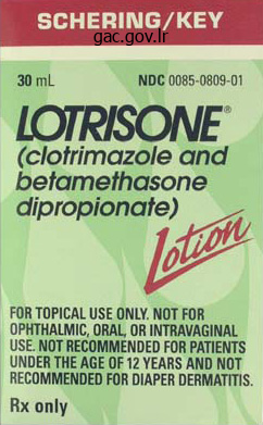
Lotrisone 10mg line
Dysbarism outcomes from barotrauma when gasoline compression or enlargement happens in elements of the body which would possibly be noncompressible or have restricted compliance. Pulmonary overinflation syndrome is probably certainly one of the most severe and probably deadly outcomes of barotrauma. Pulmonary overinflation syndrome is due to an inappropriately fast ascent inflicting alveoli rupture and air bubble extravasation into tissue planes or even the cerebral circulation. Decompression illness happens when the ascent is simply too rapid and gasoline bubbles kind and cause injury depending on their location (ie, coronary, pulmonary, spinal or cerebral blood vessels, joints, soft tissue). These gasoline bubbles cause harm due to mechanical disruption of tissue, local inflammatory response, occlusion of blood move, platelet activation, endothelial dysfunction, and capillary leakage. Decompression illness signs is dependent upon the dimensions and quantity and placement of gasoline bubbles released (notably nitrogen). Decompression sickness may occur in those that take hot showers after chilly dives. There have been medical case reports of delayed decompression illness presentation following post-dive train. This delayed decompression illness may be attributable to the cavitation results of gasoline trapped within the body, which is described as "vacuum phenomenon" in radiologic research. Preventive measures embody diver schooling; pre-dive medical screening and dive planning; strict adherence to dive course, timing, and depths; and a gradual and controlled ascent plus correct management of buoyancy. Conservative recommendation is to keep away from excessive altitudes (air journey or ground ascent) for a minimum of 24 hours after surfacing from the dive, particularly following a number of dives. Decompression sickness have to be thought-about if signs are temporally associated to recent diving or altitude or strain adjustments throughout the previous 48 hours. Continuous administration of 100% oxygen is indicated and useful for all patients. Hyperbaric oxygen remedy is usually beneficial for decompression sickness symptoms. Immediate consultation with a diving medicine or hyperbaric oxygen specialist is indicated even when gentle decompression sickness symptoms resolve. Opioids have to be used very cautiously, since these might obscure the response to recompression. Patent foramen ovale in recreational and professional divers: an necessary and largely unrecognized drawback. Hypothesis: the affect of cavitation or vacuum phenomenon for decompression illness. Symptom onset may be instant, inside minutes or hours (in the majority), or present up to 36 hours later. Prompt recognition and medical treatment of early symptoms of high-altitude illness could stop progression. Clinicians should assess different situations that may coexist or mimic symptoms of high-altitude illness (severe dehydration, hyponatremia, or hypoglycemia, trauma, infection). Immediate descent is the definitive treatment for high-altitude cerebral edema and high-altitude pulmonary edema. Later signs include irritability, issue concentrating, anorexia, insomnia, and elevated complications. It normally happens at elevations above 2500 meters (8250 feet) however may happen at decrease elevations. Hallmarks are altered psychological status, ataxia, extreme lassitude, and encephalopathy. Examination findings may embody confusion, ataxia, urinary retention or incontinence, focal neurologic deficits, papilledema, and seizures. High-altitude medical issues are because of hypobaric hypoxia at excessive altitudes (usually larger than 2000 meters or 6560 feet). High-altitude illness includes a spectrum of problems categorized by end-organ effects (mostly cerebral and pulmonary), and publicity period (acute and long-term). Long-term publicity to high altitude over months or years with insufficient acclimatization may find yourself in subacute mountain sickness and persistent mountain sickness (Monge disease). Acclimatization happens as a physiologic response to the rise in altitude and growing hypobaric hypoxia. Physiologic modifications include will increase in alveolar ventilation and oxygen extraction by the tissues and elevated hemoglobin stage and oxygen binding.
Discount lotrisone american express
If ulceration and white clues are absent one should attempt to make a selected prognosis based mostly on other clues. The prime case reveals the standard specific arrangement of linear vessels within the center of the clods. The hemorrhagic crust due to ulceration (seen as a single black clod) is a clue to malignancy in the absence of trauma. Top: A flat lesion with white traces on dermatoscopy requires histology to rule out malignancy. Middle: A nodule with white lines on dermatoscopy requires histology to rule out malignancy. Any tumor underneath the superficial vascular plexus including cysts can present with serpentine branched vessels � in this case a malignant peripheral nerve sheath tumor. Bottom: A nodule with out specific clues besides remnants of brown pigmentation in the periphery. Top: A flat lesion with a raised center without any specific clues and short linear vessels in the raised heart. It is determined by how assured one can diagnose a congenital nevus right here to decide if this lesion must be excised or not. The doctor who took care of this patient was assured sufficient to depart this lesion. Histopathologic diagnosis: Fibroepithelioma of Pinkus (a variant of basal cell carcinoma). Top: An ulcerated nodule with a yellow serum crust on dermatoscopy must be excised or biopsied to rule out malignancy. Bottom: A non-pigmented nodule with coiled and looped vessels but no particular clues. Differences between polarized gentle dermoscopy and immersion contact dermoscopy for the analysis of skin lesions. Structural correlations between dermoscopic and histopathological features of juvenile xanthogranuloma. The medical and dermoscopic features of invasive cutaneous squamous cell carcinoma depend on the histopathological grade of differentiation. Prediction without Pigment: a choice algorithm for non-pigmented skin malignancy. They often point in direction of a diagnosis, however only a few clues are particular for a specific analysis in all contexts. Clues should all the time be interpreted in context, in any other case a good clue may turn out to be a clich�. This article will look at a number of clues that deserve special attention, and at some common clich�s. Branched nice strains in flat acral lesions Brown or gray branched nice traces sprinkled with dots in a flat acral lesion is a very distinctive sample. As tinea nigra generally occurs on the ft where surgical procedure is technically difficult, the analysis is best confirmed by a profitable trial of therapy with topical antifungal cream. This leads to resolution of the lesion inside three weeks and avoids a biopsy to exclude melanoma. Branched serpentine vessels adjoining to keratin the presence of branched serpentine vessels adjacent to keratin is a strong clue to keratoacanthoma (7. There is an ongoing controversy among completely different colleges of dermatopathologists whether keratoacanthomas are benign lesions or highly differentiated variants of squamous cell carcinoma (3). However, even the proponents of the concept that a keratoacanthoma is a benign neoplasm agree that lesions that appear as keratoacanthomas clinically or dermatoscopically should be excised to rule out squamous cell carcinoma. Ulceration may in fact be brought on by trauma to normal pores and skin or benign lesions, but malignant neoplasms and especially basal cell carcinomas could ulcerate after trivial irritation. Adherent fiber could additionally be found on basal cell carcinomas even when ulceration was not observed prior to dermatoscopy. As ulceration is commonly an necessary clue to malignancy, so is the presence of dermatoscopically observed adherent fiber (7.
Purchase genuine lotrisone on line
When white and/or yellow clods predominate, as seen in these four lesions, the prognosis is seborrheic keratosis. White buildings � as within the two lesions in the best column � are the one exceptions to the general rule that construction is defined by pigment whereas the hypopigmented portion constitutes the background. In the 2 lesions within the left column one additionally sees the characteristic vessels of seborrheic keratosis, i. A additional clue is the pattern of vessels: in basal cell carcinoma serpentine vessels, often branched; in seborrheic keratosis, looped or coiled vessels (only rarely serpentine). The unconscious tendency is to interpret what one perceives to be essentially the most prominent features as the foreground and therefore constituting construction. It takes expertise and deliberate training of the attention to (when necessary) override unconscious rules and correctly make this distinction between foreground and background. White structures (lines, circles, dots or clods) are exceptions to the overall precept that pigment defines structure. That is, when white buildings are seen one ought to � Dies ist urheberrechtlich gesch�tztes Material. Invasive squamous cell carcinomas are usually non-pigmented however could have white circles or clods as a clue to the correct diagnosis (13). The diagnosis of non-pigmented lesions is mentioned in higher element in chapter 6. Red or purple clods are characteristic of hemangioma or vascular malformations (5. The differential analysis of blue clods is kind of different from that of purple or black clods, so this distinction have to be made fastidiously. Large, polygonal skin-colored clods are normally found in exophytic congenital nevi with a papillomatous floor, such as Unna nevus or Miescher nevus, and in addition sometimes in verrucous seborrheic keratosis (5. In all of these lesions one can also discover smaller orange clods interspersed between the skin-colored clods. When a pigmented lesion consists solely of brown clods, numerous forms of melanocytic nevi have to be considered within the diagnosis (5. Large, polygonal lightbrown clods are primarily indicators of an Unna nevus or Miescher nevus, especially when the scientific look is papillomatous. Quite often typical curved vessels are discovered within the middle of the clods, however the vascular morphology of these nevi could also be extremely polymorphous. In general, pigmented lesions ought to be diagnosed on the premise of their structure and colour. On dermatoscopy (right) one finds skin-colored clods with a looped vessel within the middle. Small to medium-sized, round and oval brown clods are attribute features of small congenital nevi, of both the "superficial" and "superficial and deep" sorts. Some pigmented Spitz nevi may also have only brown clods, or brown clods peripherally could mix with gray clods or strains centrally (5. Central hyperpigmentation can also be common in pigmented Spitz nevi, and peripheral clods are usually smaller than those within the center. While melanoma and mixed congenital nevus may also show a sample of blue clods, they nearly all the time even have clods of different colors, or another pattern in addition to blue clods. Therefore, when confronted with solely blue clods one ought to first search for clues to support or refute a prognosis of basal cell carcinoma. Top left: An Unna nevus composed of large polygonal brown clods, interspersed with a few small yellow clods. Top right: Unna nevus with large and polygonal, mostly skin-colored clods and some brown clods. Quite usually one finds gray clods or strains in the center (especially evident on the photograph on the right). If one finds solely blue clods the diagnosis is almost always basal cell carcinoma, as in these two examples.
Cheap lotrisone master card
The dengue vaccine Dengvaxia, made by Sanofi Pasteur, is accredited to be used in Mexico for persons aged 9�45 years. Preventive measures should be inspired, such as control of mosquitoes by screening and bug repellents including long-lasting insecticides, significantly throughout early morning and late afternoon exposures. Screening blood transfusions for dengue is of accelerating importance, especially in endemic areas. Stochastic models using rainfall and temperature are helpful in predicting geographic areas at increased threat for dengue. Binational dengue outbreak along the United StatesMexico border-Yuma County, Arizona, and Sonora, Mexico, 2014. Transfusion-transmitted dengue and related scientific symptoms in the course of the 2012 epidemic in Brazil. Relevance of non-communicable comorbidities for the development of the extreme types of dengue: a scientific literature evaluate. Association between dietary status and dengue infection: a scientific evaluation and meta-analysis. However, profit in the absence of bleeding may not be noticed, and hurt could additionally be brought on by delay in rely restoration. Monitoring very important indicators and blood quantity may help in anticipating the issues of dengue hemorrhagic fever or shock syndrome. Acute kidney harm in dengue shock syndrome portends an particularly poor prognosis. These differ in rodent hosts, geographic distribution, and diploma of pathogenicity. While retrospective diagnostics present that the disease occurred many years earlier in the United States, it was not until 1993 when the primary outbreak was recognized. Through January 16, 2016, 6590 circumstances were reported in 34 states and 235 (36%) had been deadly. All however 30 cases (95%) have been reported from states west of the Mississippi River, including all continental states west of the Mississippi River except Missouri and Arkansas, with over one-half the cases from the Four Corners space and one other focus recognized in Yosemite National Park, California. Nearly two-thirds of cases from a series in Panama skilled little or no pulmonary disease. Hantaviruses are ubiquitous with infections described in North and South America (New World hantaviruses) as well as Europe, Africa, and Asia (Old World hantaviruses). The Seoul viruses produce a much less severe type of illness and are found primarily in Korea and China. Cases of autochthonous Seoul hantavirus infections are described in the United States. The Puumala and Dobrava-Belgrade viruses, and the Seoul virus additionally, are present in Scandinavia and Europe and are related to a mild form of the syndrome termed "nephropathia epidemica," which presents with fever, headache, stomach ache, and impaired kidney perform. Between 16% and 48% of Dobrova-Belgrade virus cases, nonetheless, could require dialysis. Aerosols of virus-contaminated rodent urine and feces are thought to be the main automobile for transmission to people. Occupation is the main threat issue for transmission of all hantaviruses: animal trappers, forestry staff, laboratory personnel, farmers, and navy personnel are considered to be at highest threat. Climate change seems to be impacting the incidence of hantavirus infection primarily through effects on reservoir ecology. An ensuing cardiopulmonary section is characterized by the acute onset of pulmonary edema. In this stage, cough is usually current, abdominal pain and signs as above could dominate the clinical presentation, and in extreme instances, significant myocardial despair occurs. A 2- to 3-week incubation interval is adopted by a protracted scientific course, usually consisting of 5 distinct phases: febrile interval, hypotension, oliguria, diuresis, and convalescence section. Various degrees of renal involvement are normally seen, sometimes with frank hemorrhage.
Lotrisone 10 mg low cost
Laboratory Findings the organism may be recovered from cultures of blood, cerebrospinal fluid, urine, bone marrow, or different websites. Humans usually acquire the an infection by contact with animal tissues (eg, trapping muskrats, skinning rabbits) or from a tick or insect chunk. F tularensis has been categorised as a high-priority agent for potential bioterrorism use due to its virulence and relative ease of dissemination. Infection in humans typically produces a neighborhood lesion and widespread organ involvement however may be totally asymptomatic. Less widespread issues are pneumonitis with pleural effusion, hepatitis, and cholecystitis. Symptoms and Signs Fever, headache, and nausea start suddenly, and a neighborhood lesion-a papule on the site of inoculation-develops and shortly ulcerates. Pneumonia might develop from hematogenous spread of the organism or could additionally be main after inhalation of infected aerosols, that are liable for human-to-human transmission. Following ingestion of contaminated meat or water, an enteric type may be manifested by gastrointestinal symptoms, stupor, and delirium. In any type of involvement, the spleen could also be enlarged and tender and there may be nonspecific rashes, myalgias, and prostration. Regimens of doxycycline (200 mg/day orally for six weeks) plus rifampin (600 mg/day orally for six weeks) or streptomycin (1 g/day intramuscularly for two weeks) or gentamicin (240 mg intramuscularly once every day for 7 days) have the lowest recurrence charges. Longer courses of remedy could also be required to prevent relapse of meningitis, osteomyelitis, or endocarditis. For this reason and since cultures of F tularensis may be hazardous to laboratory personnel, the diagnosis is normally made serologically. A positive agglutination take a look at (greater than 1:80) develops within the second week after an infection and should persist for several years. Hematogenous unfold could produce meningitis, perisplenitis, pericarditis, pneumonia, and osteomyelitis. Gentamicin, which has good in vitro exercise in opposition to F tularensis, is usually much less toxic than streptomycin and possibly simply as effective. A number of other brokers (eg, fluoroquinolones) are energetic in vitro however their medical effectiveness is much less well established. Laboratory Findings the plague bacillus could additionally be present in smears from aspirates of buboes examined with Gram stain. In convalescing sufferers, an antibody titer rise could also be demonstrated by agglutination exams. The onset is sudden, with high fever, malaise, tachycardia, intense headache, delirium, and extreme myalgias. If pneumonia develops, tachypnea, productive cough, blood-tinged sputum, and cyanosis also occur. Axillary, inguinal, or cervical lymph nodes become enlarged and tender and may suppurate and drain. With hematogenous unfold, the affected person might rapidly become toxic and comatose, with purpuric spots (black plague) appearing on the pores and skin. Primary plague pneumonia is a fulminant pneumonitis with bloody, frothy sputum and sepsis. The systemic manifestations resemble those of enteric or rickettsial fevers, malaria, or influenza. The pneumonia resembles different bacterial pneumonias, and the meningitis is much like these caused by different micro organism. It is transmitted among rodents and to humans by the bites of fleas or from contact with contaminated animals. Following a fleabite, the organisms unfold by way of the lymphatics to the lymph nodes, which become greatly enlarged (buboes). The affected person with pneumonia can transmit the an infection to different people by droplets. Drug prophylaxis may provide short-term safety for individuals uncovered to the risk of plague infection, significantly by the respiratory route. Patients with plague pneumonia are positioned in strict respiratory isolation, and prophylactic therapy is given to any one who came involved with the affected person.
Buy lotrisone 10mg visa
Azotemia could additionally be present in a small number of cases associated with ureteral obstruction. Anemia might sometimes be because of persistent blood loss or to bone marrow metastases. Exfoliated cells from normal and abnormal urothelium may be readily detected in voided urine specimens. Cytology can be useful to detect the illness initially or to detect its recurrence. Cytology is delicate in detecting cancers of upper grade and stage (80�90%) but much less so in detecting superficial or well-differentiated lesions (50%). There are quite a few urinary tumor markers underneath investigation for screening, assessing recurrence, progression, prognosis, or response to therapy. Intravesical Chemotherapy Immunotherapeutic or chemotherapeutic agents delivered immediately into the bladder via a urethral catheter can scale back the probability of recurrence in those who have undergone complete transurethral resection. Side effects of intravesical chemotherapy embody irritative voiding symptoms and hemorrhagic cystitis. However, the presence of most cancers is confirmed by cystoscopy and biopsy, with imaging primarily used to consider the higher urinary tract and to stage extra advanced lesions. Cystourethroscopy and Biopsy errs es ook b ook b � Treatment the prognosis and staging of bladder cancers are made by cystoscopy and transurethral resection. If cystoscopy- performed normally underneath native anesthesia-confirms the presence of bladder cancer, the affected person is scheduled for transurethral resection underneath common or regional anesthesia. Random bladder and, every so often, transurethral prostate biopsies are performed to detect occult illness in the bladder or elsewhere and potentially establish patients at larger threat for cancer recurrence and progression. Partial cystectomy is indicated in chosen patients with solitary lesions or those with cancer in a bladder diverticulum. Radical cystectomy entails elimination of the bladder, prostate, seminal vesicles, and surrounding fat and peritoneal attachments in males and the uterus, cervix, urethra, anterior vaginal vault, and usually the ovaries in ladies. However, continent types of diversion keep away from the need of an exterior appliance and can be thought-about in a major variety of sufferers. Bladder most cancers staging relies on the extent (depth) of bladder wall penetration and the presence of regional or distant metastases. The natural historical past of bladder most cancers is based on two separate however associated processes: most cancers recurrence inside the bladder and development to higher-stage illness. The subset of patients with es kerrs oo k eb oo e//eb me External beam radiotherapy delivered in fractions over a 6- to 8-week period is generally well tolerated, however roughly 10�15% of sufferers will develop bladder, bowel, or rectal issues. Chemotherapy Metastatic disease is current in 15% of sufferers with newly identified bladder cancer, and metastases develop within 2 years in as a lot as 40% of patients who have been believed to have localized disease at the time of cystectomy or definitive radiotherapy. Cisplatin-based combination chemotherapy results in partial or full responses in 15�45% of sufferers (see Table 39�4). Combination chemotherapy has been used to decrease recurrence rates with both surgery and radiotherapy and to attempt bladder preservation in these handled with radiation. Neoadjuvant chemotherapy seems to benefit all sufferers with muscle-invasive illness previous to deliberate cystectomy. Chemotherapy should also be considered earlier than surgical procedure in these with bulky lesions or these suspected of getting regional disease. Chemoradiation is greatest suited for these with T2 or restricted T3 illness without ureteral obstruction. Alternatively, chemotherapy has been used after cystectomy in patients at high risk for recurrence, similar to those who have lymph node involvement or native invasion. Contemporary cost-effectiveness evaluation evaluating sequential bacillus Calmette-Guerin and electromotive mitomycin versus bacillus Calmette-Guerin alone for patients with high-risk non-muscle-invasive bladder most cancers. Carcinoma in situ is most frequently present in affiliation with papillary bladder cancers. Its presence identifies patients at elevated threat for recurrence and development. At initial presentation, approximately 50�80% of bladder cancers are superficial: stage Ta, Tis, or T1.
Order 10 mg lotrisone with visa
Other less particular dermatoscopic clues are clods or vessels as dots situated within the heart of reticular strains, curved lines (primarily in combination with reticular or branched lines), and densely arranged aggregations of small circles. Occasionally it may be difficult to distinguish between a Clark nevus and a congenital nevus on dermatoscopy, because the reticular sample may occur alone or in combination with different patterns in each types of nevus. Clues to melanoma, particularly gray dots and white reticular strains, may be present in some congenital nevi. Correlation of dermatoscopy and dermatopathology the histological correlate of reticular lines was defined within the section on Clark nevi. Brown clods correspond to nests of melanocytes on the dermo-epidermal junction, that are normally larger in congenital nevi than in the Clark nevus. Skin-colored clods arise due to flippantly pigmented or non-pigmented nests of melanocytes in the papillary dermis. The widened dermal papillae full of melanocytes trigger the dermis to protrude outward, which gives rise in metaphorical terminology of a cobblestone sample. Combined congenital nevi Combined congenital nevi are these showing features of each a "blue nevus" and either a "superficial" or "superficial and deep" congenital nevus (3. Occasionally, as an alternative of a blue structureless area one sees blue clods, which not often could also be distributed over the entire lesion. Top: More than one sample, symmetrical, clods peripherally, structureless blue within the heart. Bottom: More than one pattern, branched lines and clods peripherally, structureless blue within the middle, relatively symmetrical. Combined congenital nevi Pattern Typical: Structureless, reticular lines, clods Combinations of patterns are normally symmetrical Colors Typical: Structureless area: blue Reticular lines and clods: brown Occasional: Blue clods Clues the structureless blue area is in the middle. Recurrent nevus On dermatoscopy one sometimes sees a hypopigmented (lighter than surrounding skin) structureless zone, comparable to the scar after excision (3. Common patterns seen are peripheral radial strains, pseudopods, and brown clods of various sizes. Correlation between dermatoscopy and dermatopathology the radial lines and pseudopods correspond to fascicles of pigmented melanocytes on the dermo-epidermal junction. Spitz nevus the "classical" Spitz nevus as described by Sophie Spitz is non-pigmented or solely frivolously pigmented. On dermatoscopy one most frequently finds skin-colored or light-brown clods and perpendicular white strains (3. The patterns seen in pigmented Spitz nevi are brown clods peripherally; centrally grey or blue-gray clods or structureless (3. This central space is sometimes interspersed with thick, light-gray reticular strains and/ or polarizing-specific white traces. Clinically Spitz nevi are nodular or papular and Reed nevi are flat or only barely raised. Dermatoscopically, the patterns of established Reed nevi are pseudopods or radial lines. Correlation between dermatoscopy and dermatopathology Like the previously described nevi, brown clods correspond to pigmented melanocyte nests in the epidermis. The gray reticular strains in the middle of pigmented Spitz nevi are probably because of the combination of relatively heavily pigmented nests of melanocytes and acanthosis of the dermis. Reed nevus Serial dermatoscopic photography exhibits that an early Reed nevus consists solely of dark-brown clods. The characteristic pattern of radial lines or pseudopods at the periphery only develops throughout subsequent development (3. A Reed nevus is then identical to a darkly pigmented Clark nevus with reticular lines peripherally and a structureless hyperpigmented heart, or reticular traces solely. One believable principle suggests this is adopted by transepidermal elimination of melanocytes and the disappearance of the nevus. Correlation between dermatoscopy and dermatopathology the pseudopods and radial traces on the periphery are fascicles of pigmented melanocytes at the dermo-epidermal junction that have unfold centrifugally. As we noted in chapter 2, the term "blue nevus" includes various entities which could be distinguished by dermatopathology, however not by dermatoscopy or scientific examination. Less widespread once more are blue nevi with shades of gray and blue flanked by brown areas.
Lotrisone 10 mg on-line
In sebaceous hyperplasia, the white of the clods is duller than in milia or polarizing-specific white clods. Subsurface keratin in this pilomatrixoma appears as a white structureless zone on dermatoscopy (right). Yellow colour may also be found in nevus sebaceous, in which the increased variety of sebaceous glands is responsible for the yellow look on dermatoscopy (6. The yellow clods of initial cutaneous leishmaniasis most likely correspond to widened infundibula on the background of a granulomatous inflammation in the dermis whereas the yellow clods of lymphangioma correspond to dilated lymphatic vessels full of lymphatic fluid (19). The structureless zone within the center of this xanthogranuloma in an adult is yellow and orange (dermatoscopy on the right). This nevus sebaceous in a newborn is structureless yellow on dermatoscopy (right image). When one vessel kind predominates, that is called a "monomorphous" sample of vessels. In addition to the type of vessels, their arrangement � each how vessels are organized relative to each other, and the way vessels are distributed all through the lesion � can also be of diagnostic significance (6. Vessels as dots or coils may be arranged in straight lines (linear arrangement) or in serpentine lines (serpiginous arrangement). Vessels may be randomly distributed (A), clustered (B), serpiginous (C) linear (D), centered (E), radial (F), reticular (G), or branched (H). Straight linear vessels that intersect one another nearly at proper angles have a "reticular" arrangement. In flat lesions, most vessels are viewed end on and so seem as dots or quick curved lines. This variation means the identical vessel morphology may have different diagnostic significance in nodules compared to flat lesions. Examine "non-pigmented" lesions rigorously for any pigment earlier than utilizing a non-pigmented algorithm. On shut inspection, two areas of converging radial strains are obvious, allowing a assured analysis of basal cell carcinoma. As a matter of convenience, medical features are usually assessed earlier than dermatoscopy. These findings are then included in the diagnostic course of as one proceeds with dermatoscopy. Rather, a lesion is termed flat when the horizontal diameter greatly exceeds height. Macules, flat papules and patches are flat whereas elevated papules and nodules are raised. With severe persistent sun harm, the entire pores and skin surface may be scaly, including that overlying lesions. If keratin is present, the principle differential diagnoses include well-differentiated squamous cell carcinomas/ keratoacanthomas, seborrheic keratoses and viral warts (22). Keratin can be found in Unna or Miescher nevi (keratin plugs on the surface between papillomatous invaginations), in keratinizing adnexal proliferations corresponding to pilomatrixoma (subsurface keratin) (23), in keratinizing cysts (subsurface keratin), and in angiokeratoma (surface keratin) (24). The vascular sample is more diagnostically significant in flat lesions than in raised lesions. In raised lesions, other clues (ulceration, keratin, and white clues) take priority over vessel sample evaluation, just as pigmented buildings take priority for pigmented lesions. In addition to this spectrum of neoplasms, numerous inflammatory pores and skin illnesses should even be thought-about. Melanocytic lesions that may seem as non-pigmented skin-colored to red macules or patches are Clark nevi, "superficial" or "superficial and deep" congenital nevi, Spitz nevi and, after all, melanoma. We will focus on the dermatoscopic appearance of inflammatory lesions in greater detail in chapter eight. Rarely, even seborrheic keratosis and dermatofibroma could additionally be flat and non-pigmented. The most common flat non-pigmented lesions and their look on dermatoscopy are proven in table 6.
Real Experiences: Customer Reviews on Lotrisone
Moff, 35 years: Follow-up of melanocytic pores and skin lesions with digital dermoscopy: dangers and benefits.
Chris, 23 years: This may be due more to regional variation in patterns of histopathology reporting, quite than any true variation in incidence.
Jaffar, 55 years: Patients with plague pneumonia are positioned in strict respiratory isolation, and prophylactic therapy is given to any one that got here involved with the affected person.
8 of 10 - Review by D. Enzo
Votes: 65 votes
Total customer reviews: 65

