
Deltasone dosages: 40 mg, 20 mg, 10 mg, 5 mg
Deltasone packs: 30 pills, 60 pills, 90 pills, 120 pills, 180 pills, 270 pills, 360 pills

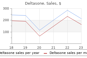
Cheap deltasone uk
A proerythroblast (center) and two intermediate erythroblasts in a bone marrow aspirate from a healthy volunteer. Early erythroblast and neutrophil in a bone marrow aspirate from a wholesome volunteer. An early erythroblast (beneath the neutrophil band type on the left) and two late erythroblasts. Four proerythroblasts (arrow) surrounded by erythroid precursors in later stages of maturation, a megakaryocyte, and a few immature granulocytes in a trephine biopsy section from a hematologically normal man. Note the sturdy amphophilic cytoplasm of the proerythroblasts and their linear or comma-shaped nucleoli, which frequently abut on the nuclear membrane. Two proerythroblasts (long arrows) and an intermediate erythroblast (short arrow) surrounded by late erythroid precursors. Disrupted erythroid island displaying early, intermediate, and late erythroblasts in a bone marrow aspirate from a wholesome volunteer. Erythroid island composed mainly of intermediate and late erythroblasts in a bit of a trephine biopsy specimen from a hematologically regular affected person. Late erythroblasts containing siderotic granules (arrows) in a bone marrow aspirate stained with Perls stain. Maturating cells of eosinophil and basophil lineage may be recognized morphologically from the myelocyte stage onward in aspirate films. Megakaryocytes and Thrombopoiesis Three levels of megakaryocyte maturation could be recognized in regular bone marrow. The smallest immature megakaryocytes measure 30 �m or extra in diameter and have a high nuclear-to-cytoplasmic ratio and basophilic, often "blebbed" cytoplasm. A myelocyte and three intermediate erythroblasts in a bone marrow aspirate from a healthy volunteer (May-Gr�nwaldGiemsa stain). The most immature cells are next to the bone, including blasts (arrows), with extra mature granulocytes deeper in the intertrabecular space. An early promyelocyte (top right) and a late promyelocyte (bottom left) in a bone marrow aspirate from a wholesome volunteer. The cell immediately above the more mature promyelocyte is a very early promyelocyte, displaying eccentricity of the nucleus and the earliest indicators of cytoplasmic maturation. Caution must be exercised in decoding cytologic features of megakaryocytes because these massive cells are very vulnerable to crushing during the spreading of a bone marrow movie; this will likely fragment a nucleus or trigger some parts of the nucleus to be partly extruded from the cell. The cytoplasm of megakaryocytes could seem to comprise intact cells of other lineages; these are literally inside the surface- linked canalicular system. This phenomenon, known as emperipolesis, is physiologic but may be exaggerated in numerous pathologic states. Late megakaryocytes are readily acknowledged as apparently naked megakaryocyte nuclei, which are bigger than the nuclei of bone marrow cells of any other lineage and are more pyknotic than other nuclei of comparable dimension. Immature (A) and mature (B) megakaryocyte in a bone marrow aspirate from a hematologically regular individual. Late megakaryocyte, which has shed nearly all its cytoplasm as platelets and seems as an almost bare nucleus, in a bone marrow movie (May-Gr�nwald-Giemsa stain). Debris-laden macrophage, eosinophil myelocyte, and two neutrophil band varieties in a bone marrow aspirate from a healthy volunteer. Megakaryocytes are irregularly distributed within the bone marrow, and determining whether or not the number of megakaryocytes in a bone marrow aspirate is regular is difficult and necessarily subjective; it usually relies on the standard of the movie in addition to the expertise of the observer. Other Myeloid Cells Monocytes, macrophages, mast cells, and osteoclasts are all of myeloid origin. Macrophages may be seen as isolated cells or in relation to erythroblasts in an erythroid island. Three megakaryocytes in a section of a trephine biopsy specimen from a hematologically regular affected person. The variation in size and the nuclear lobulation are due to sectioning throughout a large, three-dimensional megakaryocyte in the biopsy. Osteoclast adjacent to a bony spicule in a bit of a trephine biopsy specimen from a toddler. Cytologic Abnormalities in Myeloid Cells in Hematologically Normal Subjects It must be noted that the bone marrow aspirate of wholesome volunteers could present some features that might be interpreted as indicative of dysplasia, corresponding to dyserythropoietic options or the presence of non-lobulated or multinucleated megakaryocytes (Table 10-4). Normal mast cells, although rare, are readily recognizable in bone marrow aspirate movies due to their distinctive cytologic features.
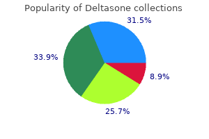
Best 40 mg deltasone
Prominent intrasinusoidal infiltration of the bone marrow by mantle cell lymphoma. Mantle cell lymphoma, in leukemic phase: characterization of its broad cytologic spectrum with emphasis on the significance of distinction from other chronic lymphoproliferative disorders. Leukemic part of B-cell lymphomas mimicking chronic lymphocytic leukemia and variants at presentation. A report of eight instances with comparable medical presentation and aggressive end result. Nucleolated variant of, mantle cell lymphoma with leukemic manifestations mimicking prolymphocytic leukemia. Nuclear expression of the non B-cell lineage Sox11 transcription issue identifies mantle cell lymphoma. Nuclear expression of sox11 is extremely associated with mantle cell lymphoma but is unbiased of t(11;14)(q13;q32) in non�mantle cell B-cell neoplasms. Clinical impact of the differentiation profile assessed by immunophenotyping in sufferers with diffuse giant B-cell lymphoma. Clinical and prognostic significance of bone marrow involvement in patients with diffuse aggressive B-cell lymphoma. Discordant bone marrow involvement in diffuse giant B-cell lymphoma: comparative molecular analysis reveals a heterogeneous group of disorders. The prognostic influence of bone marrow involvement in patients with diffuse giant cell lymphoma varies in accordance with the diploma of infiltration and presence of discordant marrow involvement. Diffuse giant B-cell lymphoma with concordant bone marrow Chapter 56 � Bone Marrow Evaluation for Lymphoma1064. Bone marrow trephine biopsy appearances of the intravascular subtype of diffuse giant B-cell lymphoma. B-cell lymphoma, unclassifiable, with options intermediate between diffuse large B-cell lymphoma and Burkitt lymphoma. Burkitt lymphoma pathogenesis and therapeutic targets from structural and practical genomics. Immunomorphologic profile and Epstein-Barr virus standing of a cohort of 35 circumstances of extranodal natural killer/T-cell lymphoma, nasal type of higher aerodigestive tract from a tertiary care middle in South India. Enteropathy-type intestinal T-cell lymphoma: medical options and treatment of 31 patients in a single heart. Enteropathy-associated T-cell lymphoma: scientific and histological findings from the worldwide peripheral T-cell lymphoma project. Bone marrow histologic and immunohistochemical findings in peripheral T-cell lymphoma: a research of 38 cases. Hepatosplenic T-cell lymphoma: a definite clinicopathologic entity of cytotoxic gamma delta T-cell origin. Hepatosplenic alphabeta T-cell lymphomas: a report of 14 cases and comparability with hepatosplenic gammadelta T-cell lymphomas. Hepato, splenic alphabeta T-cell lymphoma: an unusual case with clinical, histologic, and cytogenetic options of gammadelta hepatosplenic T-cell lymphoma. Consistent presence, of isochromosome 7q in hepatosplenic T gamma/delta lymphoma: a new cytogenetic-clinicopathologic entity. Clinicopathological features of hepatosplenic T cell lymphoma: a single centre experience from India. Hepatosplenic gam, madelta T-cell lymphoma is a rare clinicopathologic entity with poor consequence: report on a collection of 21 sufferers. Subcutaneous panniculitis-like T-cell lymphoma: clinicopathologic, immunophenotypic, and genotypic analysis of alpha/ beta and gamma/delta subtypes. Bone marrow involvement by subcutaneous panniculitis-like T-cell lymphoma: a report of three circumstances. Bone marrow histopathologic and molecular staging in epidermotropic T-cell lymphomas. Prognostic implications of a bone marrow histopathologic classification system in mycosis fungoides and the S�zary syndrome.
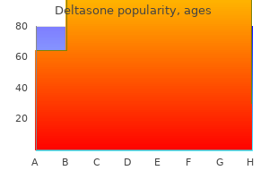
Order deltasone 20mg line
Fujisawa S, Maruta A, Motomura S, Fukawa H, Kanamori H, Ogawa K, Matsuzaki M, Miyashita H, Harano H, Murata T, Sakai R, Mohri H, Kodama F Okubo T. Prognostic significance of blood and marrow findings in acute myelogenous leukemia in remission. A attribute pattern of leukemic cell differentiation without cytoreduction throughout remission induction in 1087. Biondi A, Luciano A, Bassan R, Mininni D, Specchia G, Lanzi E, Castagna S, Cantu-Rajnoldi A, Liso V, Masera G. Kita K, Nakase K, Miwa H, Masuya M, Nishii K, Morita N, Takakura N, Otsuji A, Shirakawa S, Ueda T. Kern W, Danhauser-Riedl S, Ratei R, Schnittger S, Schoch C, Kolb H, Ludwig W, Hiddemann W, Haferlach T. Detection of minimal residual disease in unselected patients with acute myeloid leukemia using multiparameter move cytometry for definition of leukemiaassociated immunophenotypes and determination of their frequencies in normal bone marrow. Early immunophenotypical evaluation of minimal residual illness in acute myeloid leukemia identifies different patient risk groups and will contribute to postinduction therapy stratification. Venditti A, Buccisano F Del Poeta G, Maurillo L, Tam, burini A, Cox C, Battaglia A, Catalano G, Del Moro B, Cudillo L, Postorino M, Masi M, Amadori S. Level of minimal residual illness after consolidation remedy predicts outcome in acute myeloid leukemia. The function of multiparameter move cytometry for detection of minimal residual illness in acute myeloid leukemia. Karyotypic evaluation predicts consequence of preremission and postremission therapy in grownup acute myeloid leukemia: a southwest oncology group/eastern cooperative oncology group study. Minimal residual illness in acute myelogenous leukaemia and myelodysplastic syndromes: a follow-up of sufferers in medical remission. Detection of numerical aberrations in hematologic neoplasias by fluorescence in situ hybridization. Chimerism and outcomes after, allogeneic hematopoietic cell transplantation following nonmyeloablative conditioning. Real-time quantitation of minimal residual illness in inv(16)-positive acute myeloid leukemia might indicate threat for clinical relapse and should determine sufferers in a curable state. Guerrasio A, Pilatrino C, De Micheli D, Cilloni D, Serra A, Gottardi E, Parziale A, Marmont F Diverio D, Divona, M, Lo Coco F Saglio G. Next-generation sequencing-based multigene mutational screening for acute myeloid leukemia using MiSeq: applicability for diagnostics and disease monitoring. Prognostic value of early response to chemotherapy assessed by the day 15 bone marrow aspiration in grownup acute lymphoblastic leukemia: a potential evaluation of 437 instances and its utility for designing induction chemotherapy trials. Persistence of circulating blasts after 1 week of multiagent chemotherapy confers a poor prognosis in childhood acute lymphoblastic leukemia. Persistence of peripheral blood and bone marrow blasts during remission induction in adult acute lymphoblastic leukemia confers a poor prognosis depending on remedy depth. Prognostic value of day 14 blast share and absolutely the blast index in bone marrow of children with acute lymphoblastic leukemia. Persistence of lymphoblasts in bone marrow on day 15 and days 22 to 25 of remission induction predicts a dismal therapy end result in youngsters with acute lymphoblastic leukemia. High frequency of pro-B acute lymphoblastic leukemia in adults with secondary leukemia with 11q23 abnormalities. A restricted antibody panel can distinguish B-precursor acute lymphoblastic leukemia from normal B precursors with four shade flow cytometry: implications for residual disease detection. Identification of novel markers for monitoring minimal residual disease in acute lymphoblastic leukemia. Immunophenotypic evaluation of hematogones (B-lymphocyte precursors) in 662 consecutive bone 1087. Prognostic significance and modalities of flow cytometric minimal residual disease detection in childhood acute lymphoblastic leukemia. Prospective karyotype evaluation in adult acute lymphoblastic leukemia: the cancer and leukemia group B expertise. Cave H, van der Werff ten Bosch J, Suciu S, Guidal C, Waterkeyn C, Otten J, Bakkus M, Thielemans K, Grandchamp B, Vilmer E. Clinical significance of minimal residual illness in childhood acute lymphoblastic eighty four. European organization for research and treatment of cancer�childhood leukemia cooperative group.
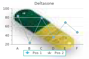
Cheap deltasone 40mg without prescription
Involvement of regional lymph nodes and distinction from angiolymphoid hyperplasia with eosinophilia. Observations in regards to the, pathogenesis of epithelioid hemangioma (angiolymphoid hyperplasia). Infrequent detection of Toxoplasma gondii genome in toxoplasmic lymphadenitis: a polymerase chain reaction examine. Lupus lymphadenitis: report of a case with immunohistologic studies on frozen sections. Lymphadenitis showing focal reticulum cell hyperplasia with nuclear debris and phagocytes. Kikuchi disease in systemic lupus erythematosus: medical features and literature evaluation. Inflammatory pseudotumor of lymph nodes: clinicopathologic and immunohistological examine of 11 Japanese cases. Hypocellular anaplastic large cell lymphoma mimicking inflammatory lesions of lymph nodes. Immunocytochemical identification of Rochalimaea henselae in bacillary (epithelioid) angiomatosis, parenchymal bacillary peliosis, and chronic fever with bacteremia. The spectrum of morphologic adjustments simulating lymphoma in lymph nodes and tonsils. Lymphadenopathy due to infectious mononucleosis: its confusion with malignant lymphoma. Epstein-barr virus infection of monocytoid B-cell proliferates: an early function of main viral infection Epstein-Barr virus latent membrane protein expression by Hodgkin and Reed-Sternberg-like 204. Cytomegalovirus as a potential cause of a illness resembling infectious mononucleosis. Concurrent herpes simplex viral lymphadenitis and chronic lymphocytic leukemia/small lymphocytic lymphoma, mimicking large-cell (Richter) transformation. Localized herpes simplex lymphadenitis: report of three circumstances and review of the literature. Lymphadenopathy induced by anticonvulsant drugs and mimicking clinically pathologically malignant lymphomas. All lymphopoietic and hematopoietic cells are in the end derived from pluripotent hematopoietic stem cells-slowly biking cells with a capacity for self-renewal. Such cells may be recognized in vitro by their capacity for self-renewal and their capacity to differentiate to produce cells of specific lineages. Cells beyond the stage of a lineage-committed progenitor could be acknowledged from cytologic in addition to useful and immunophenotypic characteristics. Some platelets are produced from megakaryocytes which have entered the circulation and lodged in the lungs. Hematopoiesis occurs in a specific bone marrow microenvironment, in cavities surrounded and traversed by bony spicules. The intertrabecular areas are occupied by stroma and hematopoietic cells, with the two parts having a dynamic interrelationship. The stroma consists of stromal cells and a matrix of proteins corresponding to laminin, thrombospondin, and fibronectin. Recognizable stromal components embody blood vessels, nerves, fat cells, different mesenchymal cells. The fiber community is detectable on a reticulin stain; if graded zero to four,2 most conventional topics have grade zero to 1 reticulin, but some have grade 2. Reticulin is deposited preferentially round arterioles and adjacent to bony spicules. The earliest recognizable granulocyte precursors-myeloblasts and promyelocytes-are situated towards the periosteum and in a band round arterioles. Myelocytes, metamyelocytes, and neutrophils are discovered progressively farther from the endosteum. Maturing erythroid cells and megakaryocytes are discovered more centrally in the intertrabecular area. Erythroblasts are clustered, forming erythroid islands by which erythroid cells of varying degrees of maturity encompass a central macrophage. Megakaryocytes are found preferentially in relation to sinusoids, and serial sections of bone marrow show that a half of the megakaryocyte cytoplasm abuts a sinusoid.
Purchase cheap deltasone on-line
Cases arise most regularly on the pinnacle and neck area, but extrafacial lesions could additionally be hardly ever observed. Morphologic features, too, are comparable, though the sample is more monomorphous. The infiltrate is confined to the dermis, generally involving the subcutaneous tissue. D, Staining for Ki67 demonstrates high proliferation of the cells; note a reactive germinal middle displaying strong nodular positivity. A, Dense, monomorphous lymphoid infiltrate in the dermis; note Grenz zone beneath an unaffected epidermis and (inset) pleomorphic nuclei of neoplastic cells. Neurologic symptoms as a sign of involvement of the central nervous system are commonly present. Histology shows a proliferation of huge lymphocytes confined to dilated blood vessels within the dermis and subcutaneous tissues. The malignant cells are large with scant cytoplasm and sometimes with distinguished nucleoli. The prognosis appears to be higher for patients with disease restricted to one organ only compared with those with illness detected in two or extra organs. Prominent epidermotropism on the edge of an ulcer with tumor necrosis surrounded by a wedge of neoplastic cells is a relatively frequent sample of cutaneous anaplastic giant cell lymphoma. Subcutaneous (lobular panniculitis-like) involvement, too, is a sample common to several types of cutaneous lymphoma. Angiocentricity or angiodestruction is another sample widespread to various sorts of cutaneous lymphomas, both of T and B phenotype. In some cutaneous diffuse giant B-cell lymphomas, the angiotropic infiltrate is composed of enormous B cells admixed with many cytotoxic T lymphocytes, representing a diagnostic pitfall. In instances with combined cell infiltrates, a staining for Ki67 permits better recognition of the proliferating cells. Although proliferation could be observed also in reactive lymphocytes, the staining is usually useful for a greater evaluation of the architecture of the infiltrate. In this context, double stainings for Ki67 and T- and B-cell antigens may be useful. Teach your surgeons to keep away from putting the biopsy specimens on gauze; drying artifacts are extraordinarily quick on pores and skin specimens, notably of the widespread, small punch biopsies. Lobular panniculitic, infiltrates with overlapping histopathologic features of lupus panniculitis (lupus profundus) and subcutaneous T-cell lymphoma: a conceptual and practical dilemma. Cutaneous gamma/ delta T-cell lymphoma during treatment with etanercept for rheumatoid arthritis. Gene expression profiling of peripheral T-cell lymphoma including gammadelta T-cell lymphoma. Subcutaneous panniculitis-like T-cell lymphoma within the pediatric age group: a lymphoma of low malignant potential. Aggressive subcutaneous panniculitis-like T-cell lymphoma with hemophagocytosis in two kids (subcutaneous panniculitis-like T-cell lymphoma). Subcutaneous panniculitis-like T-cell lymphoma in a patient receiving etanercept for rheumatoid arthritis. A case with an indolent course of subcutaneous panniculitis-like T-cell lymphoma demonstrating Epstein-Barr virus positivity and simulating dermatitis artefacta. Clinicopathological characterization and genomic aberrations in subcutaneous panniculitis-like T-cell lymphoma. The presence of clusters of plasmacytoid dendritic cells is a helpful function for differentiating lupus panniculitis from subcutaneous panniculitis-like T-cell lymphoma. Localized cutaneous small to medium-sized pleomorphic T-cell lymphoma: a report of 3 instances stable for years. Peripheral T-cell lymphomas unspecified presenting within the skin: evaluation of prognostic components in a bunch of eighty two sufferers. Primary cutaneous T-cell lymphoma localized to the decrease leg: a definite, regionally aggressive cutaneous T-cell lymphoma. Cutaneous manifestations of angioimmunoblastic T-cell lymphoma: clinical and pathological characteristics. A uncommon variant of intravascular giant cell lymphoma with frequent cytotoxic phenotype and affiliation with Epstein-Barr virus an infection. Intravascular cytotoxic T-cell lymphoma: a case report and review of the literature.
Purchase 10 mg deltasone amex
A, this 4-year-old boy has a papulovesicular eruption with vacciniform scarring of the face. B, Skin shows epidermal reticular degeneration, leading to spongiotic vesiculation. Patients present necrotic papulovesicles, nodules, or facial swelling, which may recur for years. T-cell receptor gene rearrangements of infiltrating cells in the skin are sometimes polyclonal,ninety four but they can be monoclonal. A, this 24-year-old man with recurrent necrotic papulovesicles on the face for six years finally developed systemic Epstein-Barr virus�positive T-cell lymphoma. It is often characterised by a speedy clinical development, with multiple organ failure, sepsis, and dying. The disease is usually difficult by hemophagocytic syndrome, coagulopathy, sepsis, and multiorgan failure. The illness progressed rapidly causing the demise of the patient in all cases with a median survival of seven months (1 to 13 months). The liver exhibits delicate to distinguished portal as properly as sinusoidal infiltrates of small lymphocytes with intracellular and intracanalicular cholestasis, steatosis, and focal necrosis. The spleen shows depleted white pulp and outstanding sinusoidal small lymphoid infiltrates. The B-cell areas are depleted, whereas the paracortical areas might be expanded and show a refined to putting infiltration with relatively homogeneous small, medium, or massive lymphocytes with hyperchromatic nuclei and irregular nuclear contours. The extreme scientific manifestations and the presence of hemophagocytosis normally alert one to the intense nature of the lymphoid proliferation. C, Abundant histiocytes with erythrophagocytosis intermingled with small lymphocytes lacking atypia. I, the spleen shows striking hemophagocytosis with few lymphoid cells lacking cytologic atypia. J, the liver reveals a subtle lymphoid infiltrate in the sinusoids with hemophagocytosis. The explanation for demise is usually disseminated intravascular coagulation, multiorgan failure, and sepsis. Site-Specific Features In the upper aerodigestive tract, the mucosal glands are sometimes pushed aside and destroyed by the lymphoma cells. B, In this instance, the mucosa is unbroken and densely infiltrated by lymphoma cells. A, Most lymphoma cells are small and show irregular nuclear foldings and granular chromatin. B, the small lymphoid cells present irregular nuclear foldings, however most preserve a quite rounded overall contour. B, In this example, the medium-sized lymphoma cells possess a moderate amount of clear cytoplasm. A, the big cells show distinct nucleoli, and there are many intermingled apoptotic our bodies. A Giemsa-stained contact preparation reveals medium-sized cells with pale cytoplasm. Some cells contain fantastic azurophilic granules (where cytotoxic molecules are stored). A, Numerous apoptotic our bodies (karyorrhectic debris) are found among the lymphoma cells. This qualifies for angiocentric growth as a end result of the tumor cell density is much greater within the vessel wall compared with the encompassing concerned tissue. Not uncommonly, the mobile infiltrate is polymorphous, with many intermingled acute and persistent inflammatory cells. The muscle fibers may show flocculent necrosis, invasion of the cytoplasm by lymphoma cells, or drop-out of particular person cells forsaking empty areas. The mucosal glands, which normally occur as discrete lobules, are pushed apart by the lymphomatous infiltrate. B, the interstitial lymphomatous infiltrate causes separation of the mucosal glands, which not uncommonly exhibit clear cell change. C, In this instance, the surface epithelium reveals squamous metaplasia and infiltration by lymphoma cells. A, Both the dermis and subcutaneous tissue are concerned, and there are characteristically necrotic foci (right higher field).
Buy deltasone visa
Pretreatment cytogenetics add to different prognostic factors predicting complete remission and long-term consequence in patients 60 years of age or older with acute myeloid leukemia: results from Cancer and Leukemia Group B 8461. The prognostic value of cytogenetics is bolstered by the kind of induction/consolidation therapy in influencing the outcome of acute myeloid leukemia-analysis of 848 sufferers. Individual patient, data-based meta-analysis of patients aged sixteen to 60 years with core binding issue acute myeloid leukemia: a survey of the German Acute Myeloid Leukemia Intergroup. Prognostic factors and end result of core binding issue acute myeloid leukemia sufferers with t(8;21) differ from these of sufferers with inv(16): a Cancer and Leukemia Group B study. Patients with t(8;21) (q22;q22) and acute myeloid leukemia have superior failure-free and overall survival when repetitive cycles of high-dose cytarabine are administered. Allogeneic hematopoietic stem cell transplantation for acute myeloid leukemia with t(6;9)(p23;q34) dramatically improves the affected person prognosis: a matched-pair analysis. Philadelphia chromosome-positive acute myeloid leukemia: a uncommon aggressive leukemia with clinicopathologic options distinct from persistent myeloid leukemia in myeloid blast disaster. Molecular characterization of de novo Philadelphia chromosomepositive acute myeloid leukemia. Cytogenetic, molecular genetic, and clinical traits of acute myeloid leukemia with a posh karyotype. Hyperdiploidy with 49-65 chromosomes represents a heterogeneous cytogenetic subgroup of acute myeloid leukemia with differential consequence. Adult sufferers with de novo acute myeloid leukemia and t(9; 11)(p22; q23) have a superior end result to sufferers with other translocations involving band 11q23: a Cancer and Leukemia Group B examine. Prognostic significance of 11q23 aberrations in adult acute myeloid leukemia and the role of allogeneic stem cell transplantation. Clinical relevance of mutations and gene-expression adjustments in grownup acute myeloid leukemia with regular cytogenetics: are we ready for a prognostically prioritized molecular classification Long-term prognosis of acute myeloid leukemia according to the brand new genetic risk classification of the European LeukemiaNet recommendations: analysis of the proposed reporting system. Cytogenetic abnormalities in grownup acute lymphoblastic leukemia: correlations with hematologic findings consequence. Poor prognosis of youngsters with pre-B acute lymphoblastic leukemia is related to the t(1;19)(q23;p13): a Pediatric Oncology Group study. Specific further chromosomes occur in a modal quantity dependent pattern in pediatric acute lymphoblastic leukemia. The configuration of the immunoglobulin genes in B cell continual lymphocytic leukemia. Molecular diagnostics in chronic lymphocytic leukemia-pathogenetic and clinical implications. Detection of chromothripsis-like patterns with a custom array platform for chronic lymphocytic leukemia. Patient age at analysis is related to the molecular traits of diffuse large B-cell lymphoma. The chromosomal translocation t(X;14)(q28;q11) in T-cell prolymphocytic leukaemia breaks inside one gene and prompts one other. Chromosomal breakpoints affecting immunoglobulin loci are recurrent in Hodgkin and Reed-Sternberg cells of classical Hodgkin lymphoma. Incidence and clinical significance of 6q deletions in B cell persistent lymphocytic leukemia. Identification of cytogenetic subgroups and karyotypic pathways of clonal evolution in follicular lymphomas. Clinicopathologic correlations of genomic gains and losses in follicular lymphoma. Microarraybased genomic profiling reveals novel genomic aberrations in follicular lymphoma which affiliate with affected person survival and gene expression status. Genome-wide copy-number analyses reveal genomic abnormalities involved in transformation of follicular lymphoma. Molecular pathogenesis of mantle cell lymphoma: new views and challenges with medical implications. Diffuse large B-cell lymphoma subgroups have distinct genetic profiles that influence tumor biology and improve gene-expressionbased survival prediction.

Discount deltasone express
Pediatric nodal marginal zone lymphoma in the cervical lymph node of an 11-year-old woman. The germinal middle is partially disrupted and fragmented by the IgD mantle cells, a function resembling progressive transformation of germinal centers. Recurrence developed 4 years later and shows comparable follicular disruption resembling progressive transformation of germinal facilities (IgD immunostain). Because of those features, the differential prognosis with pediatric-type follicular lymphoma may be troublesome in some circumstances (see Chapter 18). In pediatric-type follicular lymphoma, the follicles are extra usually again to again, with comparatively few interfollicular B cells. Plasmacytoid differentiation is greatest documented by stains for cytoplasmic immunoglobulin, and atypical cells displaying gentle chain restriction are seen in both the marginal zones and the colonized follicles. This novel type of marginal zone hyperplasia has been linked to a response to Haemophilus influenzae. There is a putting male predominance, with a 20: 1 male-to-female ratio for sufferers youthful than 20 years. The majority of sufferers have localized stage I illness and show a low rate of recurrence after conservative treatment. For these causes, a conservative management strategy consisting of scientific statement after surgical excision is recommended for patients with illness in a single lymph node. This area could be distinguished from main follicles or mantle cuffs because the cells are adverse for IgD by immunohistochemistry. However, some circumstances of marginal zone hyperplasia, notably in kids, can present restricted expression of lambda mild chain. This type of hyperplasia is extra frequent in girls and could additionally be associated with a background of autoimmune disease. Thus, it could be related pathogenetically to the development of some marginal zone neoplasms. The plasmacytoid cells are usually most outstanding adjoining to the lymph node sinuses, and diffuse architectural effacement could also be seen in some cases. A prominent monocytoid B-cell response is classically seen in acute acquired toxoplasmosis however also can happen in quite a lot of different reactive situations, including the response to cytomegalovirus an infection and the lymphadenopathy related to human immunodeficiency virus an infection. A, Periphery of the neoplastic follicle is composed of cells with monocytoid features. C, Monocytoid cells have ample clear cytoplasm and distinct cytoplasmic membranes. Attenuated lymphoid cuffs could additionally be seen and are more readily identified with stains for IgD. Splenic hilar lymph nodes often show intact sinuses with a small lymphocytic infiltrate that replaces pre-existent constructions, together with follicles. Nodal marginal zone lymphoma: a heterogeneous tumor: a complete evaluation of a series of 27 instances. Primary nodal marginal zone B-cell lymphoma: scientific features and prognostic evaluation of a uncommon disease. Martinez-Lopez A, Curiel-Olmo S, Mollejo M, Cereceda L, Martinez N, Montes-Moreno S, et al. Clinical presentation and therapy outcomes of nodal marginal zone B-cell lymphoma. Regression, of splenic lymphoma with villous lymphocytes after remedy of hepatitis C virus an infection. Nodal marginal zone lymphoma: present data and future instructions of an heterogeneous disease. Immunoarchitectural patterns in nodal marginal zone B-cell lymphoma: a examine of fifty one circumstances. Nodal marginal zone B-cell lymphoma with distinguished follicular colonization-difficulties in diagnosis: a examine of 15 cases. Bone marrow histology in marginal zone B-cell lymphomas: correlation with medical parameters and circulate cytometry in one hundred twenty patients. Clinical implications of nodal marginal zone B-cell lymphoma amongst Japanese: examine of 65 cases. Monocytoid B-cell lymphomas: an evaluation of diagnostic criteria and a perspective on histogenesis. Monocytoid B cells are distinct from splenic marginal zone cells and generally derive from unmutated naive B cells and less regularly from postgerminal center B cells by polyclonal transformation.