
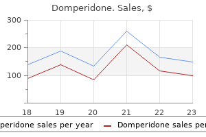
Domperidone dosages: 10 mg
Domperidone packs: 90 pills, 180 pills, 270 pills, 360 pills

Buy discount domperidone 10mg
The jugular (supraster nal) notch is palpable in the midline at the superior sternal border and sometimes lies at the level of the junction between the second and third thoracic vertebrae. The cartilaginous rings of the trachea are palpable instantly superior to the notch, utilizing a posteriorly directed finger. The manubriosternal angle is extra pronounced in the male than within the female, and is palpable at the junction of the manubrium with the sternal physique. It serves as a useful landmark as a outcome of it indicates the level of the sternal plane and the medial ends of the second costal cartilages, and so presents an accurate level at which to begin counting ribs. In nearly all of adults, the sternal angle is located between the fourth tho racic vertebra and the upper half of the fifth thoracic vertebra, with a range from the decrease half of the second thoracic vertebra to the decrease half of the sixth thoracic vertebra, a linear distance of about 8 cm; the aircraft lies at a barely larger vertebral stage in ladies. The xiphisternal joint and xiphoid process are palpable on the inferior finish of the sternum; the joint often lies on the level of the ninth thoracic vertebra. Posteriorly, the free ends of the eleventh and twelfth ribs may be Autonomicplexusesinthethorax Cardiac plexus the cardiac plexus has a superficial part inferior to the aortic arch, mendacity between it and the pulmonary trunk, and a deep half between the aortic arch and tracheal bifurcation (Ch. The superficial part is formed by the cardiac branch of the left superior cervical sympathetic ganglion and the lower of the 2 cervical cardiac branches of the left vagus. The deep half is fashioned by the cardiac branches of the cervical and upper thoracic sympathetic ganglia and of the vagus and recurrent laryngeal nerves. Branches from the cardiac plexuses additionally form the left and proper coronary and atrial plexuses. Pulmonaryplexus the pulmonary plexuses are anterior and poste rior to the other structures at the hila of the lungs (Ch. They are fashioned by cardiac branches from the second to fifth (or sixth) thoracic sympathetic ganglia and from the vagus and cervical sympathetic cardiac nerves. Vagal fibres either move by way of the plexus or are given off instantly by the vagus in the thorax. All fibres relay within the oesophageal wall and are motor to the sleek muscle in the lower oesophagus and secretomotor to mucous glands within the oesophageal mucosa. Vasomotor sympathetic fibres arise from the higher six thoracic spinal twine segments. Those from the upper segments synapse in cervical ganglia; postganglionic axons innervate the vessels of the cervical and higher thoracic oesophagus. Fibres from the lower segments pass on to the oesophageal plexus or to the coeliac ganglion, the place they synapse; postganglionic axons innervate the vessels of the distal oesophagus. The accuracy of some older descrip tions of clinically necessary floor landmarks, based on cadaveric or radiographic studies, has been questioned just lately (Hale et al 2010). The tail of the breast extends towards the axilla along the infero lateral border of pectoralis main (Ch. Key: 1, proper acromioclavicular joint; 2, clavicle and mid-clavicular line (dashed black); 3, apex of proper lung, situated posterior to the medial third of the clavicle; 4, sternal notch of manubrium sterni: the trachea could additionally be positioned here by posterior palpation; 5, sternoclavicular joint: marks the junction of the inner jugular and subclavian veins to form the brachiocephalic vein; 6, zone of formation of the superior vena cava: from first intercostal area to second costal cartilage level (78% of topics, white zone); 7, sternal angle: marks the level of the sternal plane and the second costal cartilage; 8, pectoralis main and the anterior axillary fold; 9, horizontal fissure; 10, proper indirect fissure; 11, lower anterior border of the proper lung: usually, both the sixth intercostal house or the seventh rib in the mid-clavicular line; 12, lower anterior border of the left lung: usually, either at the fifth rib or fifth intercostal space within the mid-clavicular line; thirteen, xiphisternum; 14, costal margin; 15, tenth costal cartilage, forming the lower a part of the costal margin. The levels of the spinous processes of the third, ninth and twelfth thoracic vertebrae and the primary lumbar vertebra are indicated in the midline. Key: 1, border of the left lung; 2, oblique fissure: passes anteroinferiorly from the spinous strategy of the third thoracic vertebra to cross the fifth rib within the midaxillary line. The upper lobe sits superiorly and the lower lobe inferiorly; three, decrease border of the lung: that is usually positioned at degree of the twelfth thoracic vertebra however could also be decrease, at the degree of the first lumbar vertebra, adjacent to the vertebral column at finish tidal inspiration. Dashed blue traces indicate the range of ranges for the lower border of the lung (ninth thoracic vertebra to first lumbar vertebra); 4, twelfth rib: could be traced superomedially to help identification of the spinous means of the twelfth thoracic vertebra. Posteriorly, the spinous processes of the thoracic vertebrae are palpable; the spinous strategy of the primary thoracic vertebra sits below that of the seventh cervical vertebra (vertebra prominens) and is often more outstanding. The angles of the ribs are palpable a number of centimetres lateral to the spinous processes of the vertebrae. Key: 1, inner jugular vein; 2, subclavian vein; three, formation of the brachiocephalic vein posterior to the sternoclavicular joints; four, formation of the superior vena cava, posterior to the right second costal cartilage or first intercostal area; 5, manubriosternal joint, 6, concavity of the aortic arch, usually sitting inferior to the sternal airplane, stage with the higher half of the fifth thoracic vertebra; 7, azygos vein getting into the superior vena cava: usually, it sits inferior to the sternal plane, degree with the decrease half of the fifth thoracic vertebra; eight, tracheal bifurcation: usually, it sits inferior to the sternal airplane, degree with the higher half of the sixth thoracic vertebra; 9, bifurcation of the pulmonary trunk, degree with the higher half of the sixth thoracic vertebra, roughly three cm inferior to the sternal angle. The sternal aircraft is conventionally described as lying over the tracheal bifurcation, the concavity of the aortic arch and the purpose where the azygos vein enters the superior vena cava. Not reported T6 upper (28%) (T4/5 to T7/8) Measurements relative to the sternal plane are shown as + (superior to the plane) and - (inferior to the plane). They may be modified by age, sex, stature, air flow, the posi tion of the diaphragm and posture (Macklin 1925). The projection of the cardiac borders on to the anterior thoracic wall forms a trapezoid. The proper border is a gently curved line, convex to the right, running from the third to the sixth proper costal cartilages, usually 1�2 cm lateral to the sternal edge.
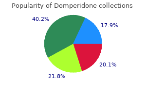
Purchase domperidone 10mg with mastercard
The radial border of the carpal groove is shaped by the tubercles of the scaphoid and trapezium. The former is distal on the anterior scaphoid floor and palpable (sometimes, also visible) as a small medial knob at the proximal border of the palmar thenar eminence, radial to the tendon of flexor carpi radialis. The carpal groove is made into an osseofibrous carpal tunnel by the flexor retinaculum hooked up to its margins. Radiocarpal, intercarpal and carpometacarpal ligaments are attached to the palmar and dorsal surfaces of the entire carpal bones, except the triquetrum and pisiform. Lunate For hamate For radius For trapezium Dorsal side For trapezoid For capitate Palmar side For capitate Distomedial facet For scaphoid Proximolateral side C. Trapezium Tubercle Tubercle For 2nd metacarpal For hamate For 2nd metacarpal For 1st metacarpal Palmar side For scaphoid For trapezoid Proximomedial side F. Trapezoid Rough space, nonarticular Dorsal floor For scaphoid For trapezium Dorsal floor For scaphoid G. The rough dorsal surface is slightly grooved, narrower than the palmar, and pierced by small nutrient foramina, which are often restricted to the distal half. The radial collateral ligament is hooked up to the lateral floor, which is also slender and tough. The radial (proximal) surface is convex, proximal and directed proximolaterally; the lunate surface is flat and semilunar, and faces medially; the capitate floor is massive, concave, distal, and directed distomedially. The proximal, palmar surface of the non-articulating portion of the scaphoid is provided by branches from the palmar carpal artery or, in Scaphoid its absence, by branches of the superficial palmar artery (Oehmke et al 2009). The center and distal thirds of the palmar surface are supplied by the superficial palmar artery and small radial rami arteries. The distal third may also receive branches from the first dorsal metacarpal artery. The dorsal floor is principally supplied by the dorsal scaphoid artery and by branches of the styloid artery. Its tough palmar floor, almost triangular, is bigger and wider than the rough dorsal surface. Its clean convex proximal surface articulates with the radius and the articular disc of the distal radio-ulnar joint. Its narrow lateral floor bears a flat semilunar side for the Wrist and hand Gelberman and Menon (1980) describe the proximal 70�80% of the scaphoid as being supplied by branches of the radial artery that enter along its dorsal ridge, either just distal to the waist (14%), immediately over the waist (59%) or just proximal to the waist (27%). They estimated that 14% of their specimens would have had vital interruption and 59% a partial interruption to the vascularity of the proximal pole of the scaphoid from a fracture via the waist, and that failure to unite would have resulted in avascular necrosis of the proximal fragment. It has been argued that an insufficient blood provide is unlikely to be the trigger of scaphoid non-union after fracture (Oehmke et al 2009), but see additionally Sendher and Ladd (2013). The scaphoid is the most regularly fractured carpal bone, sometimes because of a fall on to the outstretched hand. The distal floor is deeply concave to match the medial part of the head of the capitate. Palmar and dorsal surfaces are roughened for carpal ligaments, the dorsal being the bigger. Its medial and dorsal surfaces are confluent, and marked distally by the attachment of the ulnar collateral ligament, however smooth proximally to receive the articular disc of the distal radio-ulnar joint in full adduction. The hamate surface, lateral and distal, is concavoconvex, broad proximally and slim distally. The tendon of flexor carpi ulnaris and the distal continuations of the tendon, the pisometacarpal and pisohamate ligaments, are all hooked up to the palmar non-articular area, which surrounds and tasks distal to the articular surface. Distally, on the hamular base, a slight transverse groove may be in contact with the terminal deep branch of the ulnar nerve. The remaining palmar surface, like the dorsal, is roughened for attachment of ligaments. A faint ridge divides the distal floor into a smaller lateral side that articulates with the bottom of the fourth metacarpal, and a medial side for articulation with the bottom of the fifth. The proximal floor, the thin margin of the wedge, often bears a slender side that contacts the lunate in adduction. The medial surface is a broad strip, convex proximally, concave distally, which articulates with the triquetrum; distally, a narrow medial strip is non-articular. The lateral surface articulates with the capitate by a facet masking all however its distal palmar angle.
Syndromes
- Nausea and vomiting that continues to get worse
- Pain when doing certain activities or moving your body a certain way
- Medications such as corticosteroids, acetazolamide, and furosemide
- Tenderness and dryness of the vagina
- Have you had any recent injuries or illnesses?
- Diabetes
- Receive shots to prevent blood clots
- Wheezing
- Swelling or lumps in the neck
- Endometriosis
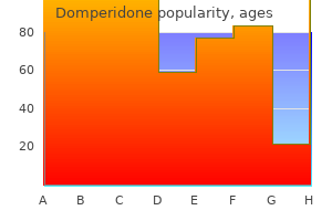
Buy domperidone 10mg overnight delivery
If the forearm is rotated into supination, the wrist will lengthen and the fingers flex. This check, the wrist tenodesis take a look at, is a useful method of analyzing the limb for tendon harm. Wrist motion is controlled principally by two wrist flexors (flexor carpi radialis and flexor carpi ulnaris) and three extensors (extensors carpi radialis longus and brevis, and extensor carpi ulnaris). It would be very restricting to have a pure hinge joint with collateral ligaments of fixed length. In this context, the wrist flexors and extensors may be regarded as variable collateral ligaments that allow the joint to be set about numerous completely different axes. It is feasible to observe and to palpate the muscle teams that are energetic in making a good fist. Flexor digitorum profundus and flexor digitorum superficialis are lively, and flexor carpi ulnaris contracts strongly, as do the wrist extensors. Palpation of the long digital extensors on the dorsum of the wrist, the primary dorsal interosseous within the thumb internet, and of the opposite interossei and the thenar and hypothenar muscles, will confirm that every one these muscles are contracting. As a agency fist is swung forwards in anger, brachioradialis stands out; at the moment of impression, virtually each muscle within the limb is active. This motion is made up of extension of the distal interphalangeal, proximal interphalangeal and metacarpophalangeal joints. The legal guidelines of mechanics would recommend that one motor could be required for each joint in a series, along with some kind of controlling mechanism to make positive that the chain of joints moved together in a coordinated fashion. In the hand, this is achieved by way of an extensor apparatus that minimizes the variety of motors required for movement by permitting the muscle tissue to act on a couple of joint, and by linking totally different ranges in the mechanism in order that the arc of motion is managed. The tendons of extensor digitorum run distally over the metacarpal heads, forming the major element of the extensor equipment. Extensor digitorum has no insertion into the proximal phalanx and, due to this fact, exerts its extensor action on the metacarpophalangeal joint indirectly via extra distal insertions. Acting at this insertion alone, extensor digitorum can prolong both metacarpophalangeal and proximal interphalangeal joints together. Thus, the hand possesses, in the proximal part of the extensor equipment, a variable mechanism that permits totally different amounts of relative metacarpophalangeal or proximal interphalangeal joint movement. A helpful analogy that has been instructed for this association is to contemplate it as two pulleys of different size on one axle. The central slip can be considered a wire that passes over the bigger wheel, and each lateral slip as a twine that passes over the smaller wheel. There is an additional mechanism by which the lateral slips transfer laterally throughout flexion of the proximal interphalangeal joint. The effect of this lateral motion is to cut back additional the gap between the lateral slips and the joint axis, thereby reducing the amount of excursion at the proximal interphalangeal joint still more and allowing extra tour on the distal joint. When the hand flexes, this mechanical linkage system permits each interphalangeal joints to flex together in a coordinated method. The extensor enlargement additionally receives contributions from the interossei and lumbricals, which method the digits from the webs and be a part of the corresponding enlargement within the proximal phase of the digit. Apart from the components of the extensor enlargement which would possibly be concerned with joint operate, the whole structure requires further anchorage. These troublesome necessities are met by transverse retinacular ligaments on the level of the joints, the transverse ligaments working to relatively fixed attachment factors within the region of the joint axis. As the expansion glides backwards and forwards, the transverse fibres move like bucket handles. Smooth gliding layers are required underneath the enlargement and retinacular ligaments to enable movement to happen without friction. Lateral inclinations of the first phalanx maximize the extent of tour of the circumduction arc. Opposition is a composite position of the thumb achieved by circumduction of the first metacarpal, internal rotation of the thumb ray, and maximal extension of the metacarpophalangeal and interphalangeal joints (see Video 50. Flexion and adduction is the position of maximal transpalmar adduction of the first metacarpal; the metacarpophalangeal and interphalangeal joints are flexed and the thumb is involved with the palm (see Video 50. The simple angular movements described above combine with rotation in regards to the lengthy axis of the metacarpal shaft. Axial rotation of the thumb metacarpal is produced by muscle exercise (which strikes the thumb via its arc of circumduction); the geometry of the articular surfaces of the trapeziometacarpal joint; and tensile forces in the ligaments (which combine with forces exerted by the muscles of opposition and retroposition to produce axial rotation). The stability of the first metacarpal is greatest after full pronation in the position of full opposition, when the tension in the ligaments, muscular contraction and joint congruence mix to maximal effect.
Discount domperidone 10 mg with amex
The trabeculated appearance is caused by a myriad of endocardial, lined, irregular muscular ridges and protrusions collectively generally known as trabeculae carneae. These protrusions and intervening grooves impart great variation in wall thickness; the protrusions range in extent from mere ridges to trabeculations, mounted at both ends but in any other case free. Other conspicuous protrusions are the papillary muscle tissue, that are inserted at one end on to the ventricular wall and are continuous on the other finish with collagenous cords, the chordae tendineae (tendinous cords), inserted on the free edge of the Heart atrioventricular valves. One protrusion in the right ventricle, the septomarginal trabeculation or septal band, is especially prominent, reinforcing the septal surface where, on the base, it divides into limbs that embrace the supraventricular crest. Towards the apex, it supports the anterior papillary muscle of the tricuspid valve and, from this level, crosses to the parietal wall of the ventricle as the moderator band (the name reflects an earlier concept that septomarginal trabeculation prevented overdistension of the ventricle). The role of the moderator band as a part of the conduction system of the heart involves the best atrioventricular bundle, as conduction cardiomyocytes move in the path of the apex of the ventricle before getting into the anterior papillary muscle. A additional series of distinguished trabeculations, the septoparietal trabeculations, prolong from its anterior surface and run on to the parietal ventricular wall. The smooth-walled outflow tract, or infundibulum, ascends to the left, superior to the septoparietal trabeculations and inferior to the arch of the supraventricular crest to the pulmonary orifice (Spicer and Anderson 2013). The connective tissues around the orifice of the atrioventricular valves separate the atrial and ventricular myocardial lots utterly, except on the point of penetration of the atrioventricular bundle; they range in density and disposition across the valvular circumference. The latter is accomplished by more tenuous, deformable fibroblastic sulcal areolar tissue. Although the extent of fibrous tissue varies with sex and age, the tissue throughout the atrioventricular junction around the tricuspid orifice is always less sturdy than similar parts discovered at the attachments of the mitral valve. The line of attachment of the leaflet is greatest appreciated within the coronary heart when examined grossly, this characteristic being extra readily discerned clinically. The leaflets are positioned anterosuperiorly, septally and inferiorly, comparable to the marginal sectors of the atrioventricular orifice named in conjunction. The inferior leaflet is usually described as being posterior, however when assessed within the attitudinally appropriate anatomical place, the leaflet is positioned inferiorly (Anderson and Loukas 2009). Each leaflet is a reduplication of endocardium enclosing a collagenous core, continuous marginally and on its ventricular facet with diverging fascicles of chordae tendineae (see below) and basally confluent with the anular connective tissue. In passing from the free margin to the inserted margin, all leaflets of the atrioventricular valves display rough, clear and basal zones. The tough zone is comparatively thick, opaque and uneven on its ventricular side where most chordae tendineae are hooked up; its atrial facet makes contact with the comparable surface of the adjacent leaflets throughout full valve closure. The clear zone is easy and translucent, receives few chordae tendineae and has a thinner, fibrous core. The basal zone, extending 2�3 mm from the circumferential attachment of the leaflets, is thicker from increased connective tissue, vascularized and innervated. The anterosuperior leaflet is the most important component of the tricuspid valve, attached mainly to the atrioventricular junction on the posterolateral facet of the supraventricular crest, and increasing along its septal limb to the membranous septum ending on the anteroseptal commissure. The attachment of the septal leaflet passes from the inferoseptal commissure on the inferior ventricular wall across the muscular septum, then angling throughout the membranous septum to the anteroseptal commissure. The inferior leaflet is wholly mural in attachment and guards the diaphragmatic surface of the atrioventricular junction, its limits being the inferoseptal and anteroinferior commissures. The zone of apposition between the inferior and the anterosuperior leaflets is supported by the septal papillary muscle of the conus. The atrioventricular valvular advanced, in both ventricles, consists of the orifice and its related anulus, the leaflets, the supporting chordae tendineae of various sorts and the papillary muscular tissues. Harmonious interaction of all of these, together with the myocardial mass, depends on the conduction tissues and mechanical cohesion provided by the cardiac skeleton. Tricuspid valvular orifice the tricuspid valve orifice is greatest seen from the atrial side. There is a transparent line of transition from the atrial wall or septum to the strains of attachment of the valvular leaflets. During diastole, the anulus dilates with proper ventricular relaxation and the large anterior and posterior leaflets move away from the airplane of the anulus into the proper ventricle. The proper heart is blue, the arrow denoting the inflow and outflow channels of the proper ventricle; the left coronary heart is handled equally in purple. The place of the letters A, P, T and M indicate the aortic, pulmonary, tricuspid and mitral auscultation areas of medical follow, respectively.
Buy cheap domperidone on line
The opening of the solitary pulmonary vein to the left of the proper pulmonary ridge is a prerequisite for atrial septation. The right lateral wall of the proper atrium, the right ventricle and the outflow tract have been removed. Retinoic acid and its downstream transcription factor, Tbx5, play a crucial role in this course of. The definitive trabeculations, coarse in the right ventricle however much finer in the left, are first observed across the fortieth day of gestation; they appear initially in the partitions of both ventricles at the degree of the atrioventricular junction and develop in the path of the apex of the heart. By the time the fetus is 10 weeks old, the trabeculations are much sparser, and are confined to the apical areas. This means of remodelling is achieved without the intervention of macrophages or inflammatory cells in the immediate interstitium. The ventricular myocardium, encompassing the trabeculations and exterior wall, possesses a chamber phenotype, the myocytes expressing, amongst other proteins, the fast-conducting gap-junctional proteins connexin forty (Cx40) and forty three, and atrial natriuretic peptide. This myocardium stops proliferating and differentiates into the fast-conducting peripheral ventricular conduction system, whereas the outer layer becomes highly proliferative and varieties the compact layer of the ventricular wall, no longer expressing Cx40 and atrial natriuretic peptide, however nonetheless expressing Cx43. It shows a attribute bend, which has a proximal half arising from the creating right ventricle, and a distal half that becomes steady with the aortic sac past the pericardial reflections. Instead, it appears probable that cells are recruited from the second heart-forming subject, and cross through the outflow tract to contribute to the definitive right ventricle. Subsequently, still more cells are recruited from pharyngeal mesenchyme, passing proximal to the pericardial reflections and forming the intrapericardial elements of the arterial trunks. There stays much disagreement as to how best to describe these morphological elements of the outflow tract. Some authors have labelled the proximal myocardial half the conus, calling the distal part the truncus, and the intrapericardial portion of the outflow tract the aortic sac. The left ventricle develops from the stem of the Y-shaped coronary heart; the right ventricle develops later, downstream relative to the left ventricle, when more myocardium has been added to the cardiac tube. As a results of the looping of the guts tube, the proper ventricle is positioned at the proper of the left ventricle, which is a prerequisite for the suitable connection with the expanding atrial element of the guts. Because the terms conus and truncus have been inconsistently utilized in literature, we prefer simply to describe proximal and distal elements of the myocardial outflow tract, and to describe, when it appears, an intrapericardial arterial portion of the outflow tract, this part interposing between the muscular part and the ventral aorta, the distal junction occurring on the pericardial reflections. The recent discovering that the stem of the center tube incorporates precursor cells solely for the left ventricle makes it important to adopt descriptions that follow dynamic occasions, somewhat than persevering with to use static names. Cells that, initially, are found in the ventricular outflow tract subsequently become cells of the best ventricle. Furthermore, working myocytes can simply be distinguished from the first myocytes of the straight coronary heart tube. The main myocardium is smoothwalled, whereas the developing myocardium of the ventricles is trabeculated, and expresses particular markers similar to atrial natriuretic peptide and Cx40 and Cx43. Moreover, within the internal curvature of the ventricular loop, the partitions of the outflow tract and atrioventricular canal fade into each other and not using a clear boundary. It is within this ventricular part of the primary coronary heart tube that the cushions of the atrioventricular canal and the outflow tract should achieve acceptable connections with the muscular ventricular septum to have the ability to divide this a part of the tube into left and proper compartments. It is, none the less, the remodelling of this inner curvature that sets the scene for the completion of cardiac septation. All areas of the early embryonic heart tube possess poorly coupled cells and show intrinsic automaticity, thus permitting slow propagation of the depolarizing impulses alongside the cardiac tube, and producing matching peristaltic waves of contraction that push the blood in an antegrade direction. This tissue has been called main myocardium, distinguishing it from the quickly conducting and well-developed working myocardium of the atrial and ventricular chambers. An grownup sort of electrocardiogram could be recorded from such hearts, displaying rapid atrial depolarization, a interval of atrioventricular delay, and speedy ventricular depolarization. The electrocardiographic tracings reflect the event of fast-conducting parts inside a slowly conducting coronary heart tube, i. The newly developed working myocardium expresses atrial natriuretic factor, along with the gap junctional proteins Cx40 and Cx43, which allow quick conduction. The slowly conducting atrioventricular canal, interposed between the atrium and the ventricle, and the outflow tract positioned between the ventricle and the great arteries, are the components that include the endocardial cushions. These are able to function as sphincteric valves, because of their extended period of contraction.
Cheap domperidone 10mg online
The phrenic nerve, chiefly derived from the ventral major ramus of C4, curves round the muscle to run down anterior to it into the thorax, lying, at first, posterior to the internal jugular vein and crossing behind the subclavian vein. The thoracic duct and the right lymphatic duct enter the brachiocephalic (innominate) vein at the subclavian�jugular (triradiate) junction on the left and proper, respectively. Bridges of muscle and fascia could pass between them, altering and diminishing the space available for the neurovascular bundles. A, the proper decrease trunk is trapped between the aponeurotic fringe of scalenus medius posteriorly and the suprapleural membrane anteriorly. The left lower trunk passes over the edge of the sickle and behind the part of scalenus anterior that passes posterior to the subclavian artery. Coote (1861) reported the removal of a left seventh cervical rib causing aneurysm of the subclavian artery. Two physicians, Lewis and Pickering (1934), pointed to the mechanism of manufacturing of the arterial lesion, particularly: intimal breakage with native thrombosis and distal embolization. The vessel is distorted by the bone and constricted by a leash fashioned by the tendon of insertion of scalenus anterior. There is commonly dilation of the artery distal to the point of constriction, and the dilation might proceed to the formation of a true aneurysm (Wickham and Martin 1962). Complete obstruction of the main vessel with distal embolization could lead to important ischaemia. The most dangerous complication is contralateral hemiplegia, doubtless from embolization from a thrombus extending proximally to the carotid vessel (Symonds 1927). The arterial form of this outlet syndrome is a situation predominantly affecting younger girls; the prevalence of such symptoms in older people must recommend a primary analysis of atherosclerosis or different systemic disorder. Doppler ultrasound examination is likely to show an abnormality: principally, a clamped, monophasic velocity sign anyplace over the arterial tree of the higher limb (Parry and Eastcott 1992). Operation is required urgently in cases of critical ischaemia, and soon in circumstances during which ischaemia is threatened. The confusion was not resolved for greater than 40 years until the applying of nerve conduction research outlined the rather more widespread disorder. Gilliatt put the annual incidence of cervical rib syndrome with muscle losing as little as 1 in 1 million in the general inhabitants. The sufferers are normally younger to middle-aged girls and the abnormalities are normally confined to one upper limb. The symptoms are insidious and sometimes gentle; presentation is delayed for some years, and is often occasioned by the sudden realization that the hand has turn out to be wasted. There is pain within the medial aspect of the forearm and there may be some blunting of sensibility in that area and in the little finger. A, Severe wasting of the intrinsic muscular tissues of the best hand in a 35-year-old lady. When the nerve was first exposed at operation, the zone of attenuation was, in reality, at the website of angulation. B, Complete occlusion on the thoracic outlet in a 40-year-old lady, a heavy smoker. Radiographs of the neck present rudimentary cervical ribs or elongated transverse processes of the seventh cervical vertebrae. The diagnostic electrophysiological findings have been characterised by Gilliatt, Le Quesne and Logue in 1970 and embrace a small or absent sensory response in the ulnar nerve and the medial cutaneous nerve of the forearm; a traditional sensory response within the median nerve; an attenuated compound motor motion potential amplitude from abductor pollicis brevis; and a low-amplitude ulnar motor response (Smith and Knight 2011). Operation is indicated in these sufferers within the expectation of alleviation of ache, enchancment in sensation and a few enchancment in power; recovery of the wasted small muscle tissue of the hand is rare. The urge to advise operation should be tempered with information of the potential of issues and the excessive fee of recurrence of symptoms as time passes (Birch 2011). Coote H 1861 Exostosis of the left transverse strategy of the seventh cervical vertebra, surrounded by blood vessels and nerves, profitable removing. Pseudoglandular phase (5�17 weeks: development of airways and blood vessels to level of acinus). Canalicular section (17�27 weeks: formation of respiratory airways and thinning of blood�gas barrier).
Azeda-Brava (Sorrel). Domperidone.
- Are there safety concerns?
- What other names is Sorrel known by?
- Dosing considerations for Sorrel.
- Inflamed nasal passage, or "sinusitis," when taken with gentian root, European elder flower, verbena, and cowslip flower (SinuComp, Sinupret).
- Fluid retention, infections, and other conditions.
- How does Sorrel work?
- What is Sorrel?
Source: http://www.rxlist.com/script/main/art.asp?articlekey=96703

Purchase domperidone overnight
Normal postnatal pulmonary arterial improvement Immediately after start, dramatic remodelling of the pulmonary vasculature occurs, to impact an abrupt reduction of pulmonary vascular resistance. This course of continues at a rapid fee throughout the primary 1�2 months, whereas the lungs adapt to extrauterine life, and then extra slowly throughout childhood. Failure to rework within the presence of an anatomically regular coronary heart leads to persistent pulmonary hypertension. Normal postnatal pulmonary arterial development in the full-term neonate could be divided into three phases. Adverse results on postnatal lung growth Stage one this lasts from birth to about postnatal day 4 and concerns the instant adaptation to extrauterine life. At start, the endothelial cells of the precapillary arteries are squat and have slim bases on the subendothelium, a low floor to volume ratio and many floor projections. The clean muscle cells show a significant reduction in diameter throughout this time. Stage two this lasts from round day four to 3�4 weeks and is the time when the cells deposit matrix round themselves to fix their new the long-term results of preterm birth and its treatment are changing over the years. Survivors from the pre-surfactant period have illness dominated by airflow obstruction. In addition, congenital anomalies of the guts and nice vessels, and of the chest and belly partitions, together with neuromuscular illness, may influence on lung growth. It is somewhat synthetic to describe airway malformations in isolation, and the potential of related vascular anomalies should all the time be thought of. A review of all features of the diagnosis, nomenclature and administration of congenital malformations of the airways, lung and chest wall. A evaluation of how early issues with lung progress and development impression on later lung disease. Bush A 2009 Prenatal presentation and postnatal administration of congenital thoracic malformations. An rationalization of how tracheal occlusion can be utilized in an animal model to improve lung progress and maturation. Tracheobronchomalacia often presents in early infancy with cough, tachypnoea, stridor and wheeze; it may even be associated with cardiac or respiratory anomalies, corresponding to absent pulmonary valve syndrome, or tracheo-oesophageal fistula, respectively. Complete cartilage rings the conventional large-airway cartilages are horseshoe-shaped, and the ends of the horseshoes are bridged by the membranous part of the trachea. Complete cartilage rings may develop in the trachea and enormous airways, and the posterior, membranous, part of the airway could also be absent in a single cartilage or in lengthy segments of the airway. This anomaly could additionally be related to vascular anomalies similar to pulmonary artery sling (where the left pulmonary artery originates from the best pulmonary artery and never from the principle pulmonary trunk). Extensive severe illness could current with neonatal respiratory misery; later presentations embody apparently steroid-resistant asthma. Agenesis and aplasia of the lungs Tracheo-oesophageal fistulae Tracheo-oesophageal fistulae are the commonest anomalies of the decrease respiratory tract and occur in about 1 in 3000 births. They often present in the new child with recurrent respiratory misery and choking spells, though late presentation in adult life has been described and could also be associated with oesophageal atresia. Normally, the oesophagus lengthens with the fast elongation of the embryo up to 7 weeks. The cellular processes that lead to separation of the trachea and oesophagus sometimes produce oesophageal atresia. Tracheo-oesophageal atresia is uncommon, with an incidence of 1 in 3000 to 1 in 4500 births. In virtually all instances, the oesophagus ends blindly and the stomach is related to the decrease end of the trachea. Because of this connection, the stomach turns into quickly distended with air as quickly as the infant is delivered and begins breathing. Prenatally, polyhydramnios may be a scientific characteristic but will not be apparent until the third trimester. Such instances are identifiable on ultrasound as a outcome of the abdomen should all the time be visible at a 20-week examination; its absence on ultrasound should prompt further analysis.
Domperidone 10mg fast delivery
The posterior rectus sheath may be incised again 1�2 cm medial to the linea semilunaris, safely preserving the laterally perforating neurovascular bundles. Delicate transection of this muscle at its most medial insertion permits access to a plane beneath transversus abdominis and superficial to the transversalis fascia, preperitoneal fats and peritoneum. This plane of dissection may be extended laterally to psoas major, inferiorly to the space of Retzius, and superiorly beneath the diaphragm. In addition to the medial mobilization of fascia aided by myofascial flaps and element separations, these myofascial planes allow for mesh reinforcement with in depth overlap without exposure to intraperitoneal contents (Heller et al 2012). Below this level, the aponeuroses of external and inside oblique and transversus abdominis cross anterior to rectus abdominis and the posterior rectus sheath ends. A Spigelian hernia is an interstitial hernia in that the hernia passes through a defect within the transversus and internal oblique aponeuroses however remains deep to the overlying external indirect aponeurosis. There is currently little consensus among surgeons as to the optimum technique of repairing a big incisional hernia and quite a few techniques are described (Cassar and Munro 2002). Needless to say, an understanding of belly wall anatomy is paramount to providing a durable repair without compromising physiological operate. A review that compares the long-term recurrence charges of open main repair with open mesh and laparoscopic repair, citing the inadequacy of open primary repair. An account of how the deep inferior epigastric artery offers the principle blood supply to the decrease belly wall. A description of how, since its unique description, the parts separation approach underwent a quantity of modifications with the ultimate word goal of decreasing the morbidity associated with the normal procedure. The frequent lack of bodily findings, together with vague associated belly complaints, makes the prognosis elusive. A retrospective evaluate of Mayo Clinic patients was carried out to discover all patients who had undergone surgical restore of a Spigelian hernia from 1976 to 1997. Although named after Adriaan van der Spieghel, the semilunar line (linea Spigeli) was solely described by Spieghel in 1645. A abstract of the outcomes in 1079 consecutive clean or cleancontaminated midline belly incisions closed with operating 0-loop nylon suture after both elective and emergency operations done between 1984 and 1991. Chopra J, Rani A, Rani A et al 2011 Re-evaluation of superficial fascia of anterior stomach wall: a computed tomographic research. Dabbas N, Adams K, Pearson K et al 2011 Frequency of belly wall hernias: is classical educating outdated Microdissection of the artery, its primary branches and the perforator vessels was undertaken in 20 cadavers and the findings summarized. The extensive subcutaneous lateral dissection had been related to ischaemia of the midline pores and skin edges, wound dehiscence, infection and seroma. Korenkov M, Beckers A, Koebke J et al 2001 Biomechanical and morphological forms of the linea alba and its possible role in the pathogenesis of midline incisional hernia. Lancerotto L, Stecco C, Macchi V et al 2011 Layers of the stomach wall: anatomical investigation of subcutaneous tissue and superficial fascia. Loukas M, Myers C, Shah R et al 2008 Arcuate line of the rectus sheath: scientific strategy. A description of the novel method for posterior component separation, which was related to a low perioperative morbidity and a low recurrence fee. Overall, transversus abdominis release may be an essential addition to the armamentarium of surgeons enterprise major belly wall reconstructions. Closure of large belly wall defects usually requires the transposition of distant myocutaneous flaps or free-tissue transfers. The objective of this study was to determine if separation of the muscle elements of the belly wall would allow mobilization of each unit over a greater distance than is feasible by mobilization of the complete abdominal wall as a block. A text that summarizes the related anatomy of the stomach wall, relating this to techniques for belly wall reconstructions. Schlenz I, Burggasser G, Kuzbari R et al 1999 External indirect stomach muscle: a brand new look on its blood provide and innervation. An account of Spigelian hernia (1�2% of all hernias), the protrusion of preperitoneal fat, peritoneal sac or organ(s) by way of a congenital or acquired defect in the Spigelian aponeurosis. Josef Klinkosch first defined the Spigelian hernia as a defect within the semilunar line in 1764.
Order domperidone 10 mg without prescription
Clusters of lymphocytes sometimes lie beneath nonciliated epithelial cells of the microfold (M-cell) type. They resemble connective tissue mast cells, and their cytoplasmic histaminecontaining granules are released in response to irritants, including inhaled allergens. Submucosal glands Tubuloacinar, seromucous glands are present in the submucosa of the trachea and bronchi and, to a lesser extent, within the larger bronchioles. They contain separate mucous and serous cells and are an necessary source of the mucus on the floor of the ciliated respiratory epithelium. Their secretions embrace mucins; bacteriostatic substances corresponding to lysozyme and lactoferrin; secretory antibodies (immunoglobulin A (IgA)) produced by plasma cells in the submucosal connective tissue; and protease inhibitors. Deficiency of 1-antitrypsin causes persistent obstructive pulmonary illness, by inducing panacinar emphysema and bronchiectasis. The secretory acini and tubules are surrounded by myoepithelial cells, which are innervated by autonomic fibres (see above). Ciliated columnar cells Goblet cells Goblet cells are present from the trachea (6000�7000 per mm2) distal to the smaller bronchi, however are normally absent from bronchioles. They comprise an apical area full of large secretory vacuoles full of mucinogen. Clara cells Clara cells are cuboidal, non-ciliated cells with apices that bulge into the lumen. Basal cells contact the basal lamina and are most frequent within the bigger conducting passages. The cilia lengthen right into a watery fluid secreted by serous cells of the submucosal glands, however their suggestions are in touch with a more superficial layer of thicker mucus secreted by surface goblet cells and mucous cells in the submucosal glands. The price of ciliary beating is often 12�16 per second; mechanical stimulation of the epithelial surface and inflammatory mediators improve the rate. In addition to tight junctions, which seal the apical intercellular space from the airway lumen, the ciliated cells are coupled by hole junctions, which permit a change in rate of beating to spread from stimulated cells to their neighbours (probably through calcium signalling) so that their metachronal coordination remains intact. This elastic framework is an important mechanical factor of the lung and is answerable for elastic recoil throughout expiration. Along the whole intrapulmonary bronchial tree, smooth muscle types two opposed helical tracts, which turn out to be thinner and eventually disappear on the degree of the alveoli. The tone of those muscle fibres is underneath nervous and hormonal management; teams of muscle cells are coupled by gap junctions to spread excitation within fascicles. Muscle cell contraction narrows the airway, while leisure permits bronchodilation. Some tone usually exists within the muscular bands, which loosen up slightly throughout inspiration and contract during expiration, thereby aiding the tidal flow of air. Numerous mast cells are current within the connective tissue of the respiratory tree, particularly in course of the bronchioles. Seromucous submucosal glands (S) are concentrated on this region and between adjacent cartilages. It enhances clearance of secretions and particulates from the airways and protects from aspiration of international materials (occurring as a consequence of aspiration or inhalation of particulate matter, pathogens, accrued secretions, postnasal drip, irritation and mediators related to inflammation). Under regular conditions, cough serves an essential protecting role within the airways and lungs, but in some conditions it might turn into extreme, non-productive and potentially harmful to the airway mucosa. An initial deep inhalation generates the quantity needed for an efficient cough and is adopted by a compression part, when forceful contraction of the muscle tissue of the chest wall, diaphragm and stomach wall in opposition to a closed glottis results in a speedy rise in intrathoracic pressure. Subsequent glottal opening causes a rapid peak expiratory airflow that dislodges and expels mucus from the airways; forceful airway compression produces the coughing sound. Occasionally, bronchoscopy also can provide some information about structures adjacent to the airways. The vagal afferents terminate centrally in largely non-overlapping areas of the caudal half of the nucleus tractus solitarius. Second-order neurones from this nucleus terminate on respiratory-related areas of the pons, medulla and spinal twine. Efferent impulses travel by way of the vagus, phrenic and spinal motor nerves to the inspiratory and expiratory muscle tissue, larynx, diaphragm and abdominal wall muscles. Airway receptors within the external auditory canals, eardrums, paranasal sinuses, pharynx, diaphragm, pleura, pericardium and stomach are in all probability mechanoreceptors, stimulated by triggers corresponding to touch or displacement (Polverino et al 2012).
Buy cheapest domperidone
These small (intrinsic) muscles have been described earlier by means of their particular person actions. All three joints are then angulated to the same diploma and the fingers form a traditional arc of flexion. As the finger flexes, the lengthy extensor tendons (extensor digitorum, extensor indicis and extensor digiti minimi) help the method by stress-free and permitting the extensor apparatus to glide distally on the dorsum of the phalanges. As the fingers wind as a lot as make a fist, the wrist tends to lengthen, notably when pressure is utilized. On its own, digital flexion would require the long tendons to move proximally in their sheaths and the flexor muscular tissues within the forearm would shorten. Extension of the wrist tends to produce a lengthening of the identical muscles, which, in normal use, is almost enough to stability the shortening due to finger flexion; the net impact is a really slight shortening (approximately 1 cm) of the long flexors within the forearm. The wrist can, due to this fact, be seen as a mechanism for maximizing drive as a result of it allows the fingers to flex while maintaining the resting length of the extrinsic muscular tissues near to the peak of the force�length curve. It is, of course, potential to wind up the fingers with the wrist held in a neutral position but the grip is somewhat weaker. Flexion of the fingers on gripping tends to end in a distal excursion of the lengthy extensors. The net impact is a very small proximal tour of the long extensor tendons on gripping, mirroring the effect on the flexor floor. If the movement of the wrist is exaggerated in order that the wrist is slightly flexed on opening the hand, and totally dorsiflexed on closing it, the online excursion of long flexors and extensors is zero, i. The reader can observe the connection between digits and wrist by performing the following manoeuvre. The wrist is held in a relaxed, mid-supinated position, with the elbow flexed at 90�. If the forearm is now rotated into pronation, the wrist will fall into flexion and the fingers will automatically lengthen. A small arterial-venous malformation proven on the index finger is appropriate for radiologically guided embolization (arrows). Key: 1, ulnar artery; 2, radial artery; three, deep palmar arch; four, princeps pollicis artery; 5, arteria radialis indicis; 6, superficial palmar arch (incomplete); 7, proper palmar digital artery (little finger); 8, frequent palmar digital artery; 9, basilic vein; 10, cephalic vein; eleven, digital vein. Position of relaxation the hand has a well-recognized place of rest, with the wrist in extension and the digits in a point of flexion. The precise position of the thumb in the position of rest appears to be rather variable. In this position, the carpometacarpal joint lies within 20� of radial abduction and 30� of palmar abduction; from clinical observations, it appears that the metacarpophalangeal joint lies inside approximately 40� of flexion and the interphalangeal joint between extension and 10� of flexion. The lateral subgroup (opposition muscles) moves the first metacarpal into palmar abduction. Radial angulation at the metacarpophalangeal joint increases the span of the hand. The metacarpophalangeal joint is stabilized principally by extensor pollicis brevis and flexor pollicis brevis. Muscles of the medial subgroup (abductor pollicis brevis and first dorsal interosseous) produce an method of the first metacarpal in direction of the palm. The intermediate subgroup consists of flexor pollicis longus, which flexes the interphalangeal or metacarpophalangeal joint. Palpating the thenar eminence throughout tip and lateral pinch provides some appreciation of the action of the pinch grip muscles. Grips From the place of rest, the tip of the thumb can strategy the radial facet of the fingers without incurring axial rotation because the palmar and dorsal trapeziometacarpal ligaments stay relaxed (see below). From completely different positions of the arc of circumduction, numerous different varieties of pinch grip are possible (see Video 50. In clinical apply, these have been categorised into two major varieties: tip pinch and lateral (or key) pinch. The thumb is activated by monoarticular muscles (abductor pollicis longus and opponens pollicis), biarticular muscular tissues (extensor pollicis brevis, adductor pollicis, abductor pollicis brevis and flexor pollicis brevis) and polyarticular muscle tissue (extensor pollicis longus and flexor pollicis longus). They place the metacarpal, an activity automatically accompanied by rotation, and in addition control the axial stability of the skeleton of the thumb. The thumb muscle tissue may be categorized into these used for retroposition, opposition and pinch grip.