Chloroquine dosages: 250 mg
Chloroquine packs: 30 pills, 60 pills, 90 pills, 120 pills, 180 pills, 270 pills, 360 pills
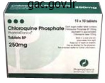
Buy on line chloroquine
Often, the morphology of the esophagus and intestine (especially with regards to the number and association of intestinal cells and their nuclear arrangement) could be diagnostically important. The reproductive tract of nematodes can be extremely variable, but with most parasitic species the male has a single testis and the feminine has paired uterine tubes. Often, eggs or microfilariae may be noticed in utero and may be diagnostically useful. The cestodes (tapeworms) are segmented worms that as adults reside in the intestinal tract of the definitive host. As such, cestodes noticed in extraintestinal tissue specimens (including all these in skin) are larval. Cestodes have complicated life cycles involving a number of hosts, and people manifesting in people as cutaneous larvae are usually zoonotic in origin. Calcareous corpuscles are an indicator of cestode morphology, and their presence in helminth tissue is pathognomonic for a cestode an infection. Larval cyclophyllidean cestodes (eg, T~nia) have 1 or extra constructions generally identified as a protoscolex, which serves because the precursor of the scolex (head) of the adult worm and consists of four suckers, 1 or extra rings of refractile booklets, and an inside spiral canal. The variety of protoscole<:es and the number and association of booklets may be diagnostically helpful 3. The trematodes (flukes) are a bunch ofparasitic flatworms that have complicated life cycles involving multiple hosts. All species parasitic to humans require a freshwater or terrestrial snail as an intermediate host Skin involvement with trematodes usually includes 1 of3 scenarios: (1) a papular erythematous rash attributable to the penetrating cercariae of avian sc. Schlstosoma species have individual female and male worms but perform as a single entity with the feminine residing in a gynecophoral canal alongside the body of the male. Beneath the tegument is a layer ofcircular muscle followed by a layer of longitudinal muscle. Between the musculature and the organ systems is the parenchyma, containing vitelline glands and interstitial fibers. Although the worms are hermaphroditic, sexual copy between 2 worms is perfect. In sexually mature adults, eggs could additionally be observed in utero, which might help with the prognosis, even in histologic sections. CiniC<11I Features Onchocercal nodules (onchocercomata) are discrete, delicate, and palpable, up to 5 cm throughout in dimension, and are sometimes situated close to bony prominences, although they might be found wherever on the body. Endemic onchocercal dermatitis is intensely itchy; predominantly papular and lichenoid; and if severe and witreated, could lead to cachexia and death from intercurrent infection. Lesions are most often generalized; much less regularly, particularly in Yemen and the savannah regions of Africa. S Depending on their period, the lesions could be spottily depigmented (leopard skin�), scaly and atrophic ("lizard skin"), or thickened and hyperkeratotic ("elephant skin"). In advanced illness, patients can also have lymphedema of the groin ("hanging groin"). Onchocerciasis acquired by travelers to endemic regions is seen as a lot as 2 years later as an itchy rash with marked peripheral eosinophilia, sometimes affecting 1 extremity only, as in sowda. In cross-section, the females measure as a lot as 400 �m in diameter, malting them one of the largest human parasitic filariae nematodes; males are markedly smaller at 200 iim. Gravid feminine worms present distinguished paired uteri containing maturing microfilariae; the reproductive tubes of the males might present spermatowa. Musculature is lowered, with only some cells per quadrant Lateral chords are brief however conspicuous. The cuticle contains transverse cuticular ridges, which are finest visualized in lateral cuts rather than cross-sections. Onchocerciasis is a chronic dermatitis that may cause extreme itching and disfiguring pores and skin lesions; ocular involvement can lead to blindness. Most human infections are related to Onchocerca volvulus, which is endemic to tropical Africa. Atlas of Fundamental Infectious Diseases Histopatholo9y: A Guide for Daily Practice.
Diseases
- Oculopalatoskeletal syndrome
- Psychosis
- Foix Chavany Marie syndrome
- Kalyanraman syndrome
- Chromosome 8, monosomy 8q
- Spastic paraparesis deafness
- Cortes Lacassie syndrome
- Frontonasal dysplasia acromelic
Generic 250mg chloroquine amex
A, Different classes of lymphocytes in the adaptive immune system recognize distinct types of antigens and differentiate into effector cells whose function is to get rid of the antigens. B lymphocytes acknowledge soluble or microbial surface antigens and differentiate into antibody-secreting cells known as plasma cells. Helper T cells acknowledge these peptides displayed on the floor of macrophages or different antigen presenting cells, and secrete cytokines that stimulate completely different mechanisms of immunity and inflammation. Cytotoxic T lymphocytes acknowledge peptides displayed by any type of infected cell sort (or tumor cell), and kill these cells. Regulatory T cells limit the activation of other lymphocytes, especially of T cells, and stop autoimmunity. Not included are T cells, pure killer cells and different innate lymphoid cells, that are discussed in Chapter 2. B cells categorical membrane-bound antibodies that function the receptors that recognize antigens and initiate the process of activation of the cells. Soluble antigens and antigens on the floor of microbes and other cells may bind to these B lymphocyte antigen receptors, resulting within the proliferation and differentiation of the antigen-specific B cells. This leads to the secretion of soluble types of antibodies with the same antigen specificity as the membrane receptors. These cells reside in and flow into between peripheral lymphoid organs and survive for a quantity of months as much as a couple of years, ready to find and respond to antigen. The effector cells within the B lymphocyte lineage are antibody-secreting cells, known as plasma cells. Plasma cells develop in response to antigenic stimulation in the peripheral T lymphocytes are answerable for cell-mediated immunity. B lymphocytes mature within the bone marrow, and T lymphocytes mature in an organ called the thymus. These websites by which mature lymphocytes are produced (generated) are known as the generative (or central) lymphoid organs. Mature lymphocytes go away the generative lymphoid organs and enter the circulation and peripheral (secondary) lymphoid organs, that are the main web site of immune responses where lymphocytes encounter antigens and are activated. Lymphocytes develop from precursors within the generative lymphoid organs (bone marrow and thymus). Mature lymphocytes enter the peripheral lymphoid organs, the place they reply to foreign antigens and recirculate in the blood and lymph. Some immature B cells leave the bone marrow and complete their maturation within the spleen (not shown). A, Naive lymphocytes recognize international antigens to provoke adaptive immune responses. The effector cells of the B lymphocyte lineage are antibody-secreting plasma cells (some of that are lengthy lived). B, the necessary traits of naive, effector, and reminiscence cells within the B and T lymphocyte lineages are summarized. The technology and features of effector cells, including modifications in migration patterns and kinds of immunoglobulin produced, are described in later chapters. When memory cells encounter the same antigen that induced their growth, the cells quickly reply to provoke secondary immune responses. Small numbers of antibody-secreting cells are additionally discovered in the blood; these are known as plasmablasts. Some of these migrate to the bone marrow, where they mature into long-lived plasma cells and continue to produce antibody years after the infection is eradicated, offering quick protection in case the infection recurs. The improvement and features of those effector cells are also discussed in later chapters. Therefore the frequency of reminiscence cells increases with age, presumably due to publicity to environmental microbes. These are the first steps in the improvement of adaptive immune responses in opposition to antigens. Dendritic cells capture protein antigens of microbes that cross epithelial obstacles and transport these antigens to regional lymph nodes, the place they display fragments of the proteins for recognition by T lymphocytes. If a microbe has invaded by way of the epithelium, it might be phagocytosed and introduced by tissue macrophages. Microbes or their antigens that enter lymphoid organs could additionally be captured by dendritic cells or macrophages that reside in these organs and introduced to lymphocytes. Dendritic cells have another essential function that offers them the ability to stimulate T cell responses.
Discount 250mg chloroquine with amex
The Hurthle cell carcinoma of the thyroid mimics primary cutaneous apocrine neoplasms in addition to oncocytic neoplasms arising from other organs such because the kidney, liver, salivary glands, and adrenal gland. The small cell variant of medullary carcinoma of the thyroid have to be differentiated from other small cell carcinomas and from malignant lymphoma. Special Stains and lmmunohistochemistry Thyroid tumors the various types of thyroid carcinoma often metastasize to the skin, especially the scalp. The uniform small cells of a carcinoid tumor might resemble these of a major adnexal tumor or glomus tumor. Keratin pearls and focal squamous, sweat gland, and sarcomatous differentiation may be seen, and mixed carcinomas, including Merkel cell carcinoma in affiliation with squamous cell, basal cell, and sweat gland carcinoma, have been reported. Differential Diagnosis Neuroblastoma and Merkel cell carcinoma have to be differentiated from different small, spherical, blue cell tumors similar to small cell neuroendocrine carcinoma from the lung and different sites, poorly differentiated sweat gland carcinoma, metastatic melanoma, and malignant lymphoma. Rhabdomyosarcomas typically specific muscle antigens such as muscle-specific actin and desmin and myogenic transcriptional regulatory proteins such as myogenin and Myo-Dl. Merkel cell carcinomas have options of each neuroendocrine and epithelial differentiation. Undifferentiated pleomorphic sarcoma (formerly generally identified as malignant fibrous histiocytoma and traditionally subclassified into storiform-pleomorphic, myxoid, big cell, inflammatory, and angiomatoid variants) is characterized by a pleomorphic spindle cell population, normally associated with hemorrhage and necrosis. Virtually all members of the broad sarcoma household of tumors have been described at least in a case report again to metastasize to the pores and skin. These just about all the time maintain the attribute histologic options, molecular options such as fusion genes, and line of differentiation of the primary tumo. However, specific light-microscopic findings continue to be important in the differential diagnosis of sarcomas and their distinction from epithelial tumors. Pleomorphic or anaplastic leiomyosarcomas with weird big cells could resemble undifferentiated pleomorphic sarcoma. Leiomyosarcoma, of soft tissue or gynecologic origin, is by far the most typical sarcoma to metastasize to skin, representing 43% of instances in a single large series. As with rhabdomyosarcomas, Ewing sarcoma raises the differential diagnosis ofsmall spherical cell tumors. The tumor c;an be associated with regional metastases to the pores and skin and subcutis with illness progression. Ewing sarc:oma may be seen as a metastasis to the pores and skin hut is also desc:ribed as a primary cutaneous twnor. Spindle cells with a fenestrated structure are embedded within a densely blue mucinous stroma. Nodular fasciitis is a benign "pseudosarcomatous� neoplasm which will arise from the fascia and infrequently from the septa of the subcutaneous fats. Nodular fasciitis is characterized by typically uniform "tissue tradition"-appearing fibroblasts and, in some instances, giant cells haphazardly organized in a highly vascular, myxoid stroma. Historic:ally, some authors have regarded atypical fibroxanthoma as a superfic:ial variant of undifferentiated pleomorphic sarc:oma (previously termed malignant fibrous histiocytoma) with much less aggressive clinkal habits. Epithelioid sarcoma is ofspecial interest as a outcome of it could be mis- taken for granulomatous inflammation. Nuclear staining for myogenic regulatory factors, similar to MyoDl and myogenin, appears to be highly specific for rhabdomyosarcomas. El expression is doc:umented in appro:rimately 80% of synovial sarc:omas (both monophas. Rates of cutaneous metastases from different inside malignancies: expertise from a Taiwanese medical center. Cutaneous metastasis; a scientific, pathological, and immunohistochemical appraisal. Clinicopathologic correlation of cutaneous metastases: expertise from a cancer middle. Zosteriform metastatic pores and skin most cancers: report of three instances and evaluate of the literature. Cutaneous zosteriform melanoma metastases arising after herpes zoster infection: a case report and review of the literature. Zosteriform metastatic transitional cell carcinoma Int J Dermatol 2005;forty four: 1028-1030. Zosteriform pores and skin involvement of nodal T-cell lymphoma: a evaluate of the revealed work of cutaneous malignancies mimicking herpes zoster. Testicular carcinoma presenting as cutaneous nasal metastasis: case report and evaluation of the literature.
250mg chloroquine for sale
The head types about one-third of a sphere and is much larger than the glenoid cavity. Its higher and posterior side is marked by three impressions-upper, center and lower. The sulcus has medial and lateral lips that symbolize downward prolongations of the lesser and greater tubercles. Borders 1 the higher one-third of the anterior border types the lateral lip of the intertubercular sulcus. Surfaces 1 the anterolateral floor lies between the anterior and lateral borders. Behind the deltoid tuberosity, the radial groove runs downwards and forwards across the surface. Lower End the lower finish of the humerus types the condyle which is expanded from side-to-side, and has articular and non-articular elements. Upper Limb 11 the brachialis arises from the decrease halves of the anteromedial and anterolateral surfaces of the shaft. Medially, the road of attachment passes between the medial epicondyle and the trochlea. The decrease fragment is generally displaced backwards, in order that the elbow is unduly distinguished, as in dislocation of the elbow joint. The three centres fuse together during the sixth 12 months to type one compound epiphysis, which fuses with the shaft in the course of the 20th year. The epiphyseal line encircles the bone at the degree of the lowest margin of the head. The centres embody one for the capitulum and the lateral flange of the trochlea (first year), one for the medial flange of the trochlea (9th year), and one for the lateral epicondyle (12th year). All three fuse in the course of the 14th year to kind another compound epiphysis, which fuses with the shaft at about sixteen years. The head and neck are free from capsular attachment and can rotate freely inside the socket. Borders 1 the anterior border extends from the anterior margin of the radial tuberosity down close to the styloid course of. It extends from the radial tuberosity above to the posterior margin of the ulnar notch under. Surfaces 1 the anterior surface lies between the anterior and interosseous borders. It has an higher rounded finish, a lower broad finish with a styloid process and a shaft. Side Determination Upper Limb 1 Upper finish is having disc-shaped head, a slim neck while decrease end is expanded with a styloid process. It has a superior concave surface which articulates with the capitulum of the humerus on the elbow joint. It matches into a socket formed by the radial notch of the ulna and the annular ligament, thus forming the superior radioulnar joint. Attachments 1 the biceps (Latin two heads) brachii is inserted into the tough posterior part of the radial tuberosity. The distal fragment is displaced upwards and backwards, and the radial styloid process comes to lie proximal to the ulnar styloid course of. The head can usually be felt in a hole behind the lateral epicondyle of the humerus. Side Determination 1 the upper finish is hook-like, with its concavity directed forwards. The annular ligament is attached to the anterior and posterior margins of the notch. The decrease a part of the lateral floor forms a depressed space to accommodate the radial tuberosity.
Schisandra sphaerandra (Schisandra). Chloroquine.
- Dosing considerations for Schisandra.
- Are there any interactions with medications?
- Improving liver function in patients with hepatitis.
- What other names is Schisandra known by?
- Improving concentration, coordination, and endurance.
- Are there safety concerns?
- What is Schisandra?
Source: http://www.rxlist.com/script/main/art.asp?articlekey=96390
Buy 250mg chloroquine fast delivery
Muscular arteries are given off to the intercostal muscular tissues, the pectoral muscle tissue and the serratus anterior. Mammary branches come up from the second, third and fourth arteries and provide the mammary gland. The proper bronchial artery arises from the right third posterior intercostal artery. Anterior Intercostal Arteries There are nine intercostal areas anteriorly as solely ten ribs attain entrance of body. The two anterior intercostal arteries finish at the costochondral junction by anastomosing with the respective posterior intercostal arteries and with the collateral branches of the posterior intercostal arteries. In the succeeding areas, they end within the venae comitantes accompanying musculophrenic artery. There is one posterior intercostal vein and one collateral vein in each intercostal house. They embrace veins from the vertebral canal, the vertebral venous plexus, and the muscular tissues and skin of the back. Vein accompanying the collateral department of the artery drains into the posterior intercostal vein. The mode of termination of the posterior intercostal veins is different on the best and left sides as given in Table 14. Their efferents unite with these of the tracheobronchial and brachiocephalic nodes to type the bronchomediastinal trunk, which joins the best lymphatic trunk on the proper side and the thoracic duct on the left side. Lymphatics from the posterior a half of the house cross to the posterior intercostal nodes which lie on the heads and necks of the ribs. Their efferents in the decrease four spaces unite to kind a trunk which descends and opens into the cisterna chyli. The artery is accompanied by two venae comitantes which unite on the degree of the fourth costal cartilage to form the interior thoracic or internal mammary vein. The vein runs upwards along the medial facet of the artery to finish in the brachiocephalic vein on the inlet of the thorax. Relations Above the primary costal cartilage, it runs downwards, forwards and medially, behind: 1 the sternal finish of the clavicle 2 the internal jugular vein three the brachiocephalic vein four the primary costal cartilage 5 the phrenic nerve. Below the primary costal cartilage, the artery runs vertically downwards up to its termination within the sixth intercostal area. Anteriorly Internal thoracic artery arises from the inferior facet of the first a half of the subclavian artery opposite the thyrocervical trunk. Beginning, Course and Termination Internal thoracic artery arises from lower border of 1st a half of subclavian artery. It descends medially and downwards behind sternal finish of clavicle, and 1st costal cartilage. Runs vertically downwards 2 cm from lateral border of sternum till sixth intercostal space. Posteriorly: 1 Lower eight thoracic vertebrae 2 Right posterior intercostal arteries Section Relations 2 1 the pericardiacophrenic artery arises within the root of the neck and accompanies the phrenic nerve to reach the diaphragm. In the feminine, the perforating branches within the second, third and fourth spaces are giant and provide the breast. Note that through its varied branches, the interior thoracic artery supplies the anterior thoracic and abdominal walls from the clavicle to the umbilicus. The vein occupies the higher a part of the posterior belly wall and the posterior mediastinum. It also connects portal venous system, caval venous system and vertebral venous system. Formation the azygos vein is fashioned by union of the lumbar azygos, proper subcostal and right ascending lumbar veins. Course the thoracic sympathetic trunk is a ganglionated chain situated one on each side of the thoracic vertebral column. Theoretically, the chain bears 12 ganglia corresponding to the 12 thoracic nerves. The first thoracic ganglion is commonly fused with the inferior cervical ganglion to kind the cervicothoracic, or stellate ganglion. The remaining thoracic ganglia usually lie on the ranges of the corresponding intervertebral discs and the intercostal nerves.
Purchase generic chloroquine online
Name the muscular tissues connected to medial border of scapula on the dorsal and costal surfaces. Name the muscles arising from the aponeurosis hooked up to the posterior border of ulna. Name the attachment of flexor digitorum superficialis and flexor digitorum profundus muscular tissues on the phalanges. Fracture of humerus at midshaft is prone to trigger damage to which of the following nerves It basically consists of constructions which connect the upper limb to the anterolateral chest wall. Medially, it articulates with the sternum on the sternoclavicular joint, and laterally with the acromion course of on the acromioclavicular joint. Laterally, on either side, the second costal cartilage joins the sternum at this level. The fossa overlies the xiphoid process, and is bounded on all sides by the seventh costal cartilage. In males, and in immature females, it often lies in the fourth intercostal area simply medial to the midclavicular line; or 10 cm from the midsternal line. Give an incision vertically down from the primary level to the second which joins the centre of the suprasternal notch to the xiphoid process in the midsagittal aircraft. From the lower end of this line, lengthen the incision In addition to fats, the superficial fascia of the pectoral region contains the following. Cutaneous nerves derived from the cervical plexus and from the intercostal nerves. Section Contents 1 the superficial fascia (Latin a band) of the pectoral area is visualised after the pores and skin has been incised. It accommodates average quantity of fat, and is continuous with that of surrounding regions. The breast, which is well developed in females, is crucial of all contents of this fascia. The fibrous septa given off by the fascia help the lobes of the gland, and the pores and skin covering the gland. Upper Limb 7 the infraclavicular fossa (deltopectoral triangle) is a triangular melancholy beneath the junction of the lateral and center thirds of the clavicle. It is bounded medially by the pectoralis major, laterally by the anterior fibres of the deltoid, and superiorly by the clavicle. The anterior end of its medial border articulates with the clavicle at the acromioclavicular joint. It varieties the rounded contour of the shoulder, extending vertically from the acromion course of to the deltoid tuberosity of the humerus. When the arm is raised (abducted), the floor of the axilla rises, the anterior and posterior folds stand out, and the space becomes extra outstanding. The anterior axillary fold contains the lower border of the pectoralis main, and posterior axillary fold contains the tendon of the latissimus dorsi winding round the fleshy teres main. The medial wall of the axilla is shaped by the upper four ribs coated by the serratus anterior. The slender lateral wall presents the upper a part of the humerus covered by the brief head of the biceps, and the coracobrachialis. Axillary arterial pulsations could be felt by urgent the artery towards the humerus. The head of the humerus can be felt by urgent the fingers upwards into the axilla. Encircle the areola and carry the incision upwards and laterally till the anterior axillary fold is reached. Continue the road of incision downwards alongside the medial border of the upper arm till its junction of upper one-third and lower two-thirds. Make one other incision horizontally from the xiphoid course of throughout the chest wall until the posterior axillary fold.
Cheap chloroquine 250 mg
Sebaceous neoplasia and the Muir-Torre syndrome: necessary connections with scientific implications. Cutaneowi sebaceous neoplasms as markers of Muir-Torre syndrome: a diagnostic algorithm. Sebaceous gland carcinoma: a delicate second malignancy following radiation therapy in patients with bilateral retinoblastoma. Sebaceous carcinomas of the ocular adnexa: a clinicopathologic study of 104 cases, with five-year follow-up data. Sebaceous carcinoma: clinicopathologic options and diagnostic function of immunohistochemistry (including androgen receptor). Primary acquired melanosis of the conjunctiva: risks for progression to melanoma in 311 eyes. Population-based assessment of clinical traits predicting outcome of conjunctiva! Clinicopathologic traits of premalignant and malignant melanocytic lesions of the conjunctiva. Lymphoid hyperplasia of the orbit and ocular adnexa: a medical pathologic evaluation. Reactive lymphoid hyperplasia of the ocular floor: clinicopathologic features and search for infectious brokers. Evidence for an affiliation between Chlamydia psittaci and ocular adnexal lymphomas. Clinicopathological options of ocular adnexal mantle-cell lymphoma in a world multicenter cohort. Extranodal marginal zone B-cell lymphoma of the lacrimal gland related to crystalstoring histiocytosis. Barnhill the mainstay of dennatopathology is the examination of pores and skin biopsies after ft:a::ation in formalin, embedding in paraffin, and marking with hematoxylin and eosin (H&:E). In the Prussian blue response, hydrochloric acid splits off the protein bond to the iron, permitting the potassium. Stains for melanin In the Fontana-Masson method, an ammoniacal silver resolution is used and not utilizing a reducing bathtub. Only substances able to Stains for microorganisms Perhaps an important ofall the particular stains in the apply ofdermatopathology are stains that assist to identify microorganisms. Silver stains, similar to according to Dieterle, Warthin-Starry, and Steiner, are generally used to establish spirochetes (syphilis, Lyme disease) and the organisms in catscratch illness and bacillary angiomatosis (Rochalimaea). Von Koua stain for calcium In this method, silver is substituted for calcium in calciwn salts; this silver is then reduced to yield a black metallic shade. Trichrome stains In this stain, phosphotungstic or phosphomolybolic acid is utilized in mixture with a number of anionic dyes. This stain is primar- Periodic acid-Schiff stain this stain demonstrates mucosubstances, basement membrane material, fibrinoid materials, and glycogen in a specific style when mixed with diastase digestion. An intensely bright purple homogeneous materials inside vessels suggests cryoglobulins. Its practical application includes alto peculiar phenomena such as the identification ofsmall globular constructions in infantile digital fibromatosis. It has traditionally been used to distinguish carcinoma (groups of cells surrounded by fibers) from sarcoma (single cells surrounded by fibers). Glemsa stain this te<:hnique could be very helpful for the demonstration of lymphoreticular cells, together with mast cells. Its main purposes are in the demonstration of arterial injury and in connective tissue nevi. The mostly used method is an immune complex methodology with an enzyme antibody conjugate. A variety of steps have been instructed to enhance the sensitivity of those procedures.
Purchase chloroquine 250mg otc
It accommodates only a skinny movie of serous fluid which lubricates the apposed surfaces and allows the guts to beat easily. Sinuses of Pericardium the epicardium at the roots of the good vessels is organized in type of two tubes. The arterial tube encloses the ascending aorta and the pulmonary trunk on the arterial finish of the center tube, and the venous tube encloses the venae cavae and pulmonary veins on the venous end of the center tube. The passage between the 2 tubes is named the transverse sinus of pericardium. As the heart will increase in measurement and these veins separate out, a pericardial reflection surrounds all of them and varieties the indirect pericardial sinus. The transverse sinus is a horizontal gap between the arterial and venous ends of the center tube. It is bounded anteriorly by the left atrium, and posteriorly by the parietal pericardium and oesophagus. On the opposite hand, cardiac pain or angina originates in the cardiac muscle or in the vessels of the heart. Note the reflections of pericardium, and the mode of formation of the transverse and oblique sinuses Make a vertical minimize via each side of the pericardium instantly anterior to the road of the phrenic nerve. Join the decrease ends of these two incisions by a transverse reduce approximately 1 cm above the diaphragm. Turn the flap of pericardium upwards and sideways to look at the pericardial cavity. Pass a probe from the right aspect behind the ascending aorta and pulmonary trunk until it seems on the left just to the proper of left atrium. Put a finger behind the left atrium right into a cul-de-sac, bounded to the proper and beneath by inferior vena cava and above and to left by lower left pulmonary vein. The coronary heart is placed obliquely behind the body of the sternum and adjoining elements of the costal cartilages, in order that one-third of it lies to the right and two-thirds to the left of the median aircraft. The course of blood circulate, from atria to the ventricles is downwards, forwards and to the left. The coronary heart measures about 12 � 9 cm and weighs about 300 g in males and 250 g in females. Right half is indirect between proper auricle and right ventricle, lodging proper coronary artery. Left part is small between left auricle and left ventricle, lodges circumflex department of left coronary artery. The coronary sulcus is overlapped anteriorly by the ascending aorta and the pulmonary trunk. The lower finish of the groove separates the apex from the relaxation of the inferior border of the heart. The posterior interventricular groove is located on the diaphragmatic or inferior surface of the guts. It is directed downwards, forwards and to the left and is overlapped by the anterior border of the left lung. It is located within the left fifth intercostal space 9 cm lateral to the midsternal line simply medial to the midclavicular line. In youngsters below 2 years, apex is located within the left fourth intercostal house in midclavicular line. Most of the sternocostal floor is roofed by the lungs, however a part of it that lies behind the cardiac notch of the left lung is uncovered. The inferior or diaphragmatic floor rests on the central tendon of the diaphragm. It is shaped in its left two-thirds by the left ventricle, and in its right onethird by the best ventricle. It is traversed by the be a half of a condition referred to as situs inversus by which all thoracic and stomach viscera are a mirror picture of normal. In relation to the base, one can see the openings of four pulmonary veins which open into the left atrium; and of the superior and inferior venae cavae (Latin, empty vein) which open into the right atrium.
Generic chloroquine 250 mg otc
One branch arises from the primary part, two branches from the second part, and three branches from the third half. Superior Thoracic Artery Thoracoacromial artery is a branch from the second part of the axillary artery. It emerges at the higher border of the pectoralis minor, pierces the clavipectoral fascia, and shortly divides into the following 4 terminal branches. The pectoral department passes between the pectoral muscles, and supplies these muscular tissues as well as the breast. The deltoid branch runs in the deltopectoral groove, together with the cephalic vein. The acromial department crosses the coracoid process and ends by joining the anastomoses over the acromion course of. The clavicular branch runs superomedially deep to the pectoralis major, and supplies the acromioclavicular joint and subclavius. Lateral Thoracic Artery Superior thoracic artery is a very small branch which arises from the first part of the axillary artery (near the subclavius). It emerges at, and runs along, the decrease border of the pectoralis minor in close relation with the anterior group of axillary lymph nodes. In females, the artery is giant and gives off the lateral mammary branches to the breast. It runs along the decrease border of the subscapularis to terminate close to the inferior angle of the scapula. It gives off a large branch, the circumflex scapular artery, which is larger than the continuation of the main artery. In order to examine bleeding from the distal a half of the limb (in injuries, operations and amputations), the artery may be successfully compressed towards the humerus in the decrease a half of the lateral wall of the axilla. It passes laterally in front of the intertubercular sulcus of the humerus, and anastomoses with the posterior circumflex humeral artery, to kind an arterial circle around the surgical neck of the humerus. Posterior Circumflex Humeral Artery the axillary vein is the continuation of the basilic vein. The axillary vein is joined by the venae comitantes of the brachial artery, slightly above the lower border of the teres main. It receives 5 out of 6 tributaries similar to the branches of axillary artery and the cephalic vein. Veins accompanying branches of thoracoacromial artery drain directly into the cephalic vein. Lateral thoracic vein of upper limb is joined to superficial epigastric vein of decrease limb by thoracoepigastric vein enabling blood to return to heart in blockage of inferior vena cava (see Flowcharts 14. It arises from the third a part of the axillary artery on the lower border of the subscapularis. It provides the shoulder joint, the deltoid, and the muscles bounding the quadrangular area. It offers off a descending branch which anastomoses with the ascending department of the profunda brachii artery. They receive lymph from the posterior wall of the upper half of the trunk, and from the axillary tail of the breast. Dorsal root is sensory and is characterised by the presence of spinal or dorsal root ganglion and enters the dorsal horn and posterior funiculus of spinal wire. The motor and sensory fibres get united in the spinal nerve which divides into quick dorsal ramus and lengthy ventral ramus. In addition, these additionally handle to get hold of sympathetic fibres by way of grey ramus communicans. Brachial plexus is shaped by ventral major rami of C5�C8 and T1 segments of spinal cord. The origin of the plexus could shift by one section either upward or downward, resulting in a prefixed or postfixed plexus, respectively. In a prefixed plexus, the contribution by C4 is large and C5 is current, T1 is small, from T2 is often absent. In a postfixed plexus, the contribution by T1 is large, T2 is all the time current, C4 is absent, and C5 is gotten smaller.

