
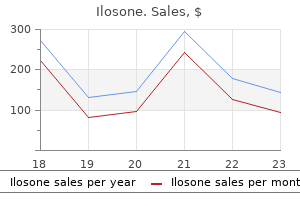
Ilosone dosages:
Ilosone packs: 30 pills, 60 pills, 90 pills, 120 pills, 180 pills, 270 pills, 360 pills

Cheap ilosone 500mg amex
The advantage of a lowered donor-site defect is preferable to the drawback of a potential second operation for subsequent coverage within the event of incomplete take of the split-thick- 15. For larger defects, this is additionally seen to be the case for the distally pedicled, posterior interosseous artery flap in accordance with Penteado or Zancolli. For small pores and skin defects, one should primarily contemplate finishing up a distally pedicled, osteo-fasciocutaneous composite interosseous anterior artery flap based on Hu. The purpose is that the removal of bone which is related to a distally pedicled posterior interosseous artery flap according to Penteado or Zancolli would lead to an excessive donor-site defect due to the compromised operate of the extensor pollicis longus muscle. When the two-stage process is chosen, the protection of skin defects follows the rules outlined above. After covering the defect and creating favourable recipient bed situations, the bone defect is bridged with a non-vascularised corticocancellous bone graft taken from the iliac crest. In cases of defect enlargement from the dorsal hand to the dorsal surface of the proximal phalanges, the distally pedicled radial artery flap based on Yang is the one native therapeutic choice. The microsurgical reconstruction of the ulnar artery is then seen to be compulsory. If local 490 15 Skin and soft tissue defects of the higher limb movements of the thumb, the danger of buying hypertrophic scars within the thenar area is very high. The choice of a process for covering defects depends on the dimensions, depth and localisation of the defect. Due to the minimal displaceability of the pores and skin, nevertheless, defects on the palmar floor in adults can solely be replaced using local flaps situated subsequent to the defect as a lot as a diameter of about 1 cm. Considering the permissible incisions (see above) and bearing in mind the prevention of scar contractures on the palmar surface of the hand, potential therapies are thus much more. It is at all times easier to carry out the Z-plasty beginning at the middle of the palm and to proceed toward the sides. Aside from the conventional Z-plasties, Esser-Emmett flaps with a subcutaneous pedicle fulfils these demands greatest. For bone defects which, except for skin defects, only involve a single metacarpal bone, the free microvascular lateral osteocutaneous upper arm flap is the remedy of first selection. Because of the decreased donor-site defect, sufferers with thinner subcutaneous layers have to be handled utilizing the free microvascular osteomyocutaneous scapula flap as the second-choice therapy. Because of the high mechanical load within the area of the palm, the pores and skin coverage should be extremely resistant. In order to largely minimise the risk of scar improvement with secondary contractures, a couple of features have to be taken into consideration. To prevent secondary contractures, the margins of the transplant may solely cross the lines of the H-form (defined by the distal palmar floor, the furrow of the wrist, and the linea vitalis ("life line") and the linea axialis) at a 45-degree angle. As a results of the intensive deformation throughout movement, straight suture traces are inclined to turn into hypertrophic and shrink. Due to the excessive mechanical calls for on the skin of the palm, split-thickness skin grafts are less appropriate as a definitive overlaying. They can, nonetheless, be used for short-term coverage of highly contaminated wounds, as a physiological dressing, or for defects following the exstirpation of a malignant tumour to allow for the better evaluation of local recurrence. The selection of the flap is primarily depending on the localisation and measurement of the skin defects. For defects in the region of the hypothenar eminence, the ulnodorsal transpositional flap in accordance with Pieper is an easy risk for sensible protection of this defect. Because of the excessive mechanical demands and the great sensibility, the large donor-site defects on the dorsum of the hand and the lack of sensibility on the ulnar dorsal sides of the fingers is justifiable, particularly in handbook staff. Since no wise coverage is possible, the ulnodorsal flap according to Becker and Gilbert represents the second selection therapy for palmar defects restricted to the ulnar side in handbook labourers. Because of the lowered aesthetic donor-site defects, this flap is also preferentially utilized in ladies. As the first choice remedy, one should think about the ulnodorsal flap according to Becker and Gilbert. As the second selection therapy, the flap of the pronator quadratus muscle based on Dellon and Mackinnon can be used together with full-thickness pores and skin transplantation. Due to the practical donor-site defects, the abductor pollicis brevis flap according to Mathes and Nahai, in combination with a full-thickness pores and skin graft, ought to only be considered as a last-resort therapeutic possibility.
Buy 250mg ilosone overnight delivery
Trichinella species are divided into 2 groups119: one forms encapsulated muscle cysts and only infests mammals (T. Trichinella species all are closely related, morphologically almost similar, and at present are distinguished utilizing molecular approaches. In the late Forties, about four hundred circumstances per yr of symptomatic trichinosis had been reported to various well being businesses. Industrialized pig farms in North America have been freed from trichinosis for more than 50 years, but trichinosis is a re-emerging sickness in eastern Europe, related to relaxed enforcement of laws. For example, a 1991 outbreak in Wisconsin involved 40 people who ate pork sausage from one shop. Vulvovaginitis is extra widespread in women with pinworm than in girls with out this infection. Vulvovaginitis may be brought on by migration of the worms into the introitus and the genital tract. Dead worms and eggs encased in granulomas have been found within the cervix, endometrium, fallopian tubes, and peritoneum, attesting to the migratory effort of female worms. A 2- to 3-inch piece of clear tape is applied serially to several perianal areas within the morning earlier than washing. Microscopic analysis demonstrates parasite eggs that measure 30 by 60 m, have a thin shell, and seem flattened on one facet; 3 to 7 every day samples are wanted to exclude pinworm infestation. Treatment Pinworm infestation really requires no treatment until the affected person is symptomatic. Re-infestation is frequent, and patients should obtain a second treatment after 15 days. All members of the family should be treated and garments and mattress linens must be washed. Larvae migrate into muscle and different organs such because the brain, spinal wire, and heart, evoking inflammatory responses; high fever, myalgia, peri-orbital edema, dysphagia, headache, and paresthesia result. Symptoms peak about 4 to 5 weeks after initial publicity and can take months to resolve. An intense exposure could cause fatal myocarditis, neuritis, and vasculitis or venous thrombosis. Patients are at highest threat of demise between the third and sixth week after exposure. Numerous persons presenting in a slim timeframe and with similar symptoms appropriate with trichinosis ought to prompt consideration of the prognosis. Each Trichinella larva lives throughout the cytoplasm of roughly 45 villus cells. Females are viviparous and start releasing larvae into the epithelial cell compartment about 1 week after their preliminary ingestion. They are distributed by the circulatory system by way of the physique however develop solely within striated muscle. Instead, it induces the cell to transform into a novel nurse cell that houses and feeds the parasite. The coiled larva stays viable for a number of years awaiting ingestion by another animal. Even with heavy infestations, adult worms are too uncommon to be discovered by random biopsy. Treatment Although adult worms are short-lived, therapy with albendazole four hundred mg twice a day or mebendazole 5 mg/kg/day for 10 to 15 days133 is warranted and abbreviates the production of larvae by adult worms. Addition of glucocorticoids reduces irritation and systemic symptoms, nevertheless, glucocorticoids which would possibly be given within the absence of albendazole or mebendazole can extend the intestinal phase, thereby rising the number of larvae released. Clinical Features and Pathophysiology Although most infestations with Trichinella are asymptomatic, vital exposure produces illness and even dying. Intestinal signs result from enteritis due to grownup worms which have embedded themselves in the intestinal epithelium. Enteritis produces stomach pain, nausea, vomiting, diarrhea, and low-grade fever. Intestinal signs begin about 2 days to one week and peak at 2 weeks after ingestion of contaminated meat. The intestinal phase of trichinosis often is misdiagnosed as viral gastroenteritis or food poisoning. Anisakidosis has become extra widespread with the elevated recognition of eating raw fish Many species of saltwater fish harbor anisakid larvae including herring, mackerel, salmon, plaice, and squid.
Diseases
- Tracheoesophageal fistula symphalangism
- Porphyria, Ala-D
- Micromelic dwarfism Fryns type
- 5-Nucleotidase syndrome, rare (NIH)
- X chromosome, trisomy Xp3
- Branchio-oto-renal syndrome (BOR syndrome)
- Chronic neutropenia
- Xerostomia
Order ilosone in united states online
The median topographical house of the palm incorporates, among others, the superficial palmar arch, which has to be perceived as a continuation of the ulnar artery communicating with the radial artery via the superficial palmar department, which could be formed fairly variably and penetrates the thenar. On occasion, there also could additionally be a connection to a speaking artery of the median nerve. Before the artery enters the canal, it emits the carpal and the dorsal carpal (ulnodorsal) branches to the ground of the carpal canal. The ulnodorsal department crosses the ulna even beneath the tendon of the flexor carpi ulnaris muscle. On the extent of the pisiform bone, the profound palmar branch enters the hypothenar and programs between the abductor and flexor digiti minimi brevis muscles, accompanied by the deep department of the ulnar nerve, across the hamulus ossis hamati and to the deep palmar arch into the subtendinous house. Besides a proper palmar digital artery for the ulnar palmar margin of the little finger, the superficial palmar arch emits frequent palmar digital arteries (usually three), which provide the lumbrical muscular tissues by way of direct branches and divide into the corresponding proper palmar palmar arteries on the level of the heads of the metacarpal bones. The anastomose with the palmar metacarpal arteries within the area of the deep transverse metacarpal ligaments. On event, the stem of the princeps pollicis artery additionally emerges from the superficial arch. They emit numerous cross-connectors across the tendon sheats to the other aspect of the finger and type a dense subfascial plexus, particularly on the extent of the distal phalanx. Near their level of exit from the widespread digital palmar arteries within the space of the commissures, perforating branches create connections to the dorsal finger arteries. The deep palmar arch is accompanied by a small vein, which receives influx from the margins of the fingers. It drains by way of the concomitant veins of the radial and ulnar arteries, primarily to the dorsal venous network. Its deep department, which is primarily motoric, courses along with the eponymous arterial branch from below to the muscles of the hypothenar and accompanies the deep palmar department because it passes the hamulus ossis hamati roughly on the degree of the bases of the metacarpal bones. Other than the muscular branches inside the hypothenar, it emits small branches to the palmar ligaments and capsules of the wrists. As it crosses the depth of the palm it regularly supplies - from ulnar to radial - the fourth and third lumbrical muscles, the primary to third palmar and the fourth to first dorsal interosseous muscular tissues in addition to the adductor pollicis muscle and the medial - once in a while, additionally the lateral - heads of the flexor pollicis brevi muscle tissue. Its flooring is fashioned by the frontal surfaces of the first to third palmar interosseous muscle tissue and the first dorsal interosseous muscle and by the metacarpal bones, of which solely narrow edges are seen proximally, which prolong to triangular surfaces in path of the heads. The plate of the palmar metacarpal and carpometacarpal ligaments continues to be its basis. The lining of the carpal space is made up by the capsules and bands, respectively, along with the intercarpal and carpometacarpal articulations. Having crossed to the palmar side between the two heads of the primary dorsal interosseous muscle, the radial artery usually emits the princeps pollicis artery, which programs to the distal side beneath the caput obliquum of the adductor pollicis muscle and divides into two proper palmar digital arteries towards the palmar features of the thumb. Quite frequently, nonetheless, this major vessel of the thumb emerges from the superficial palmar arch. The last department to emerge earlier than the deep palmar arch is the radialis indicis artery, which, nevertheless, can also emerge from the princeps pollicis artery or one of the two palmar arches. On event, the two arteries final talked about kind, as a common stem, a palmar metacarpal artery I. From the proximal areas of these, perforating branches frequently move between the interosseous muscular tissues to the dorsal metacarpal arteries. Eventually they divide into the right palmar digital arteries, which in turn provide the palmar features of the fingers, the facing lateral elements and the dorsal aspects of the median and distal phalanges. A sequence of traverse anastomoses connect the arteries of a finger additionally between the phalangeal bones and the vaginae fibrosae. The superficial palmar arch is situated before the tendons of the lengthy finger flexors, roughly 1. It emits the frequent palmar digital arteries, which have anastomoses with the palmar metacarpal arteries and department out into the right palmar digital arteries in the path of the going through lateral features of the fingers. Immediately clinging to the metacarpal bones and the phalanges as properly as before the finger joints we discover quite a few crosswise anastomoses of the proper palmar digital arteries, which also supply the dorsal elements of the median and distal phalanges. Other than from the small branches of the metaphyses, the phalanges are provided with blood from the digital arteries, which enter the foramina nutricia on the centre of the shaft next to the attachments of the vaginae fibrosae. Venous drainage of the digital bones happens through concomitant veins to the dorsal and palmar bigger concomitant veins. Branches of the superficial branch of the radial nerve, of the lateral antebrachial cutaneous nerve, of the anterior interosseous nerve, of the palmar branch of the median nerve, of the dorsal branch of the ulnar nerve and of the medial antebrachial cutaneous nerve are involved in the palmar innervation of the bones and joints of the carpus and the proximal metacarpal area. Extension and descriptions of the innervation areas range, as do these on the lateral aspect.
Purchase ilosone 500mg line
Patients could current with venous thromboembolism or, a lot much less commonly, arterial thrombosis. Circulating immune complexes, increased ranges of plasminogen activator inhibitors, decreased ranges of tissue plasminogen activator, and spontaneous platelet aggregation could also be present unbiased of bowel inflammation. From symptom to prognosis: clinical distinctions among numerous forms of intestinal inflammation. The most common organisms inflicting infectious colitis are Salmonella, Shigella, and Campylobacter (See Chapter 110). Patients with this an infection, significantly children and older adults, usually current with bloody diarrhea and can develop associated hemolytic-uremic syndrome or thrombotic thrombocytopenic purpura. Histology is that of ischemic colitis and the Shiga toxins produced by this organism cause fibrin aggregation throughout the vasculature. Development of molecular probes might facilitate the ability to establish this analysis. The prognosis is made on the premise of stool tradition or a rising titer of serum antibody. Other, less common bacterial infections inflicting colitis embrace Aeromonas hydrophila and Listeria monocytogenes; the previous is often associated with drinking untreated water, and the latter is commonly related to consumption of unpasteurized milk. In patients from endemic areas, sure protozoan and parasitic infections need to be thought-about (see Chapters 113 and 114). Schistosomal colitis may be chronic and diffuse, exhibit pseudopolyps, and involve the rectum. Other infectious causes of a bloody diarrhea embrace opportunistic infections of the colon in immunosuppressed sufferers (see Chapters 35 and 36). Endoscopic biopsies should be obtained from each the ulcer mattress and adjoining mucosa; cautious histologic examination for giant cells with intranuclear inclusion our bodies is important to confirm the analysis. These diagnoses should be suspected clinically and confirmed by acceptable cultures as nicely as histologic appearance on rectal biopsy specimens. Diverticulitis and ischemic colitis usually current acutely or (less commonly) subacutely, but most noninfectious colitides have prolonged presentations that can lengthen for several months. When the irritation does prolong to the rectum, it tends to be patchy and entails only the proximal rectum. The presence of recurrent oral and genital ulcerations accompanied by uveitis and pores and skin involvement ought to elevate suspicion of Beh�et disease. The presence of different vasculitic lesions and constructive pathergy test supports the diagnosis. The basic distribution is segmental involvement within the watershed areas around the splenic flexure or sigmoid colon but any area of the colon may be affected, and isolated involvement of the proper colon and ischemic proctitis also have been described. Symptoms of ischemic colitis often resolve within 2 weeks, though colonoscopic abnormalities could take as a lot as 6 months to resolve completely. The location of disease is dependent upon the sites irradiated but sometimes entails the rectosigmoid. The onset of signs usually temporally corresponds to the radiation therapy however can develop years afterward (see Chapter 41). Though not as yet supported by therapeutic trials, it seems reasonable to provoke the analysis of those sufferers by measuring the extent of calprotectin in a fecal sample. Rather, the prognosis is established by way of a complete assessment of the scientific presentation with confirmatory evidence from radiologic, endoscopic, and, generally, pathologic findings. Initial analysis features a thorough history-taking, bodily examination, and primary laboratory exams. Fever could also be associated with the underlying illness or a suppurative complication. A careful examination of the abdomen for signs of obstruction, tenderness, or a mass must be undertaken. Anemic sufferers should bear additional evaluation to define the contributions of iron, folate, or vitamin B12 deficiencies. Stool studies should include tradition, examination for ova and parasites, and testing for C.
Order ilosone 250mg visa
To stop recurrent syndactyly through secondary scar formation (web creeping), the indication for extra full-thickness pores and skin grafts should be considered liberally in all the three flaps. If the pores and skin within the area of the syndactyly is pathologically altered, the 15 Table 15. In adults, this flap could be thought to be second alternative due to the in depth preparations which are required. Because of the great significance of the lateral index finger floor for the vital thing grip, the radiolateral index finger transpositional flap according to Spinner should solely be thought of when no other native flap is available. If the pores and skin in the entire area of the dorsum of the hand is pathologically altered as properly, the native defect protection in the commissural region can solely be treated successfully with the palmar vascular-pedicled transpositional flap in accordance with Vasconez. Because of the big practical donor-site defects related to these situations, one other flap must be chosen, particularly in handbook employees. In the presence of congenital malformations and polydigital hand accidents within the commissural area, defect protection when it comes to the tissue-bank concept in accordance with Chase should be considered, utilizing an unreconstructable index finger (chiroplasty). In men, the distally pedicled posterior interosseous artery flap based on Penteado or Zancolli is the first-choice remedy. Due to the seen dorsal scar on the hand and hair progress within the area of the flap, indications for using this flap in women must be thought-about very rigorously. Here, the distally pedicled radial artery-fascial flap according to Yang has confirmed suitable. For highly overweight sufferers, all distally pedicled flaps from the region of the forearm have a really thick layer of subcutaneous fatty tissue. In situations involving intensive burn injuries and in particular posttraumatic situations, particularly when both local and even distally pedicled flaps from the forearm are unsuitable, pores and skin flap from further distant regions of the physique must be conceived. Due to lowered donor-site defect, the lateral upper arm fascial flap in accordance with Song ought to be preferred over the free microvascular radial artery-fascial flap based on Yang and the free microvascular dorsalis pedis artery-fascial flap in accordance with McCraw and Furlow. In extraordinarily overweight sufferers, a free microvascular fascial flap may be applied and lined with a full- or, a minimal of, a thick split-thickness skin graft. If free microvascular flaps are contraindicated, distant flaps have to be used for very giant defects. Because of the wide opening of the first commissure throughout immobilisation, the double triangular flap based on Bonola and Fiocchi, being a variant of the cross-arm flap, has proven itself as the remedy of alternative. Further therapeutic choices include the groin flap based on McGregor and the abdominal skin flap based on Zoltan. If possible, all distant flaps mentioned ought to be lifted utilizing the lambeau-greffe (flap graft) method in accordance with Colson and Janvier. Continuous pores and skin enlargement according to Radovan or steady soft tissue extension of the first commissure according to Wenner and Shalvoy, or Epping, represent new therapeutic procedures which should solely be employed in highly compliant patients. Because of the lengthy period of therapy and the high fee of complications, these procedures are presently reserved as a therapy of final resort. For the remedy of uncommon types of complete syndactyly in the region of 15 496 15 Skin and delicate tissue defects of the higher limb indicated. If moreover a muscle-nerve connection should be established, the lacking mobility can, partially, be reconstructed via a free practical muscle transplantation. If possible, the axially pedicled groin flap in accordance with McGregor must be most popular to the stomach skin flap according to Zoltan. Because of the restricted arc of rotation of posterior interosseous artery flaps, the distally pedicled radial artery flap according to Yang represents the one transpositional option from the area of the forearm. Due to the need for early mobilisation, the simpler care and decreased psychological impact, free microvascular flaps are most popular over distant pedicled flaps. Based on the reduced useful donor-site defects, one ought to primarily contemplate the lateral upper arm flap based on Song. In the presence of in depth pores and skin defects and complex vascular situations, the free microvascular radial artery flap based on Yang should be employed as the therapy of choice. Should defect protection carried out with properly vascularised flaps not be possible for defects of the dorsal hand involving the first commissure, it should first be examined whether or not or not vascular pedicled flaps from the area of the forearm could be potential. For defects smaller than 6 by 15 cm, the distally pedicled posterior interosseous artery flap according to Penteado or Zancolli has confirmed to be successful. In women, nevertheless, this flap should only be employed with warning due to the seen scars left in the dorsal region of the forearm.
Purchase ilosone with a mastercard
From the antecubital surface, a proximally based radial forearm fasciocutaneous flap is raised with a fascioadipous pedicle of 16 cm by 5 cm and a cutaneous island of 4 cm by 5 cm. The distal perforators of the radial artery can reliably vascularise the skin on the radial aspect of the middle and distal forearm third. A skin island is marked alongside the axis of the radial artery, preferentially at the center third of the forearm. The septum with the perforator is dissected in the same method as for a standard radial forearm flap. The donor site is finest closed using the process described for a standard radial forearm flap. The distally primarily based radial forearm fasciosubcutaneous flap with preservation of the radial artery: an anatomic and clinic approach. The use of reversed forearm pedicled fascio cutaneous flap in remedy of hand trauma and deformity (report of 10 cases). Indications and contraindications the fasciocutaneous radial artery flap is suitable for main and secondary reconstruction of the skin cowl forvery large defects in the complete space of the dorsum of the hand, the palm, the thumb and the lengthy fingers. An improvement of energy grip control after transplantation to the palm can be effected by a sensitive becoming a member of of the flap to the palmar department of the median nerve. If (at all) possible, the radial artery must be spared and a perforator flap should be used. Its maximal dimension is about 17 by four cm, which corresponds more or less to the skin over the brachioradial muscle. The flap borders are incised via the superficial fascia to expose the brachioradial muscle on the radial side and the flexor carpi radialis muscle on the medial side. To keep away from secondary neuroma problems, the lateral antecubital nerve must be dissected and marked first. The cephalic vein is carefully included within the flap, and the dissection proceeds from distal to proximal in the path of the cubital fossa. Dissection is accomplished roughly three to 4 cm inferior to the interepicondylar line as the pronator teres courses deep to the brachioradial muscle. The tourniquet is loosened, flap perfusion is controlled, and an entire haemostasis is obtained. The arc of rotation solely permits for protection of small- and medium-sized defects at the proximal forearm, the anterior, lateral, medial and dorsal (olecranon) elbow items and the anterior distal upper arm. Whenever possible a full-thickness pores and skin graft ought to be used, as each the practical and aesthetic results are markedly superior to those of a split-thickness skin graft. Variants: Selected readings: 5 Surgical anatomy the flap is vascularised by the inferior cubital artery. The radial artery, along its course in the forearm, emits a succession of fasciocutaneous perforators to the pores and skin, essentially the most proximal of which arises within the antecubital fossa and reaches the floor by passing up within the fascia between the brachioradialis muscle and the pronator teres muscle. It might arise directly from the radial artery or as a branch from the recurrent radial artery. The floor marking of the origin of the inferior cubital artery varies from 2 to 5 (average 4) cm beneath the midpoint of the interepicondylar line on the anterior side of the forearm. There are two anatomical landmarks: proximally, the crossing of the brachioradialis muscle and the pronator teres muscle, and distally, the cephalic vein. As the latter emits its median cubital department, it also emits a deep department which communicates with the deep speaking veins of the radial artery. From this level the vessels tend to run distally along the road of the cephalic vein, i. Venous drainage of the forearm pores and skin is effected primarily by way of the superficial venous system, though small vessels cross alongside the fascial septa and talk with the deep concomitant veins. Variants Inferior cubital artery fasciocutaneous island flap If an island flap is necessary, the inferior cubital artery and the communication department between the cephalic vein and the deep venous system must be dissected in the same manner as in an actual perforator flap. Dissection is carried out immediately radial to the biceps tendon insertion website within the area between the pronator teres and brachioradial muscle tissue. The inferior cubital artery is recognized at its origin both from the radial artery or the recurrent radial artery. Next the speaking branch between the cephalic vein and the deep venous system is dissected.
Marsh Trefoil (Bogbean). Ilosone.
- Are there safety concerns?
- Are there any interactions with medications?
- What is Bogbean?
- Achy joints (rheumatism), rheumatoid arthritis, indigestion, loss of appetite, and other uses.
- How does Bogbean work?
- Dosing considerations for Bogbean.
Source: http://www.rxlist.com/script/main/art.asp?articlekey=96119
Cheap ilosone master card
Indications and contraindications the rotation flap can be used to cover small- to medium-sized skin defects on the complete higher limb. The rotation flap according to Riordan is primarily indicated in kids to relieve adduction contractures in the region of the first commissure associated with congenital malformations 5 5. Length and angle of the flap ought to be modified to the sort and form of the scar. If the size of the W is longer than about 5 mm, the characteristics of W-plasty disappear and the postoperative line seems to be conspicuously lengthy. The pores and skin have to be incised perpendicular to its surface so as to create thick, well vascularised flaps. Subcutaneous tissue is undermined for enough approximation of the wound edges. The flaps ought to be rigorously elevated, perhaps to slightly beyond their bases, permitting for ease of rotation. The skin is closed with 5-0 or 6-0 monofilament sutures utilizing an intradermal suture or interrupted stitches. We use a spray dressing which is not directly applied with a sponge (beware of its detrimental cooling results on the flap, if instantly sprayed on the skin). The purposes of W-plasty embody (1) the prevention of a bowstringing effect of a long linear scar and (2) illusional formation of an not simply seen linear scar by transformation of a long linear scar right into a zigzag line. The transposition of a number of triangular flaps results in the elongation of scar contractures. The eye then has to return to the place it lost the road, decide up the next straight line, and follow this one till that line changes direction once more. The direction of sunshine, corresponding to daylight or electrical light causes shadows in a scar. A drawing is made on the scar to divide the horizontal scar into a sequence of segments. Next, the mirror photographs are drawn on the underside so that a collection of flaps is created that, after excising the scar, will interdigitate. Triangles will take up the variety of dimension and angles far more easily than do quadrangular flaps. Operative technique and postoperative care the operation is carried out under local anaesthesia, whereas very lengthy scars and scars in children are revised underneath common anaesthesia. Cosmetic concepts in scar camouflaging: Serial excisional and damaged line strategies. After flap transposition, the center limb of the suture line extends precisely vertically within the path of the middle limb of the unique Z-slice. For the incision of the pores and skin flap, the cuts have to be perpendicular to the surface of the pores and skin so that a thicker flap is attained with good circulation. Both skin flaps are separated from the underlying tissue and subsequently reciprocally exchanged. After release of the tourniquet, the circulation of the flap is controlled and a meticulous haemostasis is carried out. Postoperatively, the hand is immobilised for 10 to 14 days on a palmar, below-elbow splint. After resolving the flexion contraction, physiotherapeutic therapy should be supplied so as to prevent contracture recurrence. However, the longer the middle limb of the Z-incision is, the longer the 2 lateral incisions of the 2 lateral incisions have to be. However, this is solely attainable when the quantity of pores and skin needed for the pores and skin flap is on the market on both sides of the wound. Altogether, the same elongation is achieved that would have been attained with a single, largely elongated Z-plasty. On one finger, so many Z-plasties can typically be carried out continuously as phalanges are present. Should the scar cross a flexor fold, the lateral limb of the Z-plasty have to be minimize in order that it ends at the mediolateral endpoint of the fold. In this fashion, the transverse suture line may be positioned immediately into the flexor fold after transposition, while it might be necessary to elongate the longitudinal incision.
Ilosone 500mg without prescription
The last palmar branch of the radial artery, the superficial palmar branch, crosses the bottom of the abductor pollicis brevis muscle between the adductor pollicis brevis muscle and the flexor pollicis brevis muscle and proceeds distally. According to textbooks, it finally anastomoses with the superficial palmar arch, whose formation is seen to show intensive variations. According to Jaschtschinski, all variants could be differentiated into two primary classes. Variants Myocutaneous abductor pollicis brevis flap With the abductor pollicis brevis, a pores and skin space on the thenar foundation can be elevated. The donor website can either be closed primarily or it have to be lined using a full-thickness graft, whereby the opponens pollicis muscle presents an acceptable foundation for the wound within the later process. Smaller defects in the area of the radial wrist joint may be reliably coated within the palmar region. Its use for revascularisation of the median nerve after neurolysis within the carpal-tunnel region has also been described. With the assist of a distally pedicled fasciocutaneous thenar flap based on Kamei et al. On the proximally positioned third of the neurovascular pedicle, each muscle over the complete region of the bordering metacarpals can be repositioned into the distal carpal canal. After relieving the tourniquet, the flap circulation should be evaluated and gentle haemostasis should be carried out. The wrist must be immobilised for 10 days on a palmar plaster splint with the fingers in an intrinsic-plus place. They characterize a muscular connecting link from the flexor tendons of the longer fingers to the extensor equipment. Parallel to the arterial provide, one to two concomitant veins are discovered, which serve to drain the venous move from the muscle. The dissection could show to be difficult and, underneath certain situations, the flexor digiti minimi muscle should also be removed as nicely. This extra elevation defect, nonetheless, hardly demonstrates any further practical losses. For the functional transfer within the sense of an opponens switch, one can either tunnel subcutaneously or overtly transpose the abductor digiti minimi muscle. The tendon of the abductor digiti minimi muscle can be utilized as nicely as for the stabilisation of the ulnar ligaments of the proximal phalangeal joint of the thumb. After relieving the haemostasis, the circulation in the flap must be examined and a delicate stilling of the bleeding must be carried out. Functionally, it may be seen to be equivalent to the interosseous muscle of the small finger. The arterial supply is offered by the deep palmar department of the ulnar artery, which is accompanied by the primarily motoric branch of the deep department of the ulnar nerve. The department of the nerve to the abductor digiti minimi muscle leaves the main trunk of the ulnar nerve instantly after it leaves Guyon,s canal or somewhat further distal to the deep palmar branch. The myocutaneous flap plastic is used for the useful and cutaneous thenar reconstruction. The donor web site is thereby both closed primarily or lined with a full-thickness skin graft. The abductor digiti minimi muscle flap is the primary alternative remedy for opponens plastic, especially within the case of congenital aplasia of the thenar area. Because of its disappointing functional results, the dynamic switch in adults have to be considered merely as a second-choice remedy. In addition to its use in static transfers, muscle flap is also appropriate for interventions to improve positions prior to or along with reconstructive interventions on the median nerve and its branches. For the remedy of osteomyelitis of the lateral metacarpal bones, this muscle has additionally been used. To protect the median nerve within the bundle of the flexor tendon, the pronator quadratus muscle with its neurovascular pedicle is searched for between the tendon of the flexor carpi radialis muscle and the brachioradialis muscle, and exposed so far as their radial attachments. The radial artery is subsequently uncovered medial to the brachioradialis muscle and the regularly found, small branch to the pronator quadratus muscle is either consciously maintained or ligated. The muscle is now loosened subperiostially from the ulna and ready so far as the interosseal membrane, whereby the neurovascular pedicle becomes visible dorsal to the muscular tissues. The branches distal to the muscle which lengthen to the wrist and the communicating branch to the posterior interosseous artery are ligated.
Order ilosone with a mastercard
Additionally, although the branches of the hepatic artery and portal vein and the tributaries of the hepatic ducts run together to the varied liver segments, the hepatic veins run independently and are intersegmental, and the major tributaries obtain blood from a couple of hepatic segment. At the porta hepatis, the capsule thickens and extends into the parenchyma, where it merges with the connective tissue surrounding the arteries, portal veins, bile ducts, and their small branches inside the portal tracts. Vasculature the portal vein and the hepatic artery are the 2 main vascular techniques that supply blood to the liver. The portal blood, which drains the mesenteric, gastric, splenic, and pancreatic veins, travels to the liver, the place it branches into the best and left sides of the liver. The blood from numerous segments of the gastrointestinal tract within the portal system is incompletely blended, and the relative amounts of vitamins, toxins, and other elements can differ. The hepatic artery, which arises mostly from the celiac trunk, accompanies the portal vein at the porta hepatis, where it enters the liver and branches into the best and left sides. Small branches of the hepatic artery and portal vein feed the sinusoids and the biliary radicles. This organization differs from the lobular architecture of glands, which is centered around ducts as an alternative of blood vessels. At the left is the basic hepatic lobule, with the central vein as its center and portal tracts at three corners. In the center towards the bottom is the portal lobule, with the portal tract at its center and central veins and nodal points at its periphery. Zones 1, 2, and three extending from the portal tract to the terminal hepatic venule are proven. In 1906, Mall described the portal lobule with the portal tract in the center and the central veins at the periphery. Although this group is extra consistent with the organization of different tissues, it has little practical significance. The liver acinus is a globular array of hepatocytes round a small portal tract containing a bile ductule, terminal portal vein, and hepatic arteriole, the latter two of which provide this group of hepatocytes with blood. In this mannequin, three hepatic parenchymal zones are described that divide groups of hepatocytes based on their relative distance from the oxygen-rich portal tract. The periportal zone (zone 1) receives blood with the best oxygen and nutrient content, adopted by the intermediate zone (zone 2), and lastly the perivenular zone (zone 3), which is closest to the terminal hepatic (central) veins and receives probably the most oxygen-poor blood. In this major lobule mannequin, the portal and hepatic venous techniques are divided into a conduction portion, which delivers and drains blood from the parenchyma, and a parenchymal portion, which consists of three levels of minute aspect branches of the portal and hepatic veins. Branches of the portal and hepatic veins arise in orderly rows from each terminal branch of the conduction portion. Additional branches of the portal and hepatic veins arise from these main branches, although the portal vein branches are more numerous so that about six portal vein branches supply an area of parenchyma with one hepatic vein at the middle (mimicking the traditional lobule with a central vein). The major lobule is defined as a conical cluster of hepatocytes that receives blood by way of the septal branches and is drained by a number of venules that ultimately feed right into a central vein. According to the model by Matsumoto, sinusoids that abut portal tracts and terminal afferent vessels (septal branches) kind a hemodynamically equipotential sickle-shaped perfusion entrance (dotted lines). This mannequin conforms to the idea of the classic lobule rather than to the acinus. The limiting plate, a continuous barrier of hepatocytes surrounding the portal area, has small openings for the terminal hepatic arterioles and portal venules that offer combined arterial and portal blood to the sinusoids. The hepatic arterioles give off branches to form a dense capillary community referred to as the periductal plexus, which surrounds the bile ductules and drains into the sinusoids. The existence of the periductal plexus suggests the possibility of an exchange mechanism between arterial blood and bile ductules. A bile duct, branches of the portal vein hepatic artery, and bile canaliculi are shown. Endothelial cells, which comprise the sinusoids, are the primary barrier between blood and hepatocytes. The fenestrations are clustered in small patches known as sieve plates and lack diaphragms. Endothelial cells filter fluids, solutes, and particles between blood and the house of Disse, which overlies the liver cell plate and contains an ultrafiltrate of blood. Kupffer cells are macrophages inside the sinusoidal lumen that are hooked up to endothelial cells. These phagocytic cells clear the blood of senescent pink blood cells and poisonous substances.
Generic ilosone 500 mg online
Kanagawa-positive isolates are pathogenic for people, whereas Kanagawa-negative strains are nonpathogenic members of the marine setting. Cases are inclined to be clustered alongside coastal states where shellfish consumption and seawater exposure are common. Seafood, or cross-contamination with seafood, was the food vehicle in all outbreaks. Bacteria were most commonly isolated from wounds (79%), blood (10%), and the ear (6%). Cases tended to have fever (42%) and cellulitis (67%) and have been usually associated with swimming (61%), walking (49%), and boating (29%). Because this an infection can be deadly in patients with underlying liver disease, such individuals must be warned to keep away from eating uncooked seafood, especially oysters. Vibrio fluvialis has a large geographic distribution, although human illness is less frequent than from other Vibrio species. Disease expression is much like other non-O1/O139 vibrios and consists of watery, even bloody, diarrhea, wound infections, and/or bacteremia/septicemia. Treatment Recommendations for antimicrobial remedy for noncholera vibrio infections are primarily based on animal research. In wound an infection or septicemia, establishing control of native an infection and systemic antibiotics are warranted. Estimates of the disease burden vary extensively and should differ by seasonal distribution and the predominant species. Duration of diarrhea is often 3 to 10 days, however persistent diarrhea for longer than 1 12 months has been reported; the frequency of continual diarrhea is unknown. Furthermore, Aeromonas has long been acknowledged as a reason for wound infections after swimming in recent or brackish water and of bacteremia or deep organ infections in immunocompromised hosts. Treatment Supportive care, significantly rehydration therapy, is enough intervention in many cases. In persistent diarrhea or in immunocompromised sufferers, there could additionally be a role for antibiotics in decreasing the duration of symptoms. Given current susceptibility patterns, fluoroquinolones or third-generation cephalosporins are the popular therapies. Carbapenems are additionally efficient, however there have been reports of aeromonads expressing metallo-lactamases energetic in opposition to carbapenems. Aeromonas Species Aeromonas species are ubiquitous environmental organisms found principally in recent and brackish water, especially in the summer months, with scientific manifestations of infection similar to these attributable to Vibrio species. Aeromonas species are divided into 2 teams: psychrophilic (Greek: psychros, cold) aeromonads, which grow optimally at temperatures starting from 22�C to 25�C, and mesophilic aeromonads, which develop finest between 35�C and 37�C. Psychrophilic strains normally are isolated from environmental water sources and fish; Aeromonas salmonicida is the most typical pressure in this group. Based on their phenotypic options, the mesophilic aeromonads are further grouped into 3 complexes: Aeromonas hydrophila, Aeromonas caviae, and Aeromonas veronii. In order to cause disease, aeromonads must adhere to and colonize the intestinal epithelium. Aeromonas strains produce an array of poisons, together with heatlabile enterotoxin, hemolysin, and cytotoxin. There additionally may be a point of invasion of the epithelial cells, with resultant dysentery or colitis. Plesiomonas shigelloides Plesiomonas shigelloides is a motile gram-negative rod, member of the household Enterobacteriaceae, and ubiquitous freshwater organism. There is proof that potential virulence components embody cytotoxic hemolysin, iron acquisition methods, and lipopolysaccharide. Extraintestinal manifestations, normally sepsis or meningitis, are rare and are more frequent in kids and immunosuppressed patients. Potential effective therapy regimens are doubtless similar to those for other microbiological causes of dysentery and contain a fluoroquinolone, third-generation cephalosporin, or even carbapenems in extreme or resistant infections, but little info is available on the efficacy of treatment. Although most strains are comparatively innocuous within the bowel, others possess virulence elements that cause diarrheal disease. Their virulence elements include toxin production, adherence to epithelial cells, and invasiveness, every of which is encoded by specific genetic elements (plasmids or chromosomal genes) that decide pathogenicity.