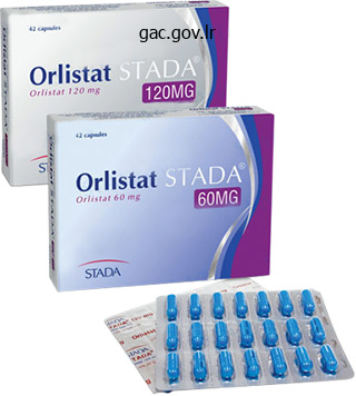
Orlistat dosages: 120 mg, 60 mg
Orlistat packs: 10 caps, 30 caps, 60 caps, 90 caps, 120 caps, 180 caps, 270 caps, 360 caps
Generic orlistat 60mg visa
A direct subclavian-axillary puncture or cephalic cutdown for venous entry is carried out. A 2-cm to 3-cm horizontal incision is made in the medial third of the inframammary crease. The incision is carried all the means down to the level of the pectoralis fascia, and blunt dissection is used to create a pocket superficial to the pectoralis fascia behind the breast. A 20-cm, 18-gauge pericardiocentesis needle is directed from the inframammary pocket to the infraclavicular incision. A 145-cm J wire is then passed from the submammary pocket through the needle and to the infraclavicular incision. The needle is removed, and with use of the retained-guidewire method, two introducer dilators of applicable size are passed consecutively over the guidewire. This progressive tunneling technique has been extraordinarily profitable and well-tolerated. Shefer et al138 described a retropectoral transaxillary percutaneous technique for optimal beauty impact. Venography is used to affirm the relationship of the axillary vein to the surface anatomy. The axillary vein is punctured when the tip of the needle touches and moves the marker wire. The pectoralis fascia is then uncovered with blunt dissection and the retropectoral area opened. At the completion of the operation, the momentary pacing lead and guidewire marker are eliminated. With any of these methods, the pulse generator and leads are connected and the incisions closed. A polyester (Dacron) Parsonnet pouch can be used to forestall rotation of the heartbeat generator and leads in the tissue. To keep away from the problems of diaphragmatic stimulation, a bipolar system must be used, particularly with the inframammary pocket. Vital to the submuscular pectoral approach is a complete understanding of the regional anatomy. The pectoralis major muscle has two main subdivisions: the clavicular head hooked up to the clavicle and the sternal head connected to the lateral border of the sternum. Inset, the deltopectoral groove and the clavicular and sternal heads of the pectoralismajormuscle. Tearing the artery or vein might result in pocket hematoma, and interruption of the nerve could result in pectoral muscle dysfunction. The deltopectoral groove is outlined by the lateral border of the clavicular head of the pectoralis major muscle. Currently, with the lively can and the dual-coil lead system, the submuscular pectoral method may be performed from both the best or the left pectoral area. The anterior strategy is just like that for a subcutaneous pacemaker pocket creation. It requires the creation of a dissection airplane between the sternal and clavicular parts of the pectoralis muscle. The lateral method requires dissection of the pectoralis major muscle along the deltopectoral groove. Similarly, the axillary approach requires dissection and institution of a aircraft on the anterior axillary fold, and the system tends to drift into the axilla and cause discomfort. Both the lateral and the axillary approach also carry the danger of interrupting the lengthy thoracic nerve, leading to "wing" scapula. The skin incision for the anterior approach is much like that used for a permanent pacemaker pocket. The incision is initiated simply medial to the coracoid course of and is carried from the extent of the coracoid course of inferomedially and perpendicular to the deltopectoral groove for about three inches (7. With a Weitlaner retractor for retraction and electrocautery, the incision is carried down to the floor of the pectoralis fascia.
Order cheap orlistat
In sufferers who exhibit hemodynamic impairment and insufficient collateral circulation, more well timed intervention is indicated. The patient is observed overnight in the intensive care unit with frequent neurological examinations. Postoperative antiplatelet regimens differ, but sometimes patients are instructed to proceed dual antiplatelet remedy for 6 months and then aspirin 325 mg day by day indefinitely. Complications and Management Perioperative complication rates for intracranial artery stenting range from 5% to 10%. Ischemic issues are mostly as a outcome of perforating vessel occlusion, either from arterial dissection or from atherosclerotic plaque migration. Clinical implications can vary from asymptomatic to everlasting neurologic deficit depending on the territory of the occluded vessel. This complication could be minimized by submaximal (rather than maximal) angioplasty and by deciding on the shortest attainable stent size to address the stenotic segment. Management varies, however it usually involves systemic anticoagulation with heparin in the acute postoperative interval. Risk elements for ischemic events are nonsmokers, prior non-perforator strokes, and old infarcts on baseline imaging. Post-procedure hemorrhage can be potentially devastating as a end result of the dual antiplatelet remedy, which likely exacerbates the hemorrhage. However, dual antiplatelet remedy is critical to preserve patency of the stent and keep away from stent thrombosis. Whereas some cases of hemorrhage can be managed expectantly with blood pressure control, some are extreme and should require surgical decompression. Risk factors for post-procedure hemorrhage are higher grade stenosis, speedy clopidogrel loading, and intraprocedural activated clotting time >300 seconds. Given the comparatively high fee of complications, stenting for intracranial artery stenosis should be restricted to medically refractory, high-grade lesions with a excessive threat of recurrent stroke and ought to be performed by skilled operators. Angioplasty ought to be carefully tailored to the diameter of the target vessel, and barely submaximal balloon inflation is recommended to scale back the incidence of perforator occlusion and dissection. Maintenance of a distal wire beyond the stenosis over which the balloon or stent may be navigated ensures that if angioplasty-related occlusion occurs, distal wire access (and thus subsequent rescue stenting) may be performed. This seminal study highlighted the significance of medical management in intracranial artery stenosis. More lately, the Vitesse Intracranial Stent Study for Ischemic Stroke Therapy trial confirmed the prevalence of medical administration over stenting by finding that 36% of the stented sufferers reached the primary consequence of stroke or dying throughout the first year compared to solely 15. Such intervention ought to be thought of on a case-by-case foundation, and patient selection as well as operator expertise are important for good outcomes. Stenting ought to be thought-about solely in patients for whom medical management has failed and symptoms continue or in uncommon instances during which quickly progressive signs occur because of an unstable atherosclerotic plaque. Failure of extracranial�intracranial arterial bypass to scale back the chance of ischemic stroke: Results of a world randomized trial. Extracranial�intracranial bypass surgical procedure for stroke prevention in hemodynamic cerebral ischemia: the Carotid Occlusion Surgery Study randomized trial. Charbel Case Presentation 19 A 23-year-old female offered to the emergency department with intermittent recurrent episodes of proper arm numbness lasting a few minutes, occurring as a lot as 3 times per day through the previous month. She denies another neurological symptoms, headache, or seizure activity, and she or he reports no present signs at the time of evaluation. The patient has a previous medical historical past of surgical restore for ventricular septal defect in infancy. A pattern of acute/subacute or persistent infarcts, particularly in watershed areas, factors toward a hemodynamic mechanism of stroke from stenosis or occlusion of a significant intracranial vessel of the anterior circulation. Differential diagnosis for subacute�chronic unilateral facial, arm, and leg numbness features a broad spectrum of disease associated with completely different underlying etiologies: a. Headache, seizure, cognitive impairment, and involuntary movements are other potential clinical manifestations.
Order discount orlistat on-line
Most pneumothoraces are asymptomatic and detected on routine postimplantation chest x-ray. In the case of sizable and/or symptomatic pneumothorax, high-flow oxygen ought to be administered, and chest tube insertion could also be required. Tension pneumothorax is on the listing of differential diagnoses for sudden hemodynamic collapse during or instantly following device implantation, and ought to be handled with prompt chest tube insertion. Hemothorax is a uncommon complication of gadget implantation, mostly attributable to damage to the nice vessels. In the event of mistaken arterial access, pressure must be applied for several minutes. Surgical intervention for inadvertent arterial entry could also be needed, depending on the degree of harm and hemodynamic stability of the affected person. Axillary access is all the time extrathoracic and due to this fact amenable to manual compression. More medial entry, especially if arterial, can outcome in uncontrollable bleeding, significantly if the site is intrathoracic. The source of bleeding may be from within the pocket itself or backbleeding from the vein at the entry site. These are applicable use of electrocautery to ensure hemostasis during pocket creation, and prevention of backbleeding from the entry website. The risk of hematoma is higher in anticoagulated sufferers, particularly within the case of heparin. Both antiplatelet therapy and anticoagulation have been related to an elevated threat of hematoma. The incidence of pocket hematoma has been reported to be approximately 4% in patients not taking any antiplatelet or anticoagulation brokers, as compared with nearly 18% in patients receiving uninterrupted clopidogrel, and approximately 7% in patients taking warfarin. Evacuation should be avoided whenever potential as a outcome of it represents the potential to introduce an infection. On event, continued bleeding, extreme rigidity, or pain will necessitate evacuation. The optimal method to sufferers being handled with one of many novel oral anticoagulants stays to be fully delineated. It has been our expertise that perforated leads which have been confirmed to be connected to the pericardium when instantly visualized in the working room may sometimes have normal sensing, impedance, and pacing threshold values. When a patient develops a persistent perforation, days to months after implantation, extraction must be performed with surgical backup on the ready in the unlikely occasion of acute tamponade. Although this can be a threat, it has been our expertise, and that reported by others, that generally transvenous extraction and reimplantation is typically uneventful. The management and prevention of central venous stenosis is particularly important amongst patients with, or at risk for, endstage renal disease. The therapy of central venous stenosis involves a quantity of therapeutic choices together with venoplasty, stenting, and lead extraction when essential. For occasion, trapping a transvenous lead towards the vein wall when stenting open a vein must be averted every time possible because it eliminates the option of future transvenous extraction if necessary. Achannel is now seen via the world of superior vena cava stenosis (white arrow). This can result in early mechanical failure of the leads and sometimes lead dislodgement. It is clear that the lead has utterly retracted into the gadget pocket at 1 week. It is completely possible that this complication may have been prevented with a single suture securing the gadget in the pocket. After infected gadget extraction, mortality remained high, with 1-year mortality after removal of 17%. This increased mortality at 1 yr was vital in each the pocket and endovascular infection teams (12% and 25%, respectively). Even with correct skin preparation and periprocedural antibiotics, the 6-month infection fee during substitute procedures was 1. The supply of this ache could also be nerve entrapment, irritation of scar tissue, migration of the device, or harm to the musculoskeletal tissues. As shall be outlined in the following part, persistent pain on the implant website should improve suspicion for system an infection. Definitive therapy for severe, persistent ache is system extraction and reimplantation on the contralateral aspect if clinically indicated.
Orlistat 120 mg line
It is vital for blood stress to be maintained in the normal vary during periods of planned momentary vessel occlusion. Indocyanine green videoangiography avoids the drawbacks associated with intraoperative catheter angiography, and it has a reported diagnostic accuracy of roughly 90%, with probably the most generally missed discovering being small residual aneurysm neck remnants. At a minimal, circulate within the mother or father vessel and all branches must be confirmed with a microvascular Doppler probe. When patients current with an intracerebral hemorrhage from a ruptured aneurysm, the choice of whether or not to proceed with surgical clipping or endovascular coiling is influenced by the dimensions of the hematoma. For patients with massive, life-threatening hematomas, surgery for evacuation of the hematoma should be mixed with clipping of the aneurysm. When sufferers present with a large or advanced aneurysm, it may be very important contemplate choices for revascularization, corresponding to extracranial�intracranial and intracranial�intracranial bypass procedures. When intraoperative inspection reveals that one or more of the M2 branches arises from the aneurysm dome, bypass of the involved vessels ought to be carried out earlier than aneurysm occlusion or attempts at clip reconstruction. Fluid and electrolyte abnormalities are frequent and necessitate careful monitoring, with the objective of sustaining normovolemia and avoiding hyponatremia. The administration of oral nimodipine is strongly really helpful for the prevention and limitation of vasospasm. Treatment should start as quickly as attainable after hemorrhage and ought to be continued for 21 days or till the affected person is discharged. Nimodipine is a calcium channel blocker that was originally developed for the treatment of high blood pressure; not surprisingly, hypotension is certainly one of the unwanted side effects that may limit its use. The recommended dose is 60 mg each four hours, but when hypotension is a matter, 30 mg each 2 hours may be higher tolerated, and further downward dose titration could also be needed for some patients. They must be performed a minimum of as quickly as per hour for the first 24 hours after surgical procedure and then at 2- to 4-hour intervals. Typical causes of neurological deterioration include hydrocephalus, hyponatremia, an infection, and vasospasm. For patients with aneurysmal hematoma, a mass effect from swelling and edema may necessitate emergency decompressive craniotomy, if it was not carried out as a half of the preliminary operative treatment. Persistent hydrocephalus requires placement of a ventriculoperitoneal shunt in 20�30% of patients. Most surgeons advocate quick postoperative angiography to document aneurysm occlusion and patency of the mother or father vessels and branches. At a minimum, all sufferers ought to be treated with elastic compression hose and intermittent pneumatic compression units. If a decompressive craniectomy is performed, subsequent cranioplasty ought to be performed as quickly as potential upon resolution of swelling and edema. The patient has a programmable shunt valve, which may be adjusted to reduce the risks of extra-axial fluid collection. Angiographic proof of vasospasm, which is recognized in 40� 70% of sufferers on routine imaging, follows a time-dependent course after hemorrhage. Clinically, vasospasm refers to the ischemic complications related to angiographic vasospasm that impacts 20�30% of sufferers. The course of medical vasospasm follows the angiographic adjustments; onset is rare before day 5 or more than 2 weeks after a hemorrhage. Initial treatment consists of shortly ruling out different causes of neurological deterioration and initiating normovolemic hypertensive remedy. One of the harbingers of vasospasm is hyponatremia, and daily monitoring of sodium ranges should be considered for the primary 2 weeks after hemorrhage. The infusion of 3% sodium chloride resolution is cheap in the acute management of hyponatremia and may be supplemented with oral sodium chloride tablets. Fludrocortisone has been reported to be effective in limiting the severity of hyponatremia, and demeclocycline may be helpful in resistant instances. Doses as small as 1 mg of tissue plasminogen activator once or twice per day have been reported to be efficient in serving to clear intraventricular hemorrhage. Bacterial ventriculitis is a clinically significant danger in sufferers who require extended extraventricular drainage. The threat of an infection can be limited by cautious consideration to sterile method throughout placement and by the use of antibiotic-impregnated catheters (Codman Bactiseal).
Generic orlistat 60mg with visa
Calculating Ventricular Rate Counting intervals for the aim of fulfilling the speed criterion is distinct from calculation of fee. The median price is extra sensitive to oversensing than undersensing; one oversensed event ends in two brief intervals, whereas one undersensed event ends in one long interval. In ZipesD,JalifeJ,editors:Cardiac electrophysiology: from cell to bedside, ed6,Philadelphia,2014,Elsevier. After 8/10 beats meet price criterion, 6/10 beats required to meet price criterion throughout "duration" delay. Confirmation/Reconfirmation After detection, charging the shock capacitors to most power takes 6 to 12 seconds. Shocks after the first shock (or shocks following aborted shocks) could also be either committed (delivered without confirmation) or noncommitted, depending on the manufacturer. It may be programmable independently of the duration for initial detection and typically is shorter. Discriminators are particular person, algorithm components, or building blocks that provide a partial or complete rhythm classification for a subset of rhythms. Sudden (abrupt) onset was one of the first single-chamber, interval-based discriminators. The dual-chamber marker channel shows atrial intervals above and ventricular intervals under the line. These dualchamber discriminators are utilized only to 1: 1 tachycardias and enhance both sensitivity and specificity compared with singlechamber onset. Methods for measuring regularity in a gaggle of R-R intervals embody imply variability, the maximum uncooked difference between a check interval and any of the other intervals, and the maximum fraction of intervals in a pair of bins of a histogram of intervals. Nevertheless, physicians ought to verify that carried out baseline beats match the template each at implant and through follow-up. In patients recognized to have rate-related aberrancy, the template must be acquired during sinus tachycardia and automatic template updating should be disabled. They differ based on methods of filtering and alignment, and details of quantitative representations. In four of those 5 dual chamber detection algorithms, step one is explicit or implicit comparability of atrial versus ventricular price. Swerdlow Pierre Bordachar Discussion Points Analysis of Stored Electrogram-Ventricular Tachycardia or Supraventricular Tachycardia Acquire the template throughout sinus tachycardia or atrial pacing with aberrancy and disable automatic template updating. B shows interval plot of atrial(open squares) and ventricular(closed circles) intervals. The required high atrial sensitivity will increase the danger of atrial oversensing, particularly of far-field R waves. Programming to forestall atrial oversensing of far-field R waves is the most important explanation for atrial undersensing. However, scientific trials that compared single versus dual-chamber discrimination algorithms have reported inconsistent outcomes. Some trials164-167 and two meta-analyses168,169 discovered no benefit to dual-chamber discrimination. On marker channel, upward vertical marks denote atrial sensed occasions and downward marks denote ventricular sensed events. Reliable atrial sensing is critical for correct performance of each of these tasks. The trade-offs in velocity, sensitivity, and specificity among algorithms have been reviewed. R wave synchronized atrial shocks are restricted to R-R intervals higher than four hundred to 500 msec to reduce the risk that a therapeutic atrial shock may be delivered into the ventricular weak interval of the previous cardiac cycle. Undersensing attributable to unexplained, transient decreases in R-wave amplitude was common with early models that used a set sensing threshold. This permits for the possibility of sensing confirmation strategies that require delays of as a lot as several seconds. The sensing vector is chosen through an computerized process at implant, topic to manual override. Integrated Enhanced Sensing Process and Detection Algorithm this integrated algorithm contains three phases: (1) Sensed Event ("Detection") Phase, (2) Certification Phase, and (3) Decision Phase. The Certification Phase certifies that the sensed events are cardiac quite than oversensed alerts.
Galbanum Gum Resin (Galbanum). Orlistat.
- Dosing considerations for Galbanum.
- What is Galbanum?
- Are there safety concerns?
- Digestive problems, intestinal gas (flatulence), reducing spasms, coughs, healing wounds, and other conditions.
- How does Galbanum work?
Source: http://www.rxlist.com/script/main/art.asp?articlekey=96655
Buy orlistat with amex
The most typical endovascular therapy option entails deployment of detachable coils to induce aneurysm thrombosis. With ruptured aneurysms, balloon-assisted coiling is favored because stent placement requires twin antiplatelet therapy-most generally aspirin and clopidogrel-to keep stent patency. In sufferers with unruptured aneurysms and no contraindications to twin antiplatelet remedy, stent-assisted coiling with one or more stents is commonly most popular and is associated with decrease charges of aneurysm recanalization and wish for retreatment. Even with complete aneurysm obliteration, endovascular coiling continues to be extra more likely to lead to residual aneurysm or aneurysm recurrence requiring re-treatment, and aneurysms in the basilar tip are at greater risk for recanalization in comparability with different aneurysm locations. With long-term follow-up, large case collection have shown that 26% of coiled basilar tip aneurysms require re-treatment. Rates of re-treatment are highest within the first 12 months of therapy, but recanalization may happen in delayed style and annual charges of re-treatment are roughly 2. Despite the shift toward endovascular remedy of basilar artery aneurysms, microsurgical clipping stays a safe and sturdy treatment choice for appropriately chosen basilar tip aneurysms. Aneurysms with extensive or complicated neck configurations, smaller dome-to-neck ratios, and smaller facet ratios (the ratio of the peak of the aneurysm to the width of the neck) are less favorable for endovascular intervention and may favor clip ligation. Similarly, aneurysms which are large or giant, partially thrombosed, or with aberrant branches arising from the aneurysm dome may be higher treated with open microsurgery. Although small, risks with endovascular intervention accumulate with every treatment and surveillance angiogram, and research have constantly demonstrated higher durability with clip ligation. What aneurysm morphologic parameters favor use of either balloon-assisted or stent-assisted coiling with endovascular remedy What anatomic factors assist information method choice when microsurgically treating aneurysms of the basilar apex Surgical Procedure Open microsurgical clipping of basilar tip aneurysms is performed underneath basic anesthesia with adequate intravenous access to facilitate fast transfusion in the event of an intraoperative rupture. To ensure patient safety, the process should be carried out with neuromonitoring, together with both motor evoked potentials and somatosensory evoked potentials, and a coordinated effort between the neurophysiologist and the anesthesiologist is required to place the affected person in burst suppression prior to aneurysm manipulation to reduce the deleterious consequences of any potential ischemic occasion. For nearly all of basilar tip aneurysms, that are at or near the extent of the dorsum sellae (such as within the current case), and for high-riding (above the dorsum sellae) aneurysms, an orbitozygomatic trans-sylvian strategy is most well-liked (unless the aneurysm is totally within the third ventricle). For low-riding (below the dorsum sellae) aneurysms, the subtemporal approach is preferred. The head is affixed to a Mayfield head holder after which rotated 15�20 degrees toward the contralateral shoulder and prolonged roughly 20 levels to make the malar eminence the best point within the operative field. A curvilinear incision is made behind the hairline from the basis of the zygoma to approximately the midline. The scalp is elevated using an interfascial approach to shield the frontalis branch of the fascial nerve, and each the scalp and the temporalis are mirrored ahead. Next, the orbitozygomatic unit is released as a second piece with a reciprocating saw. This is accomplished with a stereotypical series of cuts: (1) the zygomatic root, (2) the zygomatic body, (3) from the inferior orbital fissure to minimize 2, (4) the medial orbital roof, (5) the posterior orbital roof, and (6) the lateral orbital wall. Once launched, the lesser wing of the sphenoid is fastidiously shaved right down to the superior orbital fissure, and the dura is opened. The temporal lobe is then additional released by way of cautious dissection and division of arachnoid adhesions and bridging veins alongside the middle fossa floor and the anterior temporal pole. Further dissection of the carotid and crural cisterns frees the temporal lobe and allows it to be retracted in a posterolateral dissection. Three operative corridors to the basilar apex are then recognized: (1) the supracarotid triangle, (2) the optic�carotid triangle, and (3) the carotid�oculomotor triangle. A sequence of anatomic steps are utilized to safely achieve each proximal and distal management when accessing the basilar apex. Once the basilar apex is visualized, entry to the basilar trunk have to be established for proximal vascular management. For a low-lying basilar apex, a posterior clinoidectomy or transcavernous publicity could also be necessary. Adenosine cardiac arrest can be used at the facet of proximal management of the basilar trunk or as a standalone methodology for a short period of aneurysm deflation. After obtaining proximal and distal management, care and time have to be spent to dissect free perforating arteries to prepare the neck of the aneurysm for clip application. Utilization of temporary clipping and/or intraoperative use of adenosine could soften the aneurysm dome to facilitate mobilization of the aneurysm and circumferential dissection of perforating arteries.
Syndromes
- Torn or damaged anterior cruciate ligament (ACL) or posterior cruciate ligament (PCL)
- Short height
- Fast and weak pulse
- Tonsillitis
- Change in blood pressure
- Vomiting blood
- Muscle weakness
- Estradiol, a type of estrogen
Buy orlistat 120mg lowest price
This venous channel is a good information to the left parietal border of the center in left anterior oblique view. In proper anterior indirect view the plane of the tricuspid orifice varieties an angle of 25 levels with that of the mitral orifice. Viewed from the anteroposterior perspective, the orifices of the 4 cardiac valves are like a cascade of plates. The pulmonary valve is located most superiorly in an almost horizontal airplane, whereas the aortic valve slopes rightward and posteriorly from the pulmonary valve. The aortic valve is sandwiched by the D-shaped mitral orifice, which lies leftward and posteriorly, and the tricuspid orifice, which is positioned rightward and anteriorly. Hence in the transverse bodily plane, the aircraft of the atrial septum extends obliquely rightward, from anterior to posterior, usually at an angle of roughly 60 levels to the sagittal plane. It is a misconception to consider solely the tip portion of this structure as the appendage. A "windscreen wiper" motion then assures the interventional doctor that the catheter/lead is positioned, as supposed, within the tip of the atrial appendage. Thus in frontal projection, when an interventionist has maneuvered a catheter/lead into the tip of the atrial appendage of a patient, seeing a "windscreen wiper" movement can guarantee him or her the positioning is as meant. On the endocardial facet, the terminal crest usually appears as a distinct C-shaped muscular ridge in most hearts however is flatter and fewer obvious in others. Characteristically, an array of pectinate muscles arises from the anterior and rightward margin of the crest spreading to the anterior, lateral, and inferior partitions of the atrium. Of variable thicknesses, they department into thinner and thinner muscle bundles that line the translucent parts of the appendage wall. In some hearts there may be a number of broad pectinate muscles that department near to their origin from the crest in palm-leaf fashion. The branching and criss-cross association of the pectinate muscular tissues might facilitate the information of results in be lodged. Moreover, the world the place the crest arises from the septal facet is steady with the rightward extensions of the Bachmann bundle, which run in the subepicardium. Eustachian Valve, Vestibule, Triangle of Koch, and Atrioventricular Node the eustachian valve guards the anterior and anterolateral quadrants of the entrance of the inferior caval vein. In fetal life it directs blood from the inferior caval vein towards the oval fossa. Occasionally, there are perforations within the eustachian valve, or the valve could also be a filigreed mesh (Chiari network) that could be so intensive as to stretch throughout the atrium to connect close to the orifice of the superior caval vein. Catheters passing via such a valve might become entangled and deflected from the intended course. In the area of the cavotricuspid isthmus, the vestibule occupies the anterior portion of the isthmus. Posterior to the graceful vestibule, the isthmus wall is irregular in thickness, comprising the terminal ramifications of the terminal crest and pectinate muscles and thinner fibrofatty areas in between the muscle bundles. The sinus node is located in this groove, near the superior cavoatrial junction. Its "head" portion extends subepicardially from near the crest of the appendage to cross laterally and inferiorly deep into the musculature of the terminal crest where its "tail" portion is embedded. Multiple prongs of nodal tissue prolong into strange working atrial myocytes that make up the terminal crest, enabling transmission of the nodal impulse. Because the myocytes within the crest are primarily aligned longitudinally along its length, the crest is a vital bundle for preferential conduction. B,Thefree wall has been deflected posteriorly to reveal theterminalcrest (crista), pectinate muscle tissue, and the sagittal bundle (*). Epicardially, the vestibule is roofed extensively by the fatty tissues of the atrioventricular groove by way of which the right coronary artery passes. The distance of the coronary artery from the endocardial surface of the vestibule decreases down to <3 mm when the artery is traced anticlockwise across the atrioventricular groove from the superoanterior place to inferior position as seen in a left anterior oblique view. The tendon of Todaro running in an anterosuperior direction inside the eustachian ridge is the marker for the posterior border of the triangle. It is a nice, fibrous strand, only revealed by dissecting into the ridge or on histological preparations. The anterior border is fashioned by the hinge line (annular insertion) of the septal leaflet of the tricuspid valve, whereas the inferior border is the orifice of the coronary sinus along with the vestibule anterior to it.
Safe orlistat 60mg
Occurrence of oversensing earlier than impedance improve is typical of conductor fracture. D also displays pacing and shock impedance tendencies for a similar lead, showing transient, small, simultaneous decreases in both developments. Air trapped within the header by insertion of the lead pin escapes into surrounding electrolytic body fluid as bubbles if the set-screw seal plug is damaged87 or the seal is loose. Bubbles escape when they produce a threshold strain on the seal plug producing a nonphysiologic sign. The interval between alerts depends on the time required to develop this threshold strain after a bubble escapes. It might even lower within a single episode, but slower nearly fixed frequency alerts can also occur. This displaces fluid, leading to characteristic repetitive, uniform, nonphysiologic indicators. Whereas primary sensing all the time precedes detection, enhanced sensing options may precede, mix with, or comply with detection. Algorithmic Rejection of T Waves Algorithms that determine T waves based mostly on frequency content material or morphology could forestall T-wave oversensing. Noise Rejection Algorithms As utilized in engineering, "noise" refers to random variations in a signal. It is distinguished from "interference," which refers to undesirable alerts from known sources. Pacemakers contain algorithms to protect in opposition to prolonged inhibition of ventricular pacing caused by oversensing. Typically, the top of the blanking interval (30-60 msec) is designated as a "noise sampling window. Noise reversion operation is particularly necessary for unipolar sensing, which is extra probably than bipolar sensing to exhibit oversensing of extracardiac alerts. Thus noise is defined as a sequence of uniformly speedy, sensed occasions that begin during or immediately after the ventricular blanking interval. A quickly increasing Sensing Integrity Count (>10 per day for three consecutive days) is a sensitive indicator of pace-sense lead fracture. Many counts within the quickest bin with no counts in adjacent bins recommend nonphysiologic oversensing, however this finding is nonspecific. The first lead-alert algorithm to incorporate oversensing was based on proof that monitoring both speedy oversensing and impedance developments provided earlier warning of lead failure than a fixed impedance threshold. This function has proved significantly useful for figuring out transient oversensing attributable to insulation breaches. Both algorithms might trigger false positive alerts for circumstances other than lead failure. Some false positives are fascinating, triggered by clinically important oversensing occasions that, if unrecognized, might end in inappropriate shocks. A, Diagram illustrates interaction between noise rejection algorithm and automated adjustment of sensing threshold. Noise detection is applied after ventricular cycle lengths >406msec or when 15 consecutive cycle lengths are <188msec. The noise sampling window is the final 30msec of the 125-msec ventricular blanking period. Therelative,abruptimpedance-increasecriterionismetif any impedance is 75% or <50% of an updated baseline worth. Such interactions are rare and may be recognized by correlation of delivered pulses with sensing markers. Leads in the best atrial appendage,109,110 near the tricuspid annulus,105 or high septal proper atrium, are in shut proximity to the ventricular myocardium. Leads placed in the lateral proper atrium109 or the Bachmann bundle region110 decrease far-field R waves. E4-18),114 sensing in sinus rhythm is mostly reliable during 6-month follow-up. Enhanced sensing features may be applied earlier than intervals are counted, so that solely validated intervals contribute to the depend; alternatively, they could be utilized after price and period are fulfilled. Detection normally occurs when a certain proportion (typically 70%80%) of intervals in a sliding window (usually 10-40 intervals) fulfill the programmed rate criterion. A potential advantage of this technique is that averaging minimizes the results of intermittent undersensing by providing a "smoothing impact.
Cheap orlistat 120mg free shipping
If an earlier incision has been made to facilitate lead placement and securing, anesthesia is greatest achieved through infiltration along the sting of the incision instantly into the subcutaneous tissue. The pocket is finest formed predominantly inferior and medial to the incision, although many implanters also form a small portion of the pocket superior and lateral to an incision directed from the venipuncture web site in an inferolateral vector. The benefit of this approach is that the heartbeat generator, with leads coiled deep, can then be placed instantly beneath the incision, making subsequent pacing procedures, similar to pulse generator substitute, each simple and safe with an incision on the similar site. A plane of dissection is created on the junction of the subcutaneous tissue and pectoral fascia. This is best achieved by putting the subcutaneous tissue beneath slight tension with some form of retraction to higher define the aircraft of dissection. The Senn retractor can be used initially after which is replaced by the Goulet retractor. The aircraft of dissection could be began with both the Metzenbaum scissors or the cutting operate of electrocautery. After a airplane of dissection has been established, the rest of the pocket may be created with blunt dissection. The problems with blunt dissection are the shortage of sufficient visualization and the shortage of control with respect to tissue depth. Optimally, the pocket must be as deep as attainable, proper on top of the fascia of the pectoral muscle. This association offers the optimal subcutaneous tissue thickness necessary to keep away from erosion. Unfortunately, with blunt dissection, this ideal plane could be lost, creating inconsistent pocket thickness with higher threat of abrasion. The current units are small, limiting the amount of dissection required and the pocket size needed. As previously famous, the subcutaneous tissue is held beneath mild rigidity, defining the plane of dissection. Sharp dissection with Metzenbaum scissors or the chopping perform of electrocautery (or both) is then used to type the pocket. The pocket created by exact dissection over the pectoral muscle supplies optimum tissue thickness. Thus the pocket is created underneath direct vision, and the aircraft of dissection is wellcontrolled. There is much less danger for hematoma, as a outcome of all bleeding is directly visualized and managed with electrocautery. However, if one understands the anatomy and performs the procedure correctly, little or no bleeding is encountered when the subpectoral space is entered, though sometimes an elevated danger for hematoma formation is observed. In subpectoral implantation, the incision has already been carried right down to the floor of the pectoral muscle and the leads secured. The pectoralis main muscle is inspected for a separation or seam between parallel muscle fibers. Using the blunt ends of two Senn retractors, one can gently separate the muscle bellies and raise the pectoralis major muscle off the chest wall. Once the area is entered, one can insert an index finger and gently raise the pectoralis main muscle, bluntly creating a space beneath the muscle and on top of the pectoralis minor muscle. If this pedicle is inadvertently torn, hemostasis is easily achieved with vascular staples. If one is careful to keep away from tearing of the muscle and tissue, little or no bleeding is encountered. The muscle is separated, and a airplane of dissection is established on the chest wall. As already described, considerable bleeding could be encountered with subpectoral implantations. Careful visible inspection utilizing a Goulet retractor and good lighting is important. All bleeding sources ought to be recognized and sutured, stapled, or electrocoagulated.

