
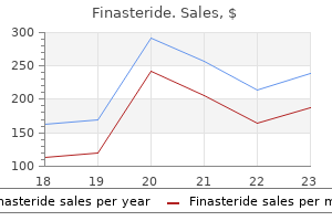
Finasteride dosages: 5 mg, 1 mg
Finasteride packs: 30 pills, 60 pills, 90 pills, 120 pills, 180 pills, 270 pills, 360 pills

Discount finasteride 1mg visa
Challenges related to hypertensive illness during being pregnant in low-income international locations. Sinusoidal fetal heart price sample after administration of nalbuphine hydrochloride: a case report. Plasma concentrations of beta-endorphin and adrenocorticotropic hormone in girls with and without childbirth preparation. Comparison of general and epidural anesthesia in elective cesarean part for placenta previa totalis: maternal hemodynamics, blood loss and neonatal end result. Lumbar epidural analgesia to improve intervillous blood flow during labor in severe preeclampsia. A comparison of paracervical block with single-shot spinal for labour analgesia in multiparous women: a randomised controlled trial. Risk components for uterine rupture and neonatal consequences of uterine rupture: a population-based research of successive pregnancies in Sweden. Comparison of automated intermittent low volume bolus with steady infusion for labour epidural analgesia. Placental transfer and neonatal results of diazepam when administered to girls simply earlier than delivery. Direct stimulation of urokinase, plasmin, and collagenase by meperidine: a possible mechanism for the power of meperidine to improve cervical effacement and dilation. A comparison of patient-controlled analgesia: fentanyl and alfentanil for labor analgesia. Clinical danger prediction for pre-eclampsia in nulliparous ladies: development of model in worldwide potential cohort. American College of Obstetricians and Gynecologists and American Society of Anesthesiologists. Labor analgesia and cesarean delivery: a person affected person meta-analysis of nulliparous girls. Breast-feeding issues after epidural analgesia for labour: a retrospective cohort research of pain, obstetrical procedures and breast-feeding practices. Secular trends in trial of labor and associated neonatal mortality and morbidity in the United States, 1995 to 2002. Use of benzodiazepines and benzodiazepine receptor agonists during pregnancy: neonatal consequence and congenital malformations. The effect of manipulation of the programmed intermittent bolus time interval and injection quantity on complete drug use for labor epidural analgesia: a randomized managed trial. Analgesia in labour and fetal acid-base steadiness: a meta-analysis evaluating epidural with systemic opioid analgesia. Labor ache administration other than neuraxial: what do we all know and where do we go subsequent Maternal and fetal results of intravenous patientcontrolled fentanyl analgesia during labour in a thrombocytopenic parturient. The impact of a fast change in availability of epidural analgesia on the cesarean supply price: a meta-analysis. Cesarean supply: a randomized trial of epidural versus patient-controlled meperidine analgesia during labor. This usually allows the clinician to reassure the mother and father that their infant looks properly and seems normal. Many severe congenital anomalies may have been recognized prenatally, their presence anticipated, and a administration plan made before delivery. If the newborn is sufficiently preterm or small for gestational age, has a major drawback identified prenatally, or is unwell. During the primary few hours after start, healthy newborns are often alert and reactive and will suck at the breast. This behavior provides an initial alternative for the mother to form an in depth attachment along with her toddler and to set up breastfeeding. It is often greatest at this stage to inquire whether there are any problems with feeding or any other worries concerning the toddler.
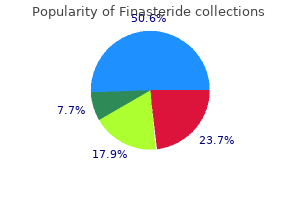
Cheap 1mg finasteride visa
Beginning posteriorly and continuing along the lateral boundary of the oor of the bony orbit is the inferior orbital ssure. Beyond the anterior end of the ssure, the zygomatic bone completes the oor of the bony orbit. Posteriorly, the orbital strategy of the palatine bone makes a small contribution to the oor of the bony orbit near the junction of the maxilla, ethmoid, and sphenoid bones. Tars al gland Superior tars al mus cle (s mooth mus cle) Eyelids the upper and lower eyelids are anterior structures that, when closed, shield the floor of the eyeball. The layers of the eyelids, from anterior to posterior, encompass skin, subcutaneous tissue, voluntary muscle, the orbital septum, the tarsus, and conjunctiva. The higher and decrease eyelids are mainly comparable in construction except for the addition of two muscles (levator palpebrae superioris and superior tarsal) within the upper eyelid. Lateral wall the lateral w all the bony orbit consists of contributions from two bones-anteriorly, the zygomatic bone and posteriorly, the greater wing of the sphenoid bone. Fractures within the orbit incessantly occur within the oor and the medial wall; nevertheless, superior and lateral wall fractures also happen. These fractures might drag the inferior indirect muscle and associated tissues into the fracture line. In these instances, sufferers may have upward gaze failure (upward gaze diplopia) in the affected eye. Medial wall fractures characteristically present air within the orbit in radiographs. This is due to fracture of the ethmoidal labyrinth allowing direct continuity between the orbit and the ethmoidal paranasal sinuses. Occasionally, sufferers feel a "full" sensation throughout the orbit when blowing their nostril. Orbicularis oculi the muscle bers encountered next in an anteroposterior direction by way of the eyelid belong to the palpebral part of orbicularis oculi. This muscle is part of the bigger orbicularis oculi muscle, which consists primarily of two parts-an orbital half, which surrounds the orbit, and the palpebral part, which is within the eyelids. The palpebral part is thin and anchored medially by the medial palpebral ligament, which attaches to the anterior lacrimal crest, and laterally blends with bers from the muscle in the decrease eyelid at the lateral palpebral ligament. Orbital septum Deep to the palpebral part of the orbicularis oculi is an extension of periosteum into each the upper and lower eyelids from the margin of the orbit. This is the orbital septum, which extends downward into the higher 468 Regional anatomy � Orbit Orbital half Palpebral part eight Orbicularis oculi mus cle Medial palpebral ligament Perios teum Orbital s eptum Tendon of levator palpebrae s uperioris mus cle Lateral palpebral ligament. Orbital s eptum eyelid and upward into the decrease eyelid and is steady with the periosteum inside and outside the orbit. The orbital septum attaches to the tendon of levator palpebrae superioris muscle within the higher eyelid and attaches to the tarsus within the decrease eyelid. Perios teum Tarsus and levator palpebrae superioris Providing major help for every eyelid is the tarsus. There is a big superior tarsus within the higher eyelid and a smaller inferior tarsus within the decrease eyelid. These plates of dense connective tissue are attached medially to the anterior lacrimal crest of the maxilla by the medial palpebral ligament and laterally to the orbital tubercle on the zygomatic bone by the lateral palpebral ligament. Associated with the tarsus in the higher eyelid is the levator palpebrae superioris muscle. In companion with the levator palpebrae superioris muscle is a group of clean muscle bers passing from the inferior surface of the levator to the upper edge of the superior tarsus. Innervated by postganglionic sympathetic bers from the superior cervical ganglion, this muscle is the superior tarsal muscle. With this membrane in place, a conjunctival sac is shaped when the eyelids are closed, and the upper and lower extensions of this sac are the superior and inferior conjunctival fornices. Blockage and in ammation of a tarsal gland is a chalazion and is on the inside floor of the eyelid.
Order finasteride no prescription
Its borders are the larger wing of the sphenoid and the maxilla, palatine, and zygomatic bones. This long ssure allows communication between: the orbit and the pterygopalatine fossa posteriorly, the orbit and the infratemporal fossa within the center, and the orbit and the temporal fossa posterolaterally. Passing via the inferior orbital ssure are the maxillary nerve [V2] and its zygomatic branch, the infra-orbital vessels, and a vein speaking with the pterygoid plexus of veins. This groove connects with the infra-orbital canal that opens onto the face at the infraorbital foramen. The infra-orbital nerve, a department of the maxillary nerve [V2], and vessels move by way of this construction as they exit onto the face. Periorbita Perios teum 8 Fas cial s heath Superior rectus mus cle Fas cial s heath Other openings Associated with the medial wall of the bony orbit are a quantity of smaller openings. The anterior and posterior ethmoidal foramina are on the junction between the superior and medial partitions. These openings provide exits from the orbit into the ethmoid bone for the anterior and posterior ethmoidal nerves and vessels. Completing the openings on the medial wall is a canal in the decrease part of the wall anteriorly. Clearly seen is the despair for the lacrimal sac fashioned by the lacrimal bone and the frontal process of the maxilla. Contained throughout the nasolacrimal canal is the nasolacrimal duct, a part of the lacrimal equipment. Orbital s eptum Sus pens ory ligament Inferior rectus mus cle Inferior oblique mus cle. Fascial specializations Periorbita the periosteum lining the bones that kind the orbit is the periorbita. It is steady at the margins of the orbit with the periosteum on the outer surface of the skull and sends extensions into the higher and decrease eyelids (the orbital septa). At the varied openings the place the orbit communicates with the cranial cavity, the periorbita is steady with the periosteal layer of dura mater. In the posterior part of the orbit, the periorbita thickens across the optic canal and the central part of the superior orbital ssure. This is the point of origin of the 4 rectus muscles and is the frequent tendinous ring. Lateral rectus mus cle Check ligament of lateral rectus mus cle Sus pens ory ligament Medial rectus mus cle Check ligament of medial rectus mus cle Inferior rectus mus cle Fascial sheath of the eyeball the fascial sheath of the eyeball (bulbar sheath) is a layer of fascia that encloses a major part of the eyeball. Additionally, as the muscles method the eyeball, the investing fascia surrounding each muscle blends with the fascial sheath of the eyeball because the muscles pass through and proceed to their point of attachment. A specialized decrease part of the fascial sheath of the eyeball is the suspensory ligament. This "sling-like" structure is made up of the fascial sheath of the eyeball and contributions from the two inferior ocular muscular tissues and the medial and lateral ocular muscular tissues. A Perios teum Inferior oblique mus cle Sus pens ory ligament Lacrimal s ac Medial check ligament Fas cial s heath Medial rectus mus cle Lateral check ligament Fas cial s heath Periorbita Lateral rectus mus cle B. These are expansions of the investing fascia masking the medial and lateral rectus muscle tissue, which attach to the medial and lateral partitions of the bony orbit and may assist in maintaining the conventional place of the eyeball: the medial examine ligament attaches immediately posterior to the posterior lacrimal crest of the lacrimal bone. The lateral check ligament attaches to the orbital tubercle of the zygomatic bone. The extrinsic muscular tissues include the levator palpebrae superioris, superior rectus, inferior rectus, medial rectus, lateral rectus, superior indirect, and inferior indirect. The intrinsic muscular tissues include the ciliary muscle, the sphincter pupillae, and the dilator pupillae. The axis of every orbit is directed barely laterally from back to entrance, but every eyeball is directed anteriorly. Therefore the pull of some muscular tissues has a quantity of results on the motion of the eyeball, whereas that of others has a single effect. The levator palpebrae superioris raises the upper eyelid and is the most superior muscle in the orbit (Table eight. Superior indirect Trochlea Levator palpebrae s uperioris Superior rectus Medial rectus Superior oblique Medial rectus Superior rectus Lateral rectus Inferior oblique A. B Lateral rectus Inferior rectus Imaging app Visualizing the muscle tissue of the eyeball Superior rectus Superior indirect Optic nerve Lateral rectus Medial rectus Inferior rectus.
Cheap finasteride 5 mg on-line
Systemic vascular resistance is decreased by isoflurane, and hypotension may end up, especially if preload is diminished. Cardiovascular results are extra much like effects with isoflurane than with halothane. The want for oxygen as part of inspired gases prevents achievement of those ranges. If ventilation is interrupted for any cause, nitrous oxide also rapidly fills the alveoli, resulting in dilution of the alveolar oxygen, producing a hypoxic mixture and fast desaturation. For these reasons, nitrous oxide is greatest limited to adjunct use throughout mask induction, with a swap to an air-oxygen mixture throughout upkeep anesthesia. As previously discussed, the entire inhalational brokers trigger neuroapoptosis in animal studies. Intravenous Agents Intravenous agents include sodium thiopental, propofol, ketamine, numerous narcotics, and benzodiazepines. Sodium thiopental is an ultrahort-acting barbiturate used primarily as an induction agent. There is a few proof that untimely neonates are extra sensitive to thiopental than older infants, presumably because of decreased binding by serum proteins. Use is limited in smaller and sicker neonates because it has negative inotropic exercise and is a peripheral vasodilator. At present, ongoing pharmacy shortages of thiopental have led to a serious decrease in utilization. Propofol is a phenol spinoff equipped as an emulsion in lipid, which in older youngsters and adults is used for induction and upkeep of anesthesia. Ketamine, a phencyclidine by-product, offers good hypnosis and amnesia and glorious analgesia. It is used rarely in adults and older children as a result of it could possibly cause a dissociative state with confusion, hallucinations, and different extreme psychological side effects. Ketamine stimulates the sympathetic nervous system and causes minimal respiratory and cardiovascular melancholy. Blood strain may enhance, and increased intracranial pressure is a priority in infants with hydrocephalus or these at risk for intraventricular hemorrhage. Ketamine may be helpful in breaking hypercyanotic spells in an infant with congenital heart illness and right-to-left shunt because it anesthetizes and will increase systemic vascular resistance. Benzodiazepines are agents that produce sedation, anxiolysis, and amnesia, however little analgesia. Midazolam is a really short-acting benzodiazepine, and it has been the most commonly used in anesthesia. Metabolism is type of entirely hepatic, and it must be anticipated that period would be prolonged by immature hepatic operate in a preterm neonate. In excessive doses, it can cause respiratory despair, though that is more common in conjunction with opioids. In the two research analyzed, infants handled with midazolam were extra sedated (as judged by varying scoring systems) in contrast with infants treated with placebo. The incidence of poor neurologic outcome was greater in the midazolam group, which no less than raises questions as to the protection of midazolam infusion in these infants. Clearance is lowest in probably the most premature infants, and it will increase with gestational age and with age after start, most likely reflecting growing hepatic maturation. Volume of distribution of fentanyl additionally seems to differ depending on gestational age and illness state. Neonates with elevated intra-abdominal pressure seem to have slower clearance of fentanyl. Slower clearance is likely a results of decreased hepatic blood circulate ensuing from the increased intra-abdominal pressure. There is a fairly extensive therapeutic vary, and even with high doses, hemodynamic stability is maintained. All narcotics are respiratory depressants, nevertheless, and with higher doses of fentanyl, prolonged respiratory despair happens, necessitating postoperative assisted air flow. Fentanyl together with a muscle relaxant has become the usual for anesthesia in premature neonates. This perception is much less properly accepted at present, and benzodiazepines or inhalational agents corresponding to isoflurane are extra generally added to fentanyl anesthesia.
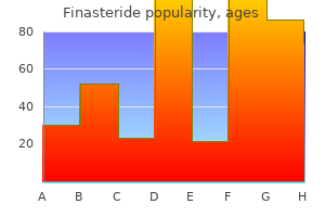
Generic finasteride 1 mg line
Because the underlying defect or genotype is commonly not recognized, phenotype in clinical apply refers to a group of particular traits, bodily findings, and the results of medical exams, corresponding to laboratory, pathologic, and radiologic studies. A major anomaly is a defect that requires important surgical or beauty intervention, such as tetralogy of Fallot or cleft lip and palate, whereas a minor anomaly has no vital surgical or cosmetic significance. The clinician should be aware that minor anomalies typically overlap with regular phenotypic variation, so a cautious seek for specific morphologic patterns is crucial. It is essential to classify an anomaly as major, minor, or regular as a outcome of implications differ for each the toddler and the household. It can additionally be important to distinguish the ideas of congenital and genetic, terms which are often confused. Congenital merely indicates that the characteristic is current at birth and might have many genetic and nongenetic causes. Anomalous external physical options are known as dysmorphisms and could be clues to the underlying cause or developmental defect. Multiple congenital anomalies in infants can be seen as a part of an association, which refers to a nonrandom occurrence of multiple malformations for which no particular or common etiology has been recognized. Although the underlying causes of congenital anomalies are heterogeneous, disruptions and isolated deformations are usually sporadic, with negligible or low recurrence risks. However, congenital malformations can even have more than one cause, typically with completely different potential related anomalies and totally different recurrence risks. Cleft lip and palate, for example, can be isolated or could be a half of dozens of various syndromes because of monogenic, multifactorial, or advanced, chromosomal, or teratogenic causes. Evaluating the newborn for a sample of main and minor anomalies will help the clinician in more efficiently figuring out the suitable tests and procedures for diagnosis and administration of a congenital anomaly, and determining future recurrence risks for family members. Most congenital malformations (86%) are isolated and not associated with different anomalies. Most isolated malformations are believed to be the consequence of multifactorial inheritance, typically referred to as complicated inheritance, occurring when a number of genetic susceptibility factors combine with environmental factors and random developmental events. From a public well being standpoint, the nongenetic results have been more durable to determine typically, though work has begun to hyperlink particular gene sequence variants to environmental factors. For example, epidemiologic studies have demonstrated that neural tube defects are related to many maternal factors, corresponding to hyperthermia, glucose ranges, and folate intake. The most typical mode of Mendelian inheritance for main malformations is autosomal dominant, with a minority of main malformations ensuing from autosomal recessive or, not often, X-linked genes. Any kind of malformation, however, could additionally be underneath the management of a single gene, including a quantity of anomalies arising in several buildings or organ methods. The mechanisms of monogenic malformation disorders are related to the dysfunction of the gene or disruption of the developmental pathway. For instance, autosomal recessive Smith-Lemli-Opitz syndrome, characterised by genital abnormalities, syndactyly of the second and third toes, ptosis, wide alveolar ridges, hypotonia, inverted nipples, and irregular fat distribution, has been discovered to be brought on by a deficiency of the enzyme 7-dehydrocholesterol reductase within the ldl cholesterol biosynthesis pathway. Neonates with these sex chromosome disorders could not have obvious malformations in the newborn interval because the phenotype might develop over time. In contrast, Turner syndrome (45,X) is present in 1 in 5000 feminine births, is usually detected prenatally, and has a phenotype in a proportion at start. Many other kinds of chromosomal aberrations have been recognized using commonplace karyotype and newer genomic applied sciences. In addition to detecting the acquire or loss of a single chromosome, routine chromosome banding strategies can determine many translocations, inversions, ring chromosomes, marker chromosomes, and deletions. In a relatively brief period of time array analysis has revolutionized the analysis of neonates and youngsters with multiple anomalies or developmental delay. Environmental Exposure and Teratogens A teratogen is anything exterior to the fetus that causes a structural or practical incapacity in prenatal or postnatal life (see Chapter 15). Teratogens could be medicine and chemical compounds, altered metabolic states within the mom, infectious agents, or mechanical forces. Known teratogenic elements trigger solely 5% to 10% of congenital anomalies despite the ever-expanding record of potential teratogens in our increasingly chemical surroundings (see Table 31-2). Dose and timing of publicity additionally alter the potential of a particular teratogenic agent. To assess for particular drug teratogenic effects, multiple sources can be found. Alcohol is thought to be the most common teratogen to which a fetus may be uncovered.
Gay-Feather (Marsh Blazing Star). Finasteride.
- What is Marsh Blazing Star?
- Kidney problems, problems with menstruation or "periods," gonorrhea, and fluid retention.
- How does Marsh Blazing Star work?
- Are there safety concerns?
- Dosing considerations for Marsh Blazing Star.
Source: http://www.rxlist.com/script/main/art.asp?articlekey=96210
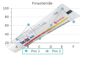
Cheap 5 mg finasteride
Principle of Operation Direct steady readings from an indwelling arterial line are thought of the gold commonplace for blood stress monitoring within the neonate. Arterial and venous pressures are normally accessed by a catheter placed within the umbilical vessels. This force can be converted to a change in voltage and calibrated to a given pressure. Indirect blood strain readings may be acquired by way of a cuff or inflatable bladder placed around the higher arm or calf. The cuff is inflated to a pressure sufficient to cause occlusion of the arterial circulate. During deflation of the cuff, measurements of diastolic and systolic blood strain can be obtained. Mean blood strain is then defined as the integrated area underneath the arterial strain waveform. The measurement and placement of the cuff can have an effect on accurate measurements of blood pressure. The American Heart Association recommends a cuff width of roughly 40% of the limb circumference. The two modes of indirect blood pressure monitoring embody the auscultatory and oscillometry methods. The auscultatory technique entails fast inflation of the cuff, followed by slow deflation whereas listening for distal Korotkoff sounds with a stethoscope. This technique, most commonly used in adults, is limited by the inaudible frequency vary of arterial sounds in neonates, intra-observer variability, and disturbance to the patient. As the pressure is slowly launched, small pulsations could be detected as the cuff approaches systolic pressure. When the cuff pressure decreases to below systolic strain the oscillations enhance in magnitude because of blood flowing into the artery. In critically sick untimely infants, oscillometric blood strain measurements have been proven to have good agreement with arterial catheter values, though accuracy is tremendously diminished in infants with a imply airway pressure less than or equal to 30 mm Hg. Blood Gas Monitoring Arterial blood fuel pattern measurements provide the most accurate estimate of arterial oxygen and carbon dioxide status. Continuous monitoring of oxygenation and carbon dioxide modalities supply an improved different by offering noninvasive, easy to use, portable, excessive resolution, and quick response options to alert the medical care provider to speedy decompensations that always occur on this high-risk toddler cohort. Even during times of supplemental oxygen making an attempt to stabilize baseline oxygenation, severity of illness compounded with immature respiratory management quite often leads to respiratory instability presenting as speedy intermittent hypoxemia occasions. A photodiode detector on the opposing facet of the electrode measures the intensity of the light passing via the extremity at every wavelength, which is equivalent to the quantity absorbed by tissue, and venous and arterial blood. Oxygen saturation values could be extrapolated from this measurement by exploiting the relatively small arterial pulsatile adjustments, also known as the plethysmogram waveform, with each heartbeat. This ratio is calculated separately for both the pink and infrared waveform alerts. The ratio of the purple (pulsatile/constant component at 660 nm) to Red 660 nm Infrared 940 nm History. The idea that pulse oximetry could be calculated from the ratios of absorption of pink and infrared mild from blood and tissue was first conceived within the early 1940s. The sign transmitted from the ear oximeter exhibited pulsatile variations prohibiting accurate measurements of cardiac output. Aoyagi devised a way to filter these oscillations by subtracting out a pulse sign detected at 900 nm similar to the infrared vary of the sunshine spectrum. In retrospect, this was most likely caused by modifications in oxygen saturation because oxygen desaturation increases infrared mild transmission while reducing red light transmission. The failure of constantly filtering out the pulsatile variations, or "noise" elements, of the dye curves led to the concept of measuring these dynamic modifications in mild transmission to compute a noninvasive estimate of arterial oxygen saturation. Initial interest and use was restricted to pulmonary operate laboratories until Jack Lloyd, founding father of Nellcor Incorporated, recognized its potential as a noninvasive technology for measuring oxygenation in unstable or severely sick patients. The principle of pulse oximetry is based on the Beer-Lambert regulation, which states that the focus of an absorbing substance in solution can be decided by the intensity of the light transmitted via the answer. The benefits of pulse oximetry are ease of use, quick response time, and steady measures of oxygen saturation. The probe requires no heating or calibration by the person and is routinely positioned on the palm of the hand or sole of the foot.
Syndromes
- Fever
- Apnea (breathing stopped)
- Fever
- CBC with differential
- Shellac
- Tuberculin skin test (also called a PPD test)
- Bloody sputum
- Bone infection
- Chest wound infection, which is more likely to happen if you are obese, have diabetes, or have already had this surgery
- Stroke
Discount generic finasteride uk
Thereafter, amniotic fluid declines by 8% per week, with a mean volume of roughly 500 mL at 40 to forty two weeks. The third nomogram found that amniotic fluid quantity continues to increase throughout gestation, confirming a imply quantity of roughly 800 mL at time period. When evaluating the well being of the amniotic fluid in twin pregnancies, singleton growth curves presently present the most effective predictors of antagonistic outcomes, and the evaluation of fluid with singleton nomograms is regularly getting used. This method appears reasonable because in the only analysis of volume in third-trimester diamniotic twin pregnancies, the amniotic fluid volume, or every sac, was just like that of regular singleton pregnancies. The incidence of main anomalies became more common in increasing polyhydramnios in each monochorionic and dichorionic pregnancies with a prevalence of simply about 20% in extreme polyhydramnios. Severe polyhydramnios was significantly associated with stillbirth in monochorionic pregnancies (27%, P <. Twin-twin transfusion syndrome carries with it a three- to fivefold improve in mortality and morbidity. Underproduction may be the end result of absent or dysfunctional kidneys, urinary tract obstruction, uteroplacental insufficiency, maternal drugs, or maternal dehydration. The clinical administration of oligohydramnios should be directed towards diagnosing and assuaging remediable underlying situations. There are conflicting information that contend that the most vital risk to the traditional fetus with incidental oligohydramnios is iatrogenic prematurity ensuing from the following rush to delivery. Late analysis of oligohydramnios in pregnancies with regular anatomy has also been discovered to be associated with undiagnosed renal anomalies as much as 9. They may also decrease uteroplacental perfusion and prematurely trigger closure of the ductus arteriosus. Although these results are reversible, sufferers maintained on indomethacin for greater than 72 hours ought to be evaluated with semiweekly amniotic fluid assessments and fetal echocardiography to assess the ductal move. Angiotensin-converting enzyme Appropriate work-up for this abnormality is a evaluation of the maternal history for evidence of rupture, anatomic evaluation of the renal system and bladder, and evaluation of placental function and fetal progress and pulmonary status. Regardless of the etiology, fetuses in pregnancies difficult by oligohydramnios are at elevated danger of antagonistic outcomes within the type of twine accidents, fetal lung hypoplasia and, if within the first and second trimesters, malformations and contractures. If only a small quantity of fluid is present, it ought to be tested with Nitrazine paper to detect the alkaline pH of amniotic fluid. Sodium chloride in the amniotic fluid crystalizes in a "ferning" sample, confirming the analysis. Cervical mucus, when applied to a slide and allowed to dry, also can create a crystalized sample, so care ought to be taken to avoid collection of cervical mucus. Frequently these examinations are negative or equivocal for causes similar to chronic leakage or contaminating blood or semen, making it difficult to conclusively diagnose persistent fluid leakage. Patients ought to be warned that if the fetus delivers within a couple of days of injection, the baby could also be stained blue. Pyridium has also been reportedly used; nonetheless, the orange to purple hue of the stained fluid is tough to distinguish from bodily fluids. The remainder of outcomes, similar to sepsis, chorioamnionitis, and abruption, was not statistically totally different amongst teams. A careful ultrasound examination with anatomic survey ought to be carried out in cases of oligohydramnios to rule out congenital anomalies, notably as a end result of renal and ureteral anomalies are the most typical anatomic reason for severe oligohydramnios in the absence of ruptured membranes. The fetal urinary system should be totally evaluated, paying shut consideration to the renal parenchyma, dimensions of the renal pelvis, and morphologic features of the fetal urinary bladder. Cardiac, skeletal, and central nervous system anomalies may coexist with major renal anomalies and should be investigated. With bilateral renal agenesis, virtual anhydramnios is current from 16 weeks onward. In addition, the fetal adrenals can become hypertrophied and resemble renal structures. In a sequence by Fisk and colleagues, suspected fetal anomalies were confirmed in 90% of patients utilizing amnioinfusion to higher visualize fetal buildings. Aspiration of a small quantity of the fluid instilled during amnioinfusion has also been used for chromosomal analysis with a 70% success price. Fortunately, unilateral renal agenesis or polycystic kidney disease not often causes important decreases in amniotic fluid. Magnetic resonance imaging can also be useful for assessing anatomy not properly seen on ultrasound. Chronic poor placental perform owing to maternal autoimmune disease, hypertension, or vasculopathy can lead to fetal progress restriction and oligohydramnios.
Purchase finasteride in united states online
The super cial temporal artery is considered one of the terminal branches and appears as an upward continuation of the exterior carotid artery; beginning posterior to the neck of mandible, it passes anterior to the ear, crosses the zygomatic means of the temporal bone, and above this level divides into anterior and posterior branches. The maxillary artery is the bigger of the 2 terminal branches of the exterior carotid artery-arising posterior to the neck of mandible, it passes through the parotid gland, continues medial to the neck of mandible and into the infratemporal fossa, and continues through this space into the pterygopalatine fossa. Veins Collecting blood from the skull, brain, super cial face, and components of the neck, the interior jugular vein. The inner jugular vein traverses the neck throughout the carotid sheath, initially posterior to the interior carotid artery, but passes to a extra lateral place farther down. It stays lateral to the frequent carotid artery by way of the rest of the neck with the vagus nerve [X] posterior and partially between the two vessels. Superficial temporal artery Pos terior auricular artery Internal jugular vein Occipital artery Internal carotid artery As cending pharyngeal artery Carotid s inus Facial artery Lingual artery External carotid artery Superior thyroid artery Thyroid gland Common carotid artery. Tributaries to each internal jugular vein include the inferior petrosal sinus, and the facial, lingual, pharyngeal, occipital, superior thyroid, and middle thyroid veins. Pharyngeal branch Nerves Numerous cranial and peripheral nerves: pass by way of the anterior triangle of the neck as they proceed to their nal vacation spot; send branches to structures in or forming boundaries of the anterior triangle of the neck; and while in the anterior triangle of the neck, send branches to close by buildings. Branches of spinal nerves in these classes include the transverse cervical nerve from the cervical plexus and the higher and lower roots of the ansa cervicalis. It begins its descent between the inner carotid artery and the internal jugular vein, lying deep to the styloid course of and the muscles related to the styloid process. Regional anatomy � Neck Hypoglos s al nerve Stylohyoid mus cle Hyoglos s us mus cle Occipital artery 8 the accessory nerve offers off no branches as it passes through the anterior triangle of the neck. As it descends, it passes outward between the interior jugular vein and inside carotid artery. At this point it passes forward, hooking around the occipital artery, across the lateral surfaces of the internal and external carotid arteries and the lingual artery, and then continues deep to the posterior belly of the digastric and stylohyoid muscles. It passes over the floor of the hyoglossus muscle and disappears deep to the mylohyoid muscle. External carotid artery Sternocleidomas toid department of occipital artery Transverse cervical nerve. The transverse cervical nerve is a branch of the cervical plexus arising from the anterior rami of cervical nerves C2 and C3. It emerges from beneath the posterior border of the sternocleidomastoid muscle, near the center of the muscle, and loops across the sternocleidomastoid to cross its anterior surface in a transverse path. Outside the cranium the vagus nerve [X] enters the carotid sheath and descends by way of the neck enclosed in this construction medial to the internal jugular vein and posterior to the interior carotid and common carotid arteries. Branches of the vagus nerve [X] as it passes via the anterior triangle of the neck embrace a motor department to the pharynx, a department to the carotid physique, the superior laryngeal nerve (which divides into exterior and internal laryngeal branches), and probably a cardiac department. It begins its descent medial to the internal jugular vein, emerging from between the internal jugular vein and internal carotid artery to cross the lateral surface of the inner jugular vein because it passes downward and backward to disappear both into or beneath the anterior border of the sternocleidomastoid muscle. These nerve bers are the superior root of the ansa cervicalis and innervate the superior stomach of the omohyoid muscle, Hypoglos s al nerve C1 C2 C3 Thyrohyoid mus cle Omohyoid mus cle (s uperior belly) Superior root of ans a cervicalis Sternohyoid mus cle Sternothyroid mus cle Omohyoid mus cle (inferior belly) Inferior root of ans a cervicalis. Internal jugular vein Pretracheal fas cia Trachea Internal jugular vein Thyrohyoid mus cle Common carotid artery Thyroid cartilage Cricoid cartilage Pyramidal lobe Thyroid gland Vagus nerve B Right recurrent laryngeal nerve Es ophagus Vertebral physique Common carotid artery 530 A. Regional anatomy � Neck and the upper parts of the sternohyoid and sternothyroid muscle tissue. Completing the loop is a direct branch from the cervical plexus containing nerve bers from the second and third cervical nerves C2 and C3. It descends either medial or lateral to the inner jugular vein before turning medially to join the superior root. At this location, the ansa cervicalis offers off branches that innervate the inferior stomach of the omohyoid, and the lower parts of the sternohyoid and sternothyroid muscles. It is continuous with the trachea below and the pharynx posterosuperiorly (see pp. Thyroid and parathyroid glands the thyroid and parathyroid glands are endocrine glands positioned anteriorly within the neck. Both glands start as pharyngeal outgrowths that migrate caudally to their nal position as development continues. The thyroid gland is a big, unpaired gland, while the parathyroid glands, often 4 in quantity, are small and are on the posterior floor of the thyroid gland. Elements of the gastrointestinal and respiratory systems the esophagus, trachea, pharynx, and larynx lie in the neck and are associated to the anterior triangles. Esophagus the esophagus is a part of the gastrointestinal system and has only a short course within the lower neck.
Finasteride 5mg visa
Services are reported on the day that the doctor sees the patient and performs the discharge companies, even when the affected person leaves the hospital on a unique day. If one other doctor is providing concurrent care, his or her providers can be reported utilizing the following hospital visit codes (99231 to 99233). Diagnosis-related groups are a kind of classification system for acute inpatient care. Patients are grouped together based mostly on an analogous medical picture and sample of useful resource use. The severity of illness is calculated from the scientific information entered into the system. Each state has established its own outlier formulation to partially compensate hospitals for these infants. These formulas differ from year to yr within every state and is usually determined by the state health care price range. This method might turn out to be extra widespread in the United States as health care reform takes maintain. Pay for efficiency (P4P) is an approach that has been used by many industries to incentivize staff and managers alike. Some question the applicability of P4P to "neonates" as a outcome of not like many sectors of the financial system in which worth is comparatively simple to measure and reward, health care is tougher, owing to scientific heterogeneity and sickness severity complexities. Nevertheless, P4P is an attractive solution for reining in health care costs that enjoys broad help across the political spectrum despite scant proof of its effectiveness. Some physicians fear the loss of autonomy beneath P4P, whereas others argue that medical apply pointers will improve autonomy by the provision of better collective knowledge upon which to base selections. The Leapfrog Group, a consortium of enormous corporations that purchases health care, has initiated many profitable P4P packages. Some research have proven that P4P improves the quality of care and affected person satisfaction, however not essentially cost effectiveness. Physicians might cherry-pick the patient or services from larger, more well-served socioeconomic groups during which outcomes are more doubtless to be higher and problems lower, due to this fact assuring greater financial rewards. The strategic design components of a successful P4P program are particular person rather than group motivators, paying the proper amount of incentive, selecting the best measures, rewarding all enhancements in high quality, and prioritizing underserved populations. If it does lead to better high quality of care, this could finally result in decrease prices. It behooves neonatologists to be proactive to ensure that P4P metrics in neonatology are developed rationally. Each is a form of capitated funds introduced and later rejected through the period of "managed care" in the Nineteen Eighties and early Nineteen Nineties. Accountable care organizations are shaped by teams of physicians and hospitals together with other health care suppliers who be a part of into an integrated community to ship high-quality care to a bunch of patients. One caveat is that any neonate may change from one category to another at any time during his or her hospitalization. The care group will usually provide care for a single illness, corresponding to diabetes or congestive heart failure. Care groups are at risk for poor performance or excessive costs, however can achieve control by lowering prices and finishing high quality measures. The group might either present the providers themselves or subcontract to different suppliers. The care group negotiates its charge from the insurer and its payments to subcontractors. It is troublesome to predict how these new fashions of health care reimbursement might work in neonatal medicine. Currently these approaches are being piloted within the care of adult patients, significantly those with chronic sickness. Neonatologists ought to stay vigilant of these newly developed fee methodologies and work intently with hospital administrators to align their interests.