Venlor dosages: 75 mg
Venlor packs: 30 pills, 60 pills, 90 pills, 120 pills, 180 pills, 270 pills, 360 pills
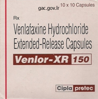
Buy venlor
Most hernias are reducible, which means they can be returned to their regular place within the peritoneal cavity by appropriate manipulation. Characteristics of direct and indirect inguinal hernias are listed and illustrated in Table B5. Normally, a lot of the processus vaginalis obliterates earlier than delivery, aside from the distal part that varieties the tunica vaginalis of the testis (see Table 5. The peritoneal part of the hernial sac of an oblique inguinal hernia is fashioned by the persisting processus vaginalis. If the whole stalk of the processus vaginalis persists, the hernia extends into the scrotum superior to the testis, forming a whole oblique inguinal hernia (Table B5. Should a hernia be present, a sudden impulse is felt against either the tip or pad of the analyzing finger when the patient is asked to cough (Swartz, 2014). With the palmar surface of the finger in opposition to the anterior abdominal wall, the deep inguinal ring could additionally be felt as a pores and skin depression superior to the inguinal ligament, 2�4 cm superolateral to the pubic tubercle. Detection of an impulse at 1036 the superficial ring and a mass on the web site of the deep ring suggests an indirect hernia. Palpation of a direct inguinal hernia is performed by inserting the palmar floor of the index and/or center finger over the inguinal triangle and asking the person to cough or bear down (strain). The finger may also be placed within the superficial inguinal ring; if a direct hernia is present, a sudden impulse is felt medial to the finger when the particular person coughs or bears down. Cremasteric Reflex Contraction of the cremaster muscle is elicited by lightly stroking the pores and skin on the medial facet of the superior part of the thigh with an applicator stick or tongue depressor. This reflex is extraordinarily energetic in kids; consequently, hyperactive cremasteric reflexes could simulate undescended testes. If the processus vaginalis remains patent in females, it might type a small peritoneal pouch (canal of Nuck), in the inguinal canal which will lengthen to the labium majus. In female infants, such remnants can enlarge and form cysts in the inguinal canal. The cysts may produce a bulge in the anterior a part of the labium majus and have the potential to develop into an oblique inguinal hernia. The fluid accumulation outcomes from secretion of an abnormal amount of serous fluid from the visceral layer of the tunica vaginalis. The measurement of the hydrocele depends on how much of the processus vaginalis persists. A congenital hydrocele of the twine and testis might talk with the peritoneal cavity. Detection of a hydrocele requires transillumination, a procedure during which a shiny gentle is utilized to the side of the scrotal enlargement in a darkened room. The transmission of light as a purple glow signifies excess serous fluid within the scrotum. Newborn male infants usually have residual peritoneal fluid of their tunica vaginalis; however, this fluid is usually absorbed through the 1st 12 months of life. Certain pathological circumstances, corresponding to damage and/or irritation of the epididymis, may produce a hydrocele in adults. Trauma could produce a scrotal and/or testicular hematoma (accumulation of blood, normally clotted, in any extravascular location). A hematocele of the testis could additionally be related to a scrotal hematocele, resulting from effusion of blood into the scrotal tissues. Torsion of Spermatic Cord Torsion of the spermatic cord is a surgical emergency because necrosis (pathologic death) of the testis may happen. The torsion (twisting) obstructs the venous drainage, with resultant edema and hemorrhage, and subsequent arterial obstruction. To prevent recurrence or occurrence on the contralateral aspect, which is in all probability going, both testes are surgically mounted to the scrotal septum. Anesthetizing Scrotum Since the anterolateral surface of the scrotum is equipped by the lumbar plexus (primarily L1 fibers through the ilio-inguinal nerve) and the postero-inferior facet is supplied by the sacral plexus (primarily S3 fibers by way of the pudendal nerve), a spinal anesthetic agent must be injected extra superiorly to anesthetize the anterolateral surface of the scrotum than is critical to anesthetize its posteroinferior surface. The appendix of the testis is a vesicular remnant of the cranial end of the paramesonephric (m�llerian) duct, the embryonic genital duct that in the female types half of the uterus.
Diseases
- Oculo tricho dysplasia
- Lagophthalmia cleft lip palate
- Capos syndrome
- Stimmler syndrome
- Hypoplastic thumbs hydranencephaly
- Glaucoma iridogoniodysgenesia
- Citrullinemia
- Metatrophic dysplasia

Buy venlor master card
Early to reasonable osteoporosis, characterized by vertical striation in the vertebral our bodies. Later, the striated pattern is lost as the continued loss of trabecular bone produces uniform radiolucency (less white, more "transparent"). Late osteoporosis within the thoracic area of the vertebral column demonstrates excessive thoracic kyphosis, a result of the collapse of the vertebral our bodies, which have turn out to be wedge formed (W), planar (P), and biconcave (B). Laminectomies are carried out surgically (or anatomically in the dissection laboratory) to gain entry to the vertebral canal, offering posterior exposure of the spinal cord (if performed above the L2 level) and/or the roots of specific 283 spinal nerves. Dislocation of Cervical Vertebrae Because of their more horizontally oriented articular sides, the cervical vertebrae are less tightly interlocked than other vertebrae. Severe dislocations, or dislocations mixed with fractures (fracture�dislocations), injure the spinal twine. The ligamentum flavum is disrupted (curved black arrow), and the spinous process is avulsed (straight black arrow). Because the taller facet of the lateral mass is directed laterally, vertical forces. Spinal cord damage is extra probably, nonetheless, if the transverse ligament has additionally been ruptured (see the scientific field "Rupture of Transverse Ligament of Atlas") indicated radiographically by broadly spread lateral masses. Fracture and Dislocation of Axis Fractures of the vertebral arch of the axis (vertebra C2) are one of the most frequent accidents of the cervical vertebrae (up to 40%) (Yochum and Rowe, 2004). In extra extreme accidents, the body of the C2 vertebra is displaced anteriorly with respect to the body of the C3 vertebra. With or without such subluxation (incomplete dislocation) of the axis, harm of the spinal twine and/or of the brainstem is likely, typically resulting in quadriplegia (paralysis of all 4 limbs) or death. Surgical treatment of lumbar stenosis may consist of decompressive laminectomy (see the medical box "Laminectomy"). This structure might differ in measurement from a small protuberance to a complete rib that happens bilaterally about 60% of the time. The supernumerary (extra) rib or a fibrous connection extending from its tip to the first thoracic rib could elevate and place stress on buildings that emerge from the superior thoracic aperture, notably the subclavian artery or inferior trunk of the brachial plexus, and may trigger thoracic outlet syndrome. Caudal Epidural Anesthesia In dwelling individuals, the sacral hiatus is closed by the membranous sacrococcygeal ligament, which is pierced by the filum terminale (a connective tissue strand extending from the tip of the spinal wire to the coccyx). In caudal epidural anesthesia or analgesia, anesthetic or analgesic agents are injected into the fat of the sacral canal that surrounds the proximal parts of the sacral nerves. The agent spreads superiorly and extradurally, the place it acts on the S2�Co1 spinal nerves of the cauda equina. The top to which the agent ascends is managed by the amount injected and the place of the affected person. Epidural anesthesia during childbirth is discussed in Chapter 6, Pelvis and Perineum. Injury of Coccyx 295 An abrupt fall onto the buttocks may cause a painful subperiosteal bruising or fracture of the coccyx, or a fracture-dislocation of the sacrococcygeal joint. Displacement is frequent, and surgical removing of the fractured bone could additionally be required to relieve pain. A troublesome syndrome, coccygodynia (or coccydynia), usually follows coccygeal trauma; ache aid is usually difficult. Abnormal Fusion of Vertebrae In approximately 5% of individuals, L5 is partly or fully included into the sacrum. When L5 is sacralized, the L5�S1 stage is powerful and the L4�L5 stage degenerates, usually producing painful signs. Longitudinal progress continues throughout adolescence, but the price decreases and is completed between ages 18 and 25. The bone loss and consequent change in form of the vertebral bodies may account partly for the slight loss in height that happens with aging. Similarly, as altered mechanics place higher stresses on the zygapophysial joints, osteophytes develop alongside the attachments of the joint capsules and accent ligaments, particularly those of the superior articular course of, whereas extensions of the articular cartilage develop around the articular sides of the inferior processes. This bony or cartilaginous progress during superior age has traditionally 298 been viewed as a disease process (spondylosis in the case of the vertebral bodies and osteoarthrosis within the case of the zygapophysial joints), but it could be extra practical to view it as an expected morphological change with age, representing normal anatomy for a specific age vary. Anomalies of Vertebrae Sometimes the epiphysis of a transverse process fails to fuse. A common delivery defect of the vertebral column is spina bifida occulta, in which the neural arches of L5 and/or S1 fail to develop normally and fuse posterior to the vertebral canal. This bony defect, current in as much as 24% of the population (Greer, 2009), normally occurs in the vertebral arch of L5 and/or S1.

Purchase venlor toronto
Addition of nutrient and microbes Sample line Valve Motor Cooling water out Impellers Temperature sensor and control unit Cooling jacket Cooling water in Valve Sparger Air in Valve Harvest line Air filter Downstream processing for mass culture of microorganisms. Such devices are geared up to add vitamins and cultures; to take away product beneath sterile or aseptic conditions; and to aerate, stir, and funky the mixture automatically. All phases of manufacturing should be carried out aseptically and monitored (usually by computer) for price of move and high quality of product. The starting uncooked substrates embrace crude plant residues, molasses, sugars, fish and meat meals, and whey. In batch fermentation, the substrate is added to the system suddenly and brought by way of a restricted run till product is harvested. In steady feed techniques, nutrients are constantly fed into the reactor and the product is siphoned off throughout the run. Some newer applied sciences make use of extremophilic archaea and their enzymes to run the processes at excessive or low temperatures or in high-salt circumstances. Hyperthermophiles have been tailored for high-temperature detergent and enzyme manufacturing. Psychrophiles are used for cold processing of reagents for molecular biology and medical exams. Pharmaceutical Products Health care merchandise derived from microbial biosynthesis embrace antibiotics, hormones, nutritional vitamins, and vaccines. The first massproduced antimicrobial was penicillin, which got here from Penicillium chrysogenum, a mildew first isolated from a cantaloupe in Wisconsin. The present pressure of this species has gone through a long time of selective mutation and screening to increase its yield. These experiences with penicillin have supplied an necessary mannequin for the manufacture of different antibiotics. Insulin is among the earliest examples of how genetic manipulation was utilized to create a bacterium that might rapidly produce mass portions of human insulin. Drugs such as Humira, Enbrel, and Remicade, used for inflammatory situations corresponding to rheumatoid arthritis, fall on this category, as do two most cancers drugs, Avastin and Herceptin. Introduction of Reactants Raw materials Pretreatment with enzymes Growth of stock culture for inoculum Nutrients added Medium sterilized Fermentation Fermentor chamber O2 pH bu er Medium collected Downstream Processing and Waste Removal Recovery of raw product Microbes recovered Miscellaneous Products Enzymes are crucial to chemical manufacturing, the agriculture and meals industries, textile and paper processing, and even laundry and dry cleansing. Mass quantities of proteases, amylases, lipases, oxidases, and cellulases are produced by fermentation expertise (see table 25. Other compounds of interest that might be mass-produced by microorganisms are amino acids, organic acids, solvents, and pure flavor compounds to be used in air fresheners and foods. Identify five industrial products made by microorganisms, and describe their applications. These general steps are followed for industrial manufacturing of drugs, enzymes, fuels, vitamins, and amino acids. The website that printed the article writes about environmental and social sustainability, and it discloses its sponsors. The intended message is that an enormous commercial firm is doing something good-providing free, clear water to millions of individuals. I would have liked more details about how the powder works or what it consists of in order that I might help explain it to others. The primary part Secondary removes bodily objects from Mixed stage Filtered the wastewater. The secondary Aerated Settled section removes the organic matter solids by biodegradation. Water high quality assays display for coliforms as indicator organisms or could assess essentially the most possible number of microorganisms. As these outcomes could additionally be deceptive, more emphasis is being positioned on identifying E. Food fermentation processes utilize micro organism or yeasts to produce desired components similar to alcohols and organic * acids in foods and drinks. Food-borne illness could be an intoxication attributable to microbial toxins produced as by-products of microbial decomposition of meals, or it could be a food an infection when pathogenic microorganisms in the meals attack the human host after being consumed.

Buy discount venlor 75mg online
However, structural rarefaction of skin capillaries, assessed with video microscopy, endured 5�15 weeks after preeclampsia, which is analogous with similar observations in essential hypertension and will offer a proof for the underlying mechanisms behind the increased long-term cardiovascular risk observed in girls who suffered from preeclampsia [12]. Therefore, prophylactic lifestyle modifications might be provided to each individuals, to scale back their cardiovascular risk [4]. Furthermore, preeclampsia itself seems to be a danger factor for several cardiovascular problems, primarily based on the evidence of impaired coronary move reserve and elevated intima-media thickness within the coronary microvasculature in ladies with out cardiovascular danger factors 5 years after preeclampsia [13]. Chronic inflammation and oxidative stress might contribute to this microvascular endothelial dysfunction in preeclampsia, assessed with transthoracic echocardiography [13]. This possibly early manifestation of inflammationdriven atherosclerosis of the coronary microvasculature might increase the risk for future coronary artery illness after preeclampsia [13]. Therefore, additional coronary threat elements Chapter 10: Microvascular Findings in Pathological Pregnancy ninety three should be modified as soon as an increased coronary artery illness danger is detected with coronary move reserve [13]. Analysis of the Microcirculation after the Onset of Preeclampsia Ex-vivo, acetylcholine-induced relaxation of subcutaneous arteries from preeclamptic ladies was attenuated, which may account for enhanced pressor sensitivity and peripheral vascular resistance and subsequently provides direct proof of endothelial dysfunction in preeclampsia [14]. This might cause endothelial cell damage within the fetal umbilical placental microcirculation with a consequent reduction in fetoplacental blood move, as a result of distinct expression of endothelial genes and adhesion molecules in addition to adenosine and nitric oxide synthase involvement [15�17]. In women diagnosed with preeclampsia, the acetylcholine-mediated microvascular response, assessed with laser Doppler flowmetry of the forearm skin, was enhanced [18]. This suggests an intact microvascular endothelial leisure response to nitric oxide in preeclampsia [18]. The elevated endothelial cell activation could relate to different elements that are released by the preeclamptic endothelium in response to acetylcholine, including enhanced vascular permeability and coagulation [18]. A comparable enhancement in capillary flux was noticed within the skin over the ankle in women with preeclampsia and gestational hypertension, using laser Doppler flowmetry without iontophoresis [1]. These postural-induced vascular modifications point out a disturbed venoarteriolar reflex, because of a persistent vasoconstriction, which is probably mediated by sympathetic overactivity [1]. Hence, the extra postural-induced vasoconstrictive stimulus may end in reflex ischemic vasodilatation, similar to vascular adjustments resulting in cutaneous edema in preeclampsia or cerebral encephalopathy in eclampsia [1, 19]. An intact venoarteriolar reflex often results in an arteriolar constriction to counteract the increased capillary filtration [19]. However, finger pores and skin laser Doppler flowmetry with out iontophoresis was not altered in preeclampsia throughout venous occlusion [20]. On the contrary, nailfold capillaroscopy with polarization spectral imaging revealed a similar disturbance in venoarteriolar reflex throughout venous occlusion, in preeclamptic ladies [20]. The discrepancy between each methods may be defined by the truth that laser Doppler flowmetry measures the move in each these nutritive capillaries and in the thermoregulatory plexus at a greater depth [20]. Several various kinds of video microscopy in numerous sites have been used to assess totally different aspects of the microcirculation in preeclamptic girls. When taking a glance at capillary density most studies utilizing intravital video microscopy discovered no or little decline in basal capillary density but a major reduction in maximal capillary density within the skin of preeclamptic ladies as in comparison with normotensive pregnant ladies [12, 21�23]. Basal capillary density represents the number of capillaries, that are open and perfused on the time of measurement, and is often referred to as useful capillary density. Maximal capillary density is obtained by moreover recruiting the nonperfused capillaries by venous ninety four Section 2: Pathological Pregnancy: Screening and Established Disease congestion. Using a noninvasive sidestream darkish subject imaging device in the sublingual microcirculation, Cornette et al. These findings are analogous to the previous observations within the skin, where variations were only observed after recruitment by venous congestion. When assessing adjustments in vessel diameters and look, refined changes in the microcirculation of preeclamptic ladies are described, corresponding to lowered conjunctival venular diameters, an increased percentage in tortuous and dilated nailfold capillaries and lowered change in venous capillary limb diameters after venous congestion [23, 25]. When assessing microvascular flow, using either capillaroscopy and polarization spectral imaging within the nailfold or sidestream darkish area imaging in the sublingual microcirculation, no differences in capillary blood cell velocity, microvascular move index or heterogeneity of flow might be noticed between preeclamptic and wholesome pregnant controls [19, 20, 24, 26]. Nevertheless, a significantly larger lower in purple cell velocity after venous occlusion suggests an impaired veno-arteriolar reflex at the nutritive stage of the skin microvasculature [20, 26]. An impaired venoarteriolar reflex of the pores and skin in ladies with preeclampsia, which apparently may be preferably demonstrated with postural laser Doppler flowmetry of the ankle or capillaroscopy of the finger, represents impaired sympathetic vasoconstriction, or may alternatively point out a down-regulation of clean muscle cell receptors by sympathetic overactivity [1, 19, 20]. The disturbed microvascular reactivity, as a outcome of an elevated peripheral resistance, seems to be a characteristic of preeclampsia, which is otherwise indicated by a delayed rise in peripheral deep body temperature after cooling of the leg [29]. Therefore, in preeclampsia, L-arginine supplementation may not be of value because of the truth that different factors, together with raised endothelin ranges, alterations in the ratio of thromboxane A2 to prostacyclin, sympathetic overactivity and elevated ranges of tumor necrosis factor-, might be responsible for the increased systemic vascular resistance, which is also reflected by lowered vasodilator response to acetylcholine [3, 30, 31].
Adderwort (Bistort). Venlor.
- Are there safety concerns?
- Are there any interactions with medications?
- How does Bistort work?
- Digestive disorders like diarrhea, mouth and throat infections, wounds, and other conditions.
- What is Bistort?
- Dosing considerations for Bistort.
Source: http://www.rxlist.com/script/main/art.asp?articlekey=96120
Order venlor cheap
Each respiratory bronchiole offers rise to 2�11 alveolar ducts, each of which gives rise to 5�6 alveolar sacs. Alveolar ducts are elongated airways densely lined with alveoli, leading to common areas, the alveolar sacs, into which clusters of alveoli open. New alveoli proceed to develop until about age eight years, by which time there are approximately 300 million alveoli. The proper and left pulmonary arteries come up from the pulmonary trunk at the level of the sternal angle; they carry lowoxygen ("venous") blood to the lungs for oxygenation. The right and left superior lobar arteries to the superior lobes arise first, earlier than entering the hilum. Continuing into the lung, the artery descends posterolateral to the primary bronchus because the inferior lobar artery of the left lung and as an intermediate artery that can divide into center and inferior lobar arteries of the best lung. The arteries and bronchi are paired in the lung, branching concurrently and working parallel programs. Likewise, a paired tertiary segmental artery and bronchus provide every bronchopulmonary phase of the lung. Usually, the artery is located on the anterior facet of the corresponding bronchus. Although the intrapulmonary relationships are accurately demonstrated, the separation of the vessels of the root of the lung has been 812 exaggerated within the hilar area to present them as they enter and leave the lung. Note that the right pulmonary artery passes beneath the arch of the aorta to attain the proper lung and that the left pulmonary artery lies completely to the left of the arch. Two pulmonary veins, a superior and an inferior pulmonary vein on all sides, carry oxygen-rich ("arterial") blood from corresponding lobes of every lung to the left atrium of the heart. Except within the central, perihilar region of the lung, the veins from the visceral pleura and the bronchial venous circulation drain into the pulmonary veins, the comparatively small volume of low-oxygen blood coming into the large quantity of oxygen-rich blood returning to the guts. Veins from the parietal pleura join systemic veins in adjacent elements of the thoracic wall. The single proper bronchial artery can also arise instantly from the aorta; however, it generally arises not directly, both by means of the proximal a part of one of the upper posterior intercostal arteries (usually the proper 3rd posterior intercostal artery) or from a standard trunk with the left superior bronchial artery. The bronchial arteries provide the supporting tissues of the lungs and visceral pleura. The bronchial veins drain the extra proximal capillary beds provided by the bronchial arteries; the remainder is drained by the pulmonary veins. Then, they sometimes move along the posterior features of the primary bronchi, supplying them and their branches as far distally as the respiratory bronchioles. The the rest of the blood is drained by the pulmonary veins, particularly the blood returning from the visceral pleura, the extra peripheral areas of the lung, and the distal components of the root of the lung. The proper bronchial vein drains into the azygos vein, and the left bronchial vein drains into the accent hemi-azygos vein or the left superior intercostal vein. The superficial subpleural lymphatic plexus lies deep to the visceral pleura and drains the lung parenchyma (tissue) and visceral pleura. Lymphatic vessels from this superficial plexus drain into the bronchopulmonary lymph nodes (hilar lymph nodes) within the region of the lung hilum. The lymphatic vessels originate from superficial subpleural and deep lymphatic plexuses. All lymph from the lung leaves alongside the root of the lung and drains to the inferior or superior tracheobronchial lymph nodes. The inferior lobe of each lungs drains to the centrally positioned inferior tracheobronchial (carinal) nodes, which primarily drain to the best aspect. The different lobes of every lung drain primarily to the ipsilateral superior tracheobronchial lymph nodes. From here, the lymph traverses a variable number of paratracheal nodes and enters the bronchomediastinal trunks. The deep bronchopulmonary lymphatic plexus is located within the submucosa of the bronchi and within the peribronchial connective tissue. It is essentially involved with draining the constructions that type the root of the lung. Lymphatic vessels from this deep plexus drain initially into the intrinsic pulmonary lymph nodes, situated along the lobar bronchi.
Purchase 75 mg venlor with amex
Pathogenesis Untreated streptococcal throat infections often may find yourself in serious problems, both instantly or days to weeks after the throat signs subside. These problems embrace scarlet fever, rheumatic fever, and glomerulonephritis. More not often, invasive and deadly conditions-such as necrotizing fasciitis, which is described in section 18. The bacterium has antigens on its floor that resemble heart, joint, and mind proteins. Eventually, though, the immune system learns to reply to these antigens and then also might attack the human analogs. Scarring and deformation change the capacity of the valves to close and shunt the blood correctly. Walker/Science Source Scarlet Fever Scarlet fever is the outcomes of an infection with an S. This lysogenic virus confers on the streptococcus the flexibility to produce erythrogenic toxin, described in the section on virulence. Scarlet fever is characterised by a sandpaper-like rash, most often on the neck, chest, elbows, and internal surfaces of the thighs. It most frequently impacts school-age youngsters and was a supply of nice suffering in the United States within the early a part of the twentieth century. Because of the worry elicited by the name "scarlet fever," the illness is usually referred to as "scarlatina" in North America. Rheumatic Fever Rheumatic fever is assumed to be due to an immunologic cross-reaction between the streptococcal M protein and coronary heart muscle. Other signs embrace arthritis in a number of joints and the looks of nodules over bony surfaces slightly below the pores and skin. Glomerulonephritis Glomerulonephritis is assumed to be the outcome of streptococcal proteins taking part in the formation of antigen-antibody complexes, which then are deposited within the basement membrane of the glomeruli of the kidney. It is characterized by nephritis (appearing as swelling in the palms and ft and low urine output), blood in the urine, increased blood stress, and occasionally heart failure. Its virulence can be enhanced by the substantial array of floor antigens, toxins, and enzymes it can generate. Specialized polysaccharides on the surface of the cell wall assist to shield the bacterium from being dissolved by the lysozyme of the host. These toxins elicit excessively robust reactions from monocytes and T lymphocytes. This is the likely mechanism for the extreme pathology of poisonous shock syndrome and necrotizing fasciitis. Extracellular Toxins Group A streptococci owe some of their virulence to the consequences of hemolysins known as streptolysins. Both hemolysins quickly injure many cells and tissues, including leukocytes and liver and heart muscle. A key toxin within the improvement of scarlet fever is erythrogenic (eh-rith-roh-jen-ik) toxin. This toxin is responsible for the brilliant purple rash typical of this disease (figure 21. Culture and Diagnosis the failure to recognize group A streptococcal infections can have devastating results. Rapid cultivation and diagnostic techniques to ensure correct therapy and prevention measures are essential. These exams are based on antibodies that react with the outer carbohydrates of group A streptococci (figure 21. A culture is generally taken concurrently the speedy swab and is plated on sheep blood agar. Group A streptococci are by far the most typical beta-hemolytic isolates in human diseases, however lately an increased variety of infections by group B streptococci (also beta-hemolytic), in addition to the existence of beta-hemolytic enterococci, have made it important to use differentiation exams. A optimistic bacitracin disc take a look at offers extra evidence for group A organisms.
Generic venlor 75mg line
The posterior and anterior nerve roots unite distal to the spinal ganglion to kind a mixed spinal nerve, which immediately divides into posterior and anterior rami. An anterior (ventral) nerve root, consisting of motor (efferent) fibers passing from nerve cell bodies in the anterior horn of spinal wire grey matter to effector organs positioned peripherally. The posterior and anterior nerve roots unite, inside or just proximal to the intervertebral foramen, to type a mixed (both motor and sensory) spinal nerve, which immediately divides into two rami (L. As branches of the mixed spinal nerve, the posterior and anterior rami carry each motor and sensory fibers, as do all their subsequent branches. The phrases motor nerve and sensory nerve are nearly always relative phrases, referring to the overwhelming majority of fiber types conveyed by that nerve. Nerves supplying muscle tissue of the trunk or limbs (motor nerves) additionally include about 40% sensory fibers, which convey pain and proprioceptive data. Conversely, cutaneous (sensory) nerves include motor fibers, which serve sweat glands and the smooth muscle of blood vessels and hair follicles. The relationship between nerves and pores and skin and muscle is established during their initial growth. After this early embryonic interval, our segmental structure is most evident in the skeleton (vertebrae and ribs) and nerves and muscle tissue of the thoracic region. Schematic illustration of the development of dermatomes (the unilateral area of skin) and myotomes (the unilateral portion of skeletal muscle) receiving innervation from single spinal nerves. Segmental distribution of myotomes 197 (B) in early limb bud stage (approximately 5 weeks) and (C) at 6 weeks. Ventrally migrating sclerotomal cells encompass the notochord, forming the beginnings of the bodies of vertebrae. Dorsally migrating sclerotomal cells surround the neural tube forming the beginnings of the neural arch of the vertebrae. The lateral side of the somites (dermatomyotomes) offers rise to the skeletal muscle tissue and dermis of the pores and skin. Cells that migrate anteriorly give rise to the hypaxial muscle tissue of the anterolateral trunk and limbs and associated dermis. Motor neurons developing throughout the anterior neural tube ship processes peripherally into the posterior and anterior regions of the dermatomyotome. Sensory neurons growing throughout the neural crests ship peripheral processes into these areas of the dermatomyotome and central processes into the posterior neural tube. Somatic sensory and motor nerve fibers which are organized segmentally alongside the neural tube become parts of all spinal nerves and some cranial nerves. The relationship between the nerves and the tissue derived from the dermatomyotome stays all through life: the unilateral space of pores and skin supplied by a single (right or left member of a pair of) spinal nerves known as a dermatome. The unilateral mass of muscle supplied by a single spinal nerve is called a myotome. Throughout life, severing a spinal nerve will denervate the realm of skin and mass of muscle it originally supplied. The strains indicating dermatomes on dermatome maps would thus be better represented by smudges or gradations of color. Dermatome maps of the physique are primarily based on an accumulation of scientific findings following spinal nerve injuries. This map relies on the research of Foerster (1933) and displays each anatomical (actual) distribution or segmental innervation and medical expertise. Another well-liked however more schematic map is that of Keegan and Garrett (1948), which is interesting for its common, extra easily extrapolated sample. Note that in the Foerster map, C5�T1 and L3�S1 are distributed almost entirely within the limbs. Posterior rami are distributed to the synovial joints of the vertebral column, deep muscles of the back, and the overlying pores and skin. The remaining anterolateral physique wall, together with the limbs, is supplied by anterior rami. Posterior (primary) rami of spinal nerves provide nerve fibers to the 202 synovial joints of the vertebral column, deep (epaxial) muscles of the again, and the overlying pores and skin in a segmental pattern. As a general rule, the posterior rami remain separate from each other (do not merge to type main somatic nerve plexuses). Anterior (primary) rami of spinal nerves supply nerve fibers to the much larger remaining area, consisting of the skin and hypaxial muscular tissues of the anterior and lateral regions of the trunk and the higher and lower limbs.
Buy venlor with paypal
Modern biotechnology includes using genetic engineering strategies to enhance or augment the naturally occurring talents of microbes. The field of biotechnology is providing lots of of applications in industry, medicine, agriculture, meals sciences, and environmental safety. Most biotechnological techniques contain the actions of micro organism, yeasts, molds, and algae that have been chosen or altered to synthesize a certain meals, drug, natural acid, alcohol, or vitamin. Many such food and industrial end merchandise are obtained via fermentation-a basic time period used right here to check with the mass, controlled culture of microbes to produce desired natural compounds. Biotechnology also contains the usage of microbes in sewage management, pollution management, metal mining, and bioremediation (Insight 25. Of course, microorganisms have lengthy been exposed to petroleum hydrocarbons that routinely seep into the surroundings. However, as we periodically witness, the sudden release of enormous amounts of oil from a supertanker accident or a nicely blowout can overwhelm the microbial biodegradative capability. In this case, far more oil washed up on the shoreline than the microbes might shortly biodegrade. In specific, there was an absence of sufficient inorganic nutrients to support the microbial development needed to eat the hydrocarbons within the oil rapidly. To overcome this limitation, inorganic nitrogen- and phosphate-containing fertilizers had been added to stimulate the expansion of the naturally occurring oildegrading microbes. No microbes have been added-they had been already there and simply wanted the added fertilizer to enable them to develop quicker. Even within the chilly, deep waters of the Gulf, microbes consumed over 90% of the oil within a month of its launch (see accompanying photo). Molecular analyses showed that various microbes, particularly Oceanospirillum and Colwellia, have been liable for the rapid biodegradation of this oil. Microbes with the correct enzymatic profile can be added to a contaminated website, or, as within the case of the Exxon Valdez, nutrients may be provided to enhance the growth of microbes that are already current and capable of degrading the contaminating compounds. Only in remote, undeveloped, or high mountain areas is that this water used in its pure type. Water supplies similar to deep wells which might be relatively clean and free of contaminants require much less treatment than these from floor sources laden with wastes. The stepwise process in water purification as carried out by most cities is proven in process figure 25. Steps 1 to 4 of the figure define what occurs to water between its natural supply and the point at which it flows by way of your faucet at residence. It entails filtration and chemical disinfection processes that make the water safe to drink. In many components of the world, the same water that serves as a source of consuming water is also used as a dump for solid and liquid wastes (figure 25. Sewage is the used wastewater draining out of homes and industries that contains a wide variety of chemicals, particles, and microorganisms. Sewage incorporates giant quantities of stable wastes, dissolved natural matter, and poisonous chemical compounds that pose a well being risk. To take away all potential well being hazards, therapy usually requires three phases: the first stage separates out giant matter; the secondary stage reduces remaining matter and can take away some toxic substances; and the tertiary stage completes the purification of the water (see the inset in course of determine 25. The latest methods use membrane bioreactors, that are combos of microbial communities and high-efficiency membranes which are rather more effective at removing contaminants. In the first part of therapy, bulkier, floating materials such as paper, plastic waste, and bottles are skimmed off. Sedimentation in settling tanks often takes 2 to 10 hours and leaves a mix wealthy in organic matter. This aqueous portion is carried right into a secondary phase of energetic microbial decomposition, or biodegradation. In this part, a various group of natural bioremediators (bacteria, algae, and protozoa) aerobically decomposes the remaining particles of wood, paper, fabrics, petroleum, and natural molecules inside a large digester tank (figure 25. This forms a suspension of fabric called sludge that tends to settle out and gradual the process.
Buy venlor 75 mg on-line
The lumbar plexus supplies innervation to the anterolateral facet of the scrotum; the sacral plexus provides innervation to the postero-inferior facet. Cremasteric fascia: derived from the investing fascia of both the superficial and deep surfaces of the interior indirect muscle. External spermatic fascia: derived from the external oblique aponeurosis and its investing fascia. The cremaster muscle reflexively draws the testis superiorly in the scrotum, particularly in response to cold. In a heat setting, such as a hot tub, the cremaster relaxes and the testis descends deeply within the scrotum. Both responses happen in an try to regulate the 1023 temperature of the testis for spermatogenesis (formation of sperms), which requires a constant temperature roughly one diploma cooler than core temperature, or during sexual exercise as a protective response. The cremaster usually acts coincidentally with the dartos muscle, easy muscle of the fatfree subcutaneous tissue of the scrotum (dartos fascia), which inserts into the skin, aiding testicular elevation because it produces contraction of the skin of the scrotum in response to the identical stimuli. The cremaster is striated muscle receiving somatic innervation, whereas the dartos is easy muscle receiving autonomic innervation. Coverings comparable to those of the spermatic cord are vague alongside the spherical ligament. Testicular artery: arising from the aorta and supplying the testis and epididymis. Pampiniform venous plexus: a network shaped by as much as 12 veins that converge superiorly as right or left testicular veins. Lymphatic vessels: draining the testis and closely associated constructions and passing to the lumbar lymph nodes. Vestige of processus vaginalis: could additionally be seen as a fibrous thread in the anterior a half of the spermatic wire extending between the belly peritoneum and the tunica vaginalis; it may not be detectable. It contains only vestiges of the lower part of the ovarian gubernaculum paralleled by remnants, if any, of the obliterated processus vaginalis. Because the dartos muscle attaches to the skin, its contraction causes the scrotum to wrinkle when cold, thickening the integumentary layer while lowering scrotal floor area and helping the cremaster muscle tissue in holding the testes nearer to the physique, all of which reduces warmth loss. The scrotum is split internally by a continuation of the dartos fascia, the septum of the scrotum, into proper and left compartments. The septum is demarcated externally by the scrotal raphe (see Chapter 6, Pelvis and Perineum), a cutaneous ridge marking the road of fusion of the embryonic labioscrotal swellings. The development of the scrotum is intently associated to the formation of the inguinal canals. The scrotum develops from the labioscrotal swellings, two cutaneous outpouchings of the anterior belly wall that fuse to form a pendulous cutaneous pouch. The lymphatic vessels of the scrotum drain into the superficial inguinal lymph nodes. Anterior scrotal nerves: branches of the ilio-inguinal nerve (L1) supplying the anterior floor. Perineal branches of the posterior cutaneous nerve of the thigh (S2, S3): supplying the postero-inferior surface. The testes are suspended in the scrotum by the spermatic cords, with the left testis normally suspended (hanging) extra inferiorly than the right testis. The distal part of the contents of the spermatic cord, the epididymis, and many of the testis are surrounded by a collapsed sac, the tunica vaginalis. The outer parietal layer traces the peritesticular continuation of the inner spermatic fascia. The tunica vaginalis is a closed peritoneal sac partially surrounding the testis, which represents the closed-off distal a part of the embryonic processus vaginalis. The visceral layer of the tunica vaginalis is closely applied to the testis, epididymis, and inferior a part of the ductus deferens. The slit-like recess of the tunica vaginalis, the sinus of the epididymis, is between the body of the epididymis and the posterolateral floor of the testis. The parietal layer of the tunica vaginalis, adjacent to the internal spermatic fascia, is more in depth than the visceral layer and extends superiorly for a short distance onto the distal part of the spermatic cord. The small quantity of fluid within the cavity of the tunica vaginalis separates the visceral and parietal layers, permitting the testis to transfer freely in the scrotum.

