
Elimite dosages: 30 gm
Elimite packs: 2 creams, 3 creams, 4 creams, 5 creams, 6 creams, 7 creams, 8 creams, 9 creams, 10 creams

30 gm elimite sale
Respiratory mucosa lining the nostril and paranasal sinuses, along with specialized olfactory epithelium at the cribriform plate, give rise to tumors of epithelial origin. About 20% of paranasal sinus malignancies are of minor salivary gland origin, of which adenoid cystic carcinomas and adenocarcinomas are commonest. Rare mesenchymal neoplasms, similar to solitary fibrous tumor, could additionally be tough to diagnose both clinically and histologically. Metastases to the cranium base from distant main tumors are uncommon, but should be thought-about, particularly when the lesion is predominantly inside the bony structures in a affected person with a prior known malignancy. Meningiomas and tumors of neural origin, such as schwannomas, also are encountered regularly, as are chordomas, which originate from notochordal remnants. In addition, a quantity of benign proliferations, such as angiofibroma, inverted papilloma, fibrous dysplasia, and pituitary adenomas, warrant dialogue. Extension laterally bows the posterior maxillary forty two Pathology of the Sinonasal Region and Anterior and Central Skull Base 549 Inverted Papilloma and Other Papillomas the histopathologic review of papillomas by Hyams remains totally valid at present,four as mentioned anew by Batsakis. Microscopically, islands of thickened epithelial cells are surrounded by edematous and inflamed stroma. The invaginated nests are composed of mature nonkeratinized squamous cells admixed with mucinous cells and mucous cysts and enveloped by a skinny basement membrane. Typical histopathologic findings embody a polypoid mass with easy or bosselated edges, and characteristically irregular delicate, thin-walled "staghorn" blood vessels embedded in a variably hyalinized collagenous stroma containing plump, stellate, or spindled fibroblasts. Pleomorphism, mitotic exercise, and necrosis are uncommon, besides in cases of infarction following preoperative embolization. The stromal cells and endothelial cells are immunoreactive with antibodies for androgen, estrogen, and progesterone receptors. Even for the ten to 20% that reach to the cranial base, endoscopic resection is usually indicated. Thus, if the affected person is near adulthood and the tumor is asymptomatic and sluggish growing, observation could additionally be preferable to irradiation. Theroundednests are enveloped by basement membranes and lack a desmoplastic hostresponse(H&E320). Note the preservation of mobile polarity, lack of cytologicatypia,andabsenceofdivisionfigures. Although histopathologic features related to the danger for malignant transformation have been sought, none have been found constantly. When the tumor is confined to the nasal cavity, ethmoid sinus, and medial maxillary wall, endoscopic surgical resection of inverted papilloma has a low recurrence price (less than 5%). Whether this enhance displays underlying tumor biologic predisposition or the technical problem of utterly resecting the attachment website is tough to discern. The recurrence fee is,35% for the few instances (10%) that stretch beyond the paranasal sinuses, corresponding to by way of the anterior skull base. Surgical resection of these, especially when recurrent, may be challenging, even with supplemental external approaches. As the anterior lobe migrates posteriorly to fuse with the infundibulum, small islands could stay alongside the trail. These ectopic foci might give rise to ectopic adenomas adjoining to the gland or throughout the sphenoid bone. They turn into symptomatic by causing either overproduction or underproduction of pituitary hormones or by compressing close by structures. Hence, with out hormonally related signs, these usually present as an alternative with signs that happen secondary to compression of adjacent constructions, such as the remaining pituitary gland (hypopituitarism), dura (headache), optic nerve or chiasm (visual area loss), oculomotor nerves (diplopia), and trigeminal divisions (facial numbness). Its cells are organized in a multilayered columnar style with ample granular eosinophilic cytoplasm. Septal Papilloma Septal papilloma occurs as an exophytic mass on the anterior nasal septum. The papillary fronds are composed of as a lot as 20 cell layers and show normal squamous maturation toward the surface epithelium.
Buy generic elimite
The mechanism and the impact on the innate immune response would set off or preactivate dendritic cells for the appropriate response. Signal 2 are the costimulatory, coinhibitory signals, and signal three are the cytokines produced by dendritic cells that guide the differentiation into the completely different T helper subtypes. This organism is believed to contribute to the pathophysiology of atopic dermatitis and nasal polyposis by its ability to induce polyclonal activation of the humoral immune response. The second signal is a group of activating or inhibiting surface molecules on dendritic cells that interact with matching receptors on T lymphocytes. Depending on the pathogen and local tissue setting encountered by the dendritic cells, different sets of cytokines are produced that set off the differentiation of the naive T lymphocyte into completely different T helper subsets. However, for Th1 and Th2, this picture has changed with the invention of the involvement of notch signaling, where it was shown that deltarelated ligands on dendritic cells contribute to the Th2 differentiation, and jagged alerts contribute to the Th1 response. We ought to stress that the last word has not but been said on this issue and that a number of the observations. Antibodies come largely in two main courses that differ by the variety of constant areas in their heavy chain (three in IgG, IgD, and IgA1 or four in IgM and IgE) and the presence or absence of a hinge area. Differences between the different antibody subtypes are in the number and site of disulfide bridges that hyperlink the 2 heavy chains or that hyperlink a heavy with a light chain. B Lymphocytes and Immunoglobulin Production Plasma cells, which are derived from activated B lymphocytes, are the supply of manufacturing of all forms of immunoglobulins. The structure of immunoglobulins is advanced and their formation even more advanced. This complexity is probably dictated by the requirement of immunoglobulins to be succesful of specifically recognize a seemingly infinite variation of epitopes in as many potential pathogens or different danger signals. Traditionally, the immunoglobulin could be seen as a Y-shaped type with two heavy chains related by disulfide bridges and two light chains additionally linked by disulfide bridges to the heavy chains. The structural parts in both heavy and light-weight chains are the constant regions, three in the heavy chain and one in the mild chain. The components that acknowledge and bind to the epitopes are contained inside the variable region of both heavy and light-weight chains. Because of a novel mechanism, this variable region is ready to recognize the pleiotropy of different epitopes. The first stage of variation stems from the totally different types of light and heavy chains. The mild chains are available two varieties, kappa () and lambda (), every with a variable and fixed domain as beforehand described. Both mild chain genes contain a number of variable domain-encoding sections and some fixed domain-encoding sections. Specific recombination involving a specialised joining region upstream of the fixed area sections permits the recombination of any of the 40 variable domain-encoding sections in a light-weight chain to mix with any of the 5 joining domain-encoding regions and single fixed encoding area, and any of the 30 variable domainencoding sections in a light-weight chain to mix with any of the four becoming a member of domain-encoding regions and matching constant domains. This second mechanism introduces a major enhance to the overall variability of immunoglobulins. Add to this a similar mechanism in the heavy chain of the immunoglobulins that mixes any of the 40 variable regions with any of the 6 joining regions and 5 constant domains, and the variation increases even further. The T cell receptor that interacts with the many different peptides presented on dendritic cells additionally has an immunoglobulin-like structure. The two chains (and) that make up the receptor are formed via recombination between a number of variable segments (70�80 for and fifty two for) and a number of joining segments (61 for and thirteen for), which together with the variation induced by faults in the becoming a member of course of introduces a range 1018. The 5 constant domains outline the totally different courses of immunoglobulins named IgG1�4, IgM, IgD, IgA1�2, and IgE. From the proximal to the more distal location, these are C, C, C three, C 1, C 1, C 2, C four, C, and C 2. Depending on what part of the immune 70 4 Immunology and the Nose: From Basic Science to Clinical I Basic Science and Patient Assessment system is activated, completely different constant regions are used and sometimes in succession from left to proper in a process called class switching. This class switching will only have an effect on the constant parts and not the epitope-binding variable domains which may be situated upstream, thus maintaining the epitope specificity of the unique immunoglobulin. It has been estimated that the general variability launched to the combination of the process described above is 1013. The constant areas of the heavy chain outline the precise subtype of immunoglobulins. As indicated above, the expression of the totally different classes of immunoglobulins is variable and depends on completely different situations.
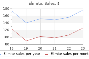
Order 30 gm elimite with mastercard
The objective of these efforts is to facilitate a recording of spontaneous sleep cycles as properly as an awake portion. Typically, nine scalp positions are used (Fl, F2, C3, C4, Cz, T3, T4, O1, O2), though others may be added. In addition, electrodes are placed at Al and A2, and a ground electrode is positioned either at mid-forehead or on a mastoid region. Because digital recordings are fundamentally referential, an extra reference electrode position may be wanted (typically noncephalic), though some devices present a so-called "inside" reference. The Fl and F2 electrode positions are 20% of the inion-nasion distance above the nasion and 10% of the circumferential measurement from the midline. Electrode placement for the neonatal electroencephalogram designated by bolded circles. After head measurement, the scalp is prepared with slight abrasion at each electrode web site with a gentle abrasive gel, utilizing the soft end of a cotton applicator. A ball of cotton is placed over every electrode, and the electrode array is lastly secured with paper tape. This is a specific problem for infants in special care models who may be in confined areas corresponding to isolettes that may focus fumes and for those infants with already compromised pulmonary function. The identical procedures are followed for electrode software as described earlier for scalp electrodes. This assists in staging sleep and within the dedication of the origin of some electrical potentials recorded in anterior cephalic electrodes that will have been generated by eye movement. In addition, limb actions may be detected and characterised by triaxial accelerometry (Frost et al. These are most frequently used in analysis protocols or particular medical circumstances. Intraarterial blood strain could be measured from an indwelling catheter already positioned for scientific P. These are finest achieved in a sustained recording through the use of a single, bipolar montage with broad coverage over the scalp. A typical montage, with sufficient protection over the scalp, including the required channels with a Cz electrode placement, is given in Table 2-2. New knowledge obtained from these recordings may lead to further pointers for neonatal recordings. Paper speed is about at 30 mm/sec for analog recordings or 10 sec/screen or "page" for digital recordings. These paper-speed settings are utilized in many laboratories within the United States and are used on this atlas. However, several laboratories, notably those with ties to the French school of recording, use a gradual pace: 15 mm/sec or 20 sec/screen or "page. However, many of the difficulties that had been present in the course of the improvement of those techniques nonetheless persist. Instrumentation, camera mount, electrode cables, and junction box must all be positioned to not intervene with the continued care of the neonate. It is also essential to maintain the video picture as free as possible from personnel and instruments that block the digital camera view. The ambient temperature of the recording environment must be controlled in addition to attainable. These considerations must be addressed with nursing workers at the onset of the recording session on the bedside and with mother and father. For a digital recording, a 7 V sign ought to produce a vertical deflection equivalent to 1/30 of the horizontal distance spanned by 1 second. A number of constraints happen in sustaining an excellent video image: Limbs with intravascular line placements may be restrained; agitated infants may be swaddled; some infants may be intubated with limited vary of head and neck movement; wound dressings could additionally be present; or infants should remain coated because of temperature instability. These and different issues have to be addressed to get hold of one of the best video picture for each research. Lighting Lighting in neonatal special care areas could be suboptimal for video recording. Ambient mild is dictated by medical needs and day/night cycles imposed for optimal toddler adaptation. When additional gentle is used to improve image high quality, extra problems may arise.
Generic elimite 30 gm free shipping
If you believe you studied acute hyponatraemia, seek urgent expert recommendation however, in an emergency, purpose toincreaseplasmaNa+by4�6mmol/Lover1�2 hours then reassess. The cause may be hypovolaemic hyponatraemia as a result of extra-renal losses (see above) but with out apparent medical manifestations of hypovolaemia. In this case, urine osmolality will be (>150 mmol/kg) because of the presence of other solutes. Consider using instruments such because the Cornell scale to help analysis and, if you suspect despair, reassess cognitive perform after a trial of anti-depressant remedy. Acute diarrhoea (<2 weeks) is normally infectious but sometimes is due to medicine or a first presentation of inflammatory bowel disease. Chronic/relapsing diarrhoea may replicate colorectal most cancers or inflammatory bowel disease, however essentially the most frequent trigger is irritable bowel syndrome. The predominant bowel habit may alternate between diarrhoea and constipation, and diagnosis relies on typical medical features (Box 9. Symptoms tend to observe a relapsing and remitting course, often exacerbated by psychosocial stress fre. Viruses and toxins predominantly have an result on the stomach and small bowel, causing large-volume, watery diarrhoea and pronounced vomiting; in toxin-mediated diarrhoea. Invasive intestinal pathogens, together with sure strains of Escherichia coli, and Shigella, may trigger bloody diarrhoea, often with extreme belly cramps and systemic upset (dysentery). Diarrhoea that persists for >10 days is unlikely to be infective, but think about protozoal infections. Supporting symptoms embody: � altered stool frequency � altered stool kind � altered stool passage (straining and/or urgency) � mucorrhoea � abdominal bloating or subjective distension. Fat malabsorption causes pale, greasy, offensive stools that float and are troublesome to flush (steatorrhoea). Other options of malabsorption embrace undigested foodstuffs in stool, weight reduction, bloating and nutritional deficiencies. Yes Stool sample for ova, cysts and parasites Discontinue drug if possible m 2 Yes k 1 Assess resuscitation necessities and illness severity. Consider dysentery, diverticulitis, ischaemic colitis, inflammatory bowel disease f C. Refer any affected person with recognized inflammatory bowel disease, extra-intestinal manifestations (see Box 9. If signs persist >10 days, search specialist advice and consider additional assessment as for chronic/relapsing diarrhoea (see below). Updated steerage on the administration and treatment of Clostridium difficile infection. No Yes Likely malabsorption disorder Investigate for trigger c No s No 5 Specific an infection risk If the stool is hard or impacted, deal with with faecal softeners and laxatives, then reassess. Expedite decrease bowel investigation to exclude colorecta cancer/inflammatory bowel disease if the affected person has persistent diarrhoea associated with any of the following: � rectalbleeding � palpablerectal/abdominalmass � weightloss � irondeficiencyanaemia � newpresentationinapatient>45 years. Consider trial discontinuation the place feasible however change only one agent at any re re. If not one of the above is current, a functional trigger, e g irritable bowel syndrome, is most likely going � notably when typical signs are current (Box 9. Faecal calprotectin ranges can assist in the differentiation between inflammatory and non-inflammatory bowel illness. Occasionally, transient alteration of consciousness or focal neurological deficit shall be described as a dizzy turn However, most sufferers with dizziness have vertigo, light-headedness/presyncope or a sensation of unsteadiness. Labyrinthine involvement (labyrinthitis) causes tinnitus and/or hearing impairment. Brief episodes of vertigo, typically lasting 10�60 seconds, are provoked by changes in head place. Acoustic neuroma this cerebellopontine angle tumour normally presents with unilateral sensorineural listening to loss. Other problems inflicting dizziness c co m Many other circumstances can cause dizziness, including hypoglycaemia, partial (temporal lobe) seizure, migraine variants, normal strain hydrocephalus, hyperventilation and anxiety. The most common presentat ons include hemiparesis, hemisensory disturbance, facial droop, speech disturbance, diplop a and monocular visible loss (amaurosis fugax). Yes Possible vertebrobasilar transient ischaemic attacks m No fre Associated migraine Consider neuroimaging if atypical options or symptoms persist >2 weeks Refer for audiometry � exclusion of acoustic neuroma Dizziness Vertigo: step-by-step assessment re the presence of any of the options in Box 10.
Order elimite in india
Its skinny bone is carefully faraway from the inside aspect of the flap, concomitantly dissecting it from the lateral mucoperiosteum and transecting it from the cranium base. Once the bone is removed, the lateral attachment of the flap to the skull base is sharply launched. Further elevation of the flap posteriorly and elimination of the basal lamella yields a posteriorly pedicled flap. The pedicle is further dissected to improve its arc of rotation and to extend its reach. It turns into much more challenging in the presence of anatomic variations, such as concha bullosa, paradoxical turbinate, or hypoplasia. The middle turbinate flap is most useful for reconstruction of small- to moderate-size defects of the planum sphenoidale, cribriform plate, or sella, however can be used to reconstruct the defects of the clivus. It is an axial flap primarily based on the supraorbital and supratrochlear arteries, which yields massive, sturdy vascularized tissue that can cover the entire skull base. However, using the pericranial flap became limited with the appearance of endoscopic cranium base techniques due to lack of ability to incorporate the externally harvested flap into the nasal cavity. By creating a method that transposes the flap through a bony window at the nasion, the pericranial flap was reintroduced into the reconstructive arsenal after endoscopic skull base surgery. A 2-cm midline incision and a 1-cm lateral port incision are made along the coronal aircraft of the scalp. The supraorbital and supratrochlear arteries are situated by Doppler ultrasound, around which a 3-cm-wide flap pedicle is marked at the level of the supraorbital rim. A subgaleal airplane dissection is performed via the vertex incisions, extending it to the vascular pedicle. The pericranium is incised using an prolonged insulated needle-tip electrocautery to the desired floor space. The pericranial flap is then elevated from the underlying bone right down to the pedicle. A subperiosteal aircraft is developed, communicating it with the subperiosteal airplane of the flap dissection. Then the bone over the nasion is drilled to form a horizontal channel that enters into the nasal cavity. Care have to be taken to not twist the flap because it passes by way of the glabellar incision and the bony window. The flap ought to be utilized to the skull base defect as it might in an open approach; the superficial surface of the flap should keep up a correspondence with the dural defect. The flap is then bolstered into place as described previously for the nasoseptal flap reconstruction. The length of the flap could be prolonged by inserting the incisions additional posterior on the scalp or by dissecting posterior to the incisions. The pericranial flap healed without any complication in three sufferers who had earlier radiation treatment. It courses via the retromandibular parotid gland, crosses the posterior root of the zygomatic course of, and becomes incorporated into the temporal fascia at the degree of the zygomatic arch. Previous incisions over the realm of the flap could alter the laterality of the harvesting. The pterygopalatine ganglion could additionally be preserved, but the vidian nerve has to be divided to permit for the displacement of the ganglion. Once sufficient surface area is uncovered, the fascia is incised at its lateral margins and elevated from the cranium and deep temporal fascia all the method down to its pedicle. The superficial layer of the deep temporal fascia is incised vertically, and the fascia is separated from the muscle following this plane of dissection inferiorly to elevate the periosteum from the floor of the zygomatic arch. This creates a large tunnel beneath the superficial layer of the deep temporalis fascia that may settle for the passage of the pedicle with out compression. This creates a tunnel that communicates the temporal, the infratemporal fossa, and the endoscopic transpterygoid strategy. This delicate tissue tunnel is sequentially dilated, advancing percutaneous tracheotomy dilators over a guidewire. After an enough tunnel is created, the dilators are removed, and the flap is tied to the external end of the guidewire. As the nasal end of the guidewire is pulled out through the nostril, it pulls the flap via the tunnel into the nasal cavity.
Buy elimite 30gm low price
If the minimize lumen of an arterial bleeder may be clearly recognized, it could also be clipped. Severe arterial hemorrhage in instances of internal carotid or internal maxillary artery damage are greatest prevented by cautious analysis of all preoperative imaging, considered use of image steerage technology, and recognizable and established endoscopic landmarks. Injury to a serious vessel, such because the carotid artery, requires quick management of the hemorrhage with packing materials in addition to placement of an intranasal Foley catheter to temporalize the bleeding. Angiography and embolization of the vessel is often thought-about the most secure and most expeditious technique of controlling the bleeding. If no arterial defect is found, repeat angiography should be carried out in 1 to 2 weeks to rule out a pseudoaneurysm. Patient components similar to hydrocephalus or weight problems, as properly as the nature of the surgical defect may contribute to a higher leak fee. Our experience suggests that almost all of low-volume leaks will resolve with conservative measures. Other authors report that these leaks hardly ever resolve with conservative measures and must be reexplored instantly. Closure of leaks might involve minor revision with autologous grafts such as fats, fascia lata, or polymers corresponding to Medpor and, very hardly ever, titanium plates. Postoperative Care Postoperative care for patients is appropriately tailored based on the pathology, extent of the method, dural involvement, reconstructive technique, and lumbar drain use. The nature of the reconstruction and packing also dictates postoperative management. Our nasal dressing consists of FloSeal for hemostasis and Telfa packs to wick minor bleeding. This contains sitting the patient up and leaning his or her torso forward for a couple of minutes to elicit any clear or serosanguinous rhinorrhea. Expanding pneumocephalus or worsening neurologic standing mandates an instantaneous endoscopic or open exploration to appropriate the pneumocephalus and seal the defect. Anticipated or minimal pneumocephalus could additionally be managed with conservative measures similar to a trial of mattress rest, head of bed elevation, stool softeners, and lumbar drainage. This is managed with postoperative serum sodium and osmolality, urine sodium, and specific gravity, in addition to cortisol levels. Perioperative antibiotics are continued for 24 hours after the surgical procedure and discontinued. Patients are typically followed by the rhinologist 1 week, 4 weeks, and three months after discharge from the hospital. At the first postoperative go to, gentle debridement of old blood, crusting, and breakdown merchandise of FloSeal and DuraSeal is performed. In addition, patients are placed on gentamicin nasal spray 3 times a day to present nasal hygiene and enhance nasal ciliary function. Patients are instructed to refrain from nostril blowing and to keep away from strenuous exercise for 10 to 14 days. Patients are evaluated for clinically important rhinosinusitis and placed on antibiotics if necessary. At the 4-four week visit, the patient undergoes extra debridement of crusting and delicate lysis of synechiae if present. Some sufferers undergo from long-term crusting due to incomplete mucociliary clearance. Careful examination of the cranium base reconstruction is also performed, and any indicators or symptoms of meningismus are elicited. The anatomic complexity of these regions represents a challenge for the endoscopic surgeon addressing pathology in these areas. A solid information of the anatomy and variations is important for profitable surgical management. Advances in endoscopic know-how, instrumentation, and anatomic knowledge have enabled surgeons to achieve successful outcomes for ailments of the pterygopalatine and infratemporal fossae. The endoscopic, endonasal, transmaxillary transpterygoid strategy to the pterygopalatine fossa, infratemporal fossa, petrous apex, and the Meckel cave. Endoscopic management of benign tumors extending into the infratemporal fossa: a two-surgeon transnasal approach. Endoscopic ligation of the sphenopalatine artery for refractory posterior epistaxis. Transoral carbon dioxide laser microsurgery for recurrent glottic carcinoma after radiotherapy.
Buy elimite 30gm on-line
Implication for Intranasal Substance Delivery Intranasal drugs are commonly utilized in treating nasal diseases and for delivering systemically appearing medicine as a outcome of the giant absorptive floor area out there in shut proximity to the nostrils. Particle trajectories and deposition websites had been calculated in the presence of steadystate inspiratory airflow at volumetric move rates of 7. When plotted against the impaction parameter, deposition efficiencies in these regions exhibited maximum values of fifty three, 20, and 3%, respectively. The deposition patterns of micron particles within the left cavity are totally different compared with the right cavity, especially within the turbinate areas. In distinction, the deposition for nanoparticles reveals a moderately even distribution of particles throughout the airway. Furthermore, the particle-releasing place clearly influences the native deposition patterns. The influence of the particlereleasing place is mainly shown near the nasal valve area for micron particle deposition, whereas for submicron particle deposition, each the nasal valve and the turbinate region are influenced. Spray pattern and plume geometry outline the form of the increasing aerosol cloud, whereas droplet size determines the probability of deposition throughout the nasal cavity by inertial impaction. A clear difference is seen in intranasal deposition between two aerosols with markedly completely different size distributions. However, not all these head positions are easily performed or nicely supported by goal knowledge. Drug penetration is elevated within the presence of inspiratory airflow versus no airflow. Physiologic Functions of the Nasal Epithelium Role of Mucociliary Functions Mucociliary clearance is the process by which cilia of the nasal epithelial cells transport the viscous mucous Physiologic Functions of the Nasal Epithelium 39 blanket of the higher airway to the gastrointestinal tract. This clearance mechanism is the first means by which the higher airway clears itself of pathogens (bacteria and viruses), allergens, particles, and toxins. Under normal situations, the cilia of the respiratory epithelial cells beat regularly in a coordinated trend. Messerklinger confirmed in his experiments how in the frontal sinus, the mucociliary circulate pattern passes superiorly along the intersinus septum, laterally along the roof, and inferomedially down the lateral wall (see Video 1, Historic Video of). Recirculation could Professor Walter Messerklinger occur within the frontal recess, with significant mucociliary flow again into the sinus. Some of the mucus, nonetheless, passes by way of the ostiomeatal pathways into either the center meatus or the superior aspect of the hiatus semilunaris. Within the maxillary sinus, mucociliary clearance spreads out in a starlike pattern from the ground, ascends alongside each wall, and passes toward the pure ostium. From right here, the drainage passes into the slender ethmoidal infundibulum, joining with the secretions from this area. Recirculation of nasal mucus may occur when secretions which have been transported out of the pure ostium return to the sinus by way of a surgically created or accent ostium. Recirculation will increase the danger of persistent sinus an infection (see Video 1, Historic Video of Professor Walter Messerklinger, and Video 2, Mucus Circulation from Inferior Meatal Antrostomy to Natural Maxillary Ostium). The flexibility of mucin fibers allows mucus gel to stick to any surface with which mucin fibers can form multiple low-affinity bonds. A modest improve in viscoelasticity can markedly inhibit the power of cilia to clear respiratory mucus. It is likely that almost all mucosal epithelia regulate the ionic surroundings, which regulates mucus hydration and therefore viscoelasticity. Other factors that contribute to the regulation of mucus viscoelasticity are secreted lipids, trefoil issue, pH, calcium, and nonmucin glycoproteins. Ciliated Columnar Cells and Cilia the respiratory mucociliary epithelium is a synchronized and extremely effective waste disposal system. It makes use of mucus as a vehicle, pushed by beating cilia, to transport unwanted particles trapped in the mucus away from the respiratory system. The capability to enhance beating in response to numerous physiologic cues is a trademark of mucociliary cells. An intricate signaling network controls ciliary activity, which relies on the interplay between calcium and cyclic nucleotide pathways. The room required for a single cilium to complete its beating cycle tremendously exceeds the spacing between neighboring cilia.
Buy elimite master card
Failure to preserve fixation on the target is evidenced by the necessity for a voluntary, corrective eye motion again in course of the goal. This indicates peripheral vestibular dysfunction on the facet to which the top was turned. Episodes might happen in bouts lasting several weeks interspersed with intervals of remission. Patients with a crescendo pattern of symptoms may have impending thrombosis and require immediate referral for imaging of the posterior circulation, e g. In these circumstances, recurrent attacks of spontaneous vertigo may be the solely presenting function. Suspect ototoxicity in any patient with newonset vertigo or different balance/hearing disturbance if gentamicin, furosemide or cisplatin has recently been prescribed. Persistent signs despite drug cessation might indicate irreversible vestibulocochlear dysfunction. Maintain this place for at least 30 seconds, asking the affected person to report any signs while observing for nystagmus. No co 104 Dizziness Assess as per Chapter 30 2 Postural symptoms + orthostatic hypotension Yes Echocardiogram Cardiac syncope sf four r Yes Document rhythm Intermittent arrhythmia No 5 Typical precipitant or prodrome No Yes Likely reflex presyncope om o Diagnosis unsure; think about other causes of dizziness If recurrent presyncope, contemplate tilt test, Holter monitor, implantable loop recorder or refer Cardiology Dizziness Light-headedness/presyncope: step-by-step evaluation ks fe Acute light-headedness may signify haemodynamic instability. Consider whether or not episodes of light-headedness occurred while the affected person was standing and had a clear precipitating set off. The key to clinching the diagnosis is to doc the rhythm during a typical episode If episodes are comparatively frequent, prepare Holter monitoring. Multiple elements could contribute to unsteadiness, particularly in the elderly, including limb weak point, sensory neuropathy, impaired proprioception, joint illness, impaired visual acuity and loss of confidence. If the patient has evidence of a couple of of those, is frail/elderly or reveals unsteadiness during strolling, assess as described for mobility issues (Ch. There is sudden onset of full dysphagia, typically with an lack of ability to swallow even saliva. The analysis is usually apparent from the history but it might be the first manifestation of an underlying stricture. Cachexia and lymphadenopathy are suggestive but physical indicators are usually absent. Unless the historical past clearly factors to a useful oropharyngeal cause, oesophageal investigation is important to rule out mechanical obstruction � specifically, malignancy. Dysphagia is of gradual onset (often years), happens for liquids and solids and may initially be intermittent. Introduction to Clinical Examination, 8th edn Edinburgh: Churchill Livingstone, 2005. To elicit the jaw jerk, ask the fe Scleroderma Oesophageal involvement is seen in ~90% of circumstances; impaired peristalsis results from alternative of muscle with fibrous tissues and extreme gastro-oesophageal reflux occurs because of incompetence of the lower oesophageal sphincter. Odynophagia is ache on swallowing and suggests oesophageal inflammation or ulceration. However, dysphagia may be the principal grievance in bulbar palsy and pseudobulbar palsy so look rigorously for the relevant medical options (Table eleven. Exclude oropharyngeal most cancers if the patient has an oropharyngeal pattern of dysphagia (see step 2) without proof of underlying neurological/ neuromuscular disorder � particularly if risk components (smoking, alcohol, human papillomavirus infection) or other suggestive scientific features. The dysphagia typically presents with rising problem in swallowing after the primary few mouthfuls and issue in chewing. Once vital pathology of the oropharynx and oesophagus is actively excluded, refer sufferers with suspected oropharyngeal dysphagia of unsure trigger to the speech and language remedy staff for an in depth swallow analysis with formal speech and � videofluoroscopy. This will verify the presence of oropharyngeal dysfunction and assist to clarify the mechanism. When evaluating a affected person with breathlessness, keep in mind that severity is extremely subjective. Some individuals could not expertise dyspnoea regardless of extreme impairment of gas change. In the initial section of evaluation, prognosis and remedy must be concurrent, and the cycle of intervention and reassessment ought to proceed until the patient is steady. The significance of respiratory signs and the prognosis of persistent bronchitis in a working inhabitants. No Yes Treat and reassess Final analysis 1 m 2 Assess effort and adequacy of oxygenation and ventilation co 114 Dyspnoea o No fr 4 Evidence of respiratory tract infection Assess the effort of respiratory by repeated medical remark of rate, depth and sample of respiration; look for use of accent muscle tissue and options of exhaustion. Distinguish acute and acute-on-chronic ventilatory failure from persistent compensated hypercapnia.