Malegra FXT Plus dosages: 160 mg
Malegra FXT Plus packs: 20 pills, 30 pills, 60 pills, 90 pills, 120 pills, 180 pills, 270 pills
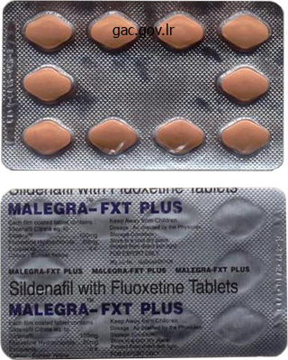
Order malegra fxt plus overnight delivery
Because lymphocytes are unable to utilize the salvage pathway of nucleotide synthesis, mycophenolate effectively blocks T- and B-cell proliferation by eliminating de novo manufacturing of guanosine monophosphate. These drugs are used as adjunctive immunosuppressant brokers, primarily with calcineurin inhibitors with or with out corticosteroids. However, mycophenolate has largely changed azathioprine on this function because of its improved security and efficacy profile. Allopurinol inhibits the metabolism of azathioprine, thereby enhancing the opposed results of azathioprine. Thus, concomitant use of allopurinol requires a big discount in azathioprine dose. Mycophenolate is available in two formulations-as a prodrug mycophenolate mofetil and as an active drug mycophenolic acid. Mycophenolate mofetil is quickly hydrolyzed within the gastrointestinal tract to mycophenolic acid. Glucuronidation of mycophenolic acid within the liver produces an inactive metabolite, however enterohepatic recirculation happens, prolonging the effect of the drug. Mycophenolic acid is an enteric-coated tablet designed to theoretically reduce the gastrointestinal upset generally experienced with mycophenolate mofetil. Corticosteroids the corticosteroids (see Chapter 26) had been the primary pharmacologic brokers to be used as immunosuppressives, both in transplantation and in various autoimmune disorders. The steroids are capable of rapidly cut back lymphocyte populations by lysis or redistribution. Consequently, efforts are being directed toward decreasing or eliminating the usage of steroids within the upkeep of allografts. Tacrolimus and cyclosporine are each calcineurin inhibitors and have the same mechanism of motion. Additionally, cyclosporine and tacrolimus are both extraordinarily nephrotoxic and when used collectively would cause harm to patients. Mycophenolate mofetil exerts its immunosuppressive motion by inhibiting inosine monophosphate dehydrogenase, thus depriving the cells of guanosine monophosphate, a key precursor of nucleic acids. Hirsutism, or extreme hair development, is a properly known opposed impact of cyclosporine. Many patients experience darkish, coarse facial or body hair growth whereas taking cyclosporine. A patient with an irregular lipid profile is a poor candidate for immunosuppression with sirolimus, since this medicine is thought to trigger or exacerbate hyperlipidemia, significantly triglycerides and whole cholesterol. Sirolimus is known to impair wound therapeutic, however a patient with a fully healed incision website might appropriately be placed on sirolimus. What further drug therapy is required for acceptable administration of this treatment Infusion-related reactions are common with the administration of antithymocyte globulins as a end result of cytokine launch. Premedication with acetaminophen, diphenhydramine, and corticosteroids should be administered 30 minutes previous to the beginning of the infusion to forestall this syndrome. Although diphenhydramine and acetaminophen are right, corticosteroids are also wanted as premedication. Overview Histamine and serotonin, along with prostaglandins, belong to a bunch of endogenous compounds known as autacoids. These heterogeneous substances have extensively differing constructions and pharmacologic actions. They all have the widespread function of being shaped by the tissues on which they act and, due to this fact, function as local hormones. The medication described in this chapter are either autacoids or autacoid antagonists (compounds that inhibit the synthesis of sure autacoids or that interfere with their interactions with receptors). Histamine, by way of multiple receptor methods, mediates a variety of cellular responses, including allergic and inflammatory reactions, gastric acid secretion, and neurotransmission in parts of the mind. Histamine has no clinical applications, but brokers that inhibit the action of histamine (antihistamines or histamine receptor blockers) have essential therapeutic applications. Release of histamine Most often, histamine is simply one of a number of chemical mediators launched in response to stimuli. The stimuli for release of histamine from tissues may embrace destruction of cells because of chilly, toxins from organisms, venoms from bugs and spiders, and trauma.
Buy generic malegra fxt plus on-line
An instance of skinny, stratified squamous epithelium with out connective tissue papillae indentation is found within the cornea of the eye; the surface underlying the epithelium is easy. This type of epithelium is only a few cell layers thick, however it has the characteristic association of basal columnar, polyhedral, and superficial squamous cells. The outermost layer of the pores and skin contains lifeless cells and known as the stratum corneum (5). Inferior to the stratum corneum (5) are the different cell layers that give rise to the stratum corneum (5). This medium-power photomicrograph illustrates the stratified squamous keratinized epithelium (1) of the palm and the cell layers stratum granulosum (6) and stratum spinosum (7) as well as the basal cell layer, stratum basale (8). The epithelium is attached to the underlying connective tissue (3) layer composed of dense collagen fibers and fibroblasts. The underlying surface of the epithelium (1) is indented by connective tissue (3) extensions referred to as papillae (2) that type the characteristic wavy boundary between the epithelium (1) and the connective tissue (3). Passing by way of the connective tissue (3) and the epithelium (1) are excretory ducts of the sweat glands (4) which may be positioned deep to the epithelium. The larger excretory ducts in the salivary glands and within the pancreas are lined with stratified cuboidal epithelium. This figure illustrates a high-power photomicrograph of a big excretory duct of a salivary gland. The luminal lining consists of two layers of cuboidal cells, forming the stratified cuboidal epithelium (1). Surrounding the excretory duct are collagen fibers of the connective tissue (2, 7) and blood vessels (3, 5) which are lined by easy squamous epithelium known as endothelium (4, 6). What modification would be finest fitted to cells transporting supplies across their surfaces This membrane, when stained with the right chemical substances, is seen with the light microscope. With the transmission electron microscope, the basement membrane consists of basal lamina and lamina reticularis. The keratin on the epithelial surface varieties a protective protect that stops abrasion and bacterial invasion. Cilia are motile constructions that line cells in respiratory tracts, uterine tubes, and efferent ducts of the testes, they usually can transfer objects across their surfaces. When the bladder is beginning to fill with urine, transitional epithelium allows for distension of the organ to accommodate extra fluid before voiding. These glands develop from epithelial cells that extend from the floor into the underlying connective tissue. Exocrine glands are linked to the surface epithelium by excretory ducts, into which their secretory products move to the exterior surface. In distinction, the endocrine glands have misplaced their connection to the floor epithelium, and their secretory merchandise are delivered immediately into the capillaries of the connective tissue that surrounds the circulatory system. The mucus-secreting goblet cells discovered within the epithelia of the small and huge intestines and in the respiratory passages are the most effective examples of unicellular glands. Multicellular glands are characterized by a secretory portion, an finish piece where the epithelial cells secrete a product, and an epithelium-lined excretory ductal portion, via which the secretion is delivered to the exterior of the gland. Larger excretory ducts are often lined by stratified epithelium, either cuboidal or columnar. Simple and Compound Exocrine Glands Multicellular exocrine glands are divided into two main classes relying on the structure of their ductal portion. A easy exocrine gland displays an unbranched duct, which can be straight or coiled. Also, if the terminal secretory portion of the gland is formed within the type of a tube, the gland is called a tubular gland. An exocrine gland that exhibits a repeated branching sample of the ducts that drain the secretory portions is known as a compound exocrine gland. Furthermore, 146 if the secretory parts of the gland are formed like a flask or a tube, the glands are referred to as acinar (alveolar) glands or tubular glands, respectively.
Purchase genuine malegra fxt plus
The microvilli are coated by a glycoprotein coat (glycocalyx), which incorporates brush border enzymes. Glands, Cells, and Lymphatic Cells and Nodules in Small Intestine Intestinal Glands Located all through the small intestine are the intestinal glands (crypts of Lieberk�hn). The easy columnar epithelium that strains the villi is continuous with that of the intestinal glands that include regenerative stem cells, absorptive cells, goblet cells, Paneth cells, and enteroendocrine cells. Intestinal Cells Absorptive cells are the most typical cells within the intestinal epithelium. A thick glycocalyx coat covers and protects the microvilli from the corrosive digestive chemical compounds. Goblet cells are interspersed among the many columnar absorptive cells of the epithelium. Duodenal (Brunner) glands are primarily discovered within the submucosa of the initial portion of the duodenum and characterize this region of the small gut. The ducts of duodenal glands penetrate the muscularis mucosae and discharge their secretory products on the base of intestinal glands positioned between the villi. Undifferentiated or stem cells are positioned at the base of intestinal glands, they usually exhibit increased mitotic exercise. These stem cells substitute all wornout columnar absorptive cells, goblet cells, and intestinal gland cells in the small gut. Lymphatic Nodules and Lymphocytic Cells Peyer patches are aggregations of carefully packed, permanent lymphatic nodules which might be discovered primarily in the wall of the terminal portion of the small intestine, the ileum. These nodules occupy a big portion of the lamina propria and submucosa of the ileum. Instead of microvilli, these cells exhibit numerous apical microfolds, hence the name "M cells. Regional Differences in Small Intestine the duodenum is the shortest section of the small intestine. Here, the villi are broad, tall, and numerous, with fewer goblet cells in the epithelium. Branched duodenal (Brunner) glands with mucus-secreting cells in the submucosa characterize this area. The jejunum is longer than the duodenum and contains the most important floor area for the absorption of the digested material. Here, the villi are tall and lined with easy columnar epithelium composed of absorptive cells and a few mucussecreting goblet cells. There are also more goblet cells in the epithelium of the jejunum than in the duodenum. The ileum contains villi which are narrow and short with the epithelium containing extra goblet cells than the duodenum or the jejunum. In addition to increased numbers of lymphocytes within the lamina propria, the aggregated lymphatic nodules (Peyer patches) are giant and most quite a few within the distal 589 ileum. Lymphatic nodules combination within the lamina propria and submucosa to kind the distinguished Peyer patches. These layers are continuous with those of the abdomen, small gut, and huge gut (colon). The small intestine is characterized by finger-like extensions, villi (7) (singular, villus); a lining epithelium (7a) of columnar cells lined with the microvilli that kind the brush borders; light-staining goblet cells (2); and short, tubular intestinal glands (crypts of Lieberk�hn) (4, 8) in the lamina propria (7b). Although the duodenal glands (3, 13) within the submucosa (13) characterize the duodenum, such glands are absent from the remainder of the small intestine (jejunum and ileum) and the big gut. Each villus (7) incorporates a core of lamina propria (7b), strands of clean muscle fibers (10) that stretch upward into the villi from the muscularis mucosae (9, 12), and a central lymphatic vessel referred to as a lacteal (11). The intestinal glands (4, 8) are located within the lamina propria (7b) and open into the intervillous areas (1). In sections of the duodenum, the submucosal duodenal glands (13) prolong into the lamina propria (3). The lamina propria (7b) also contains nice connective tissue fibers with reticular cells, diffuse lymphatic tissue, and lymphatic nodules (5). The submucosa (13) in the duodenum is almost completely full of branched, tubular duodenal glands (13).
Order generic malegra fxt plus canada
Powders are intimate mixtures of dry, finely divided drugs and/or chemical substances that might be intended for internal or external use. For instance, powdered drugs could additionally be blended with powdered fillers and other pharmaceutical elements to fabricate strong 184 dosage varieties as tablets and capsules; they may be dissolved or suspended in solvents or liquid autos to make numerous liquid dosage varieties; or they could be integrated into semisolid bases in the preparation of medicated ointments and lotions. Granules, that are prepared agglomerates of powdered supplies, could also be used per se for the medicinal worth of their content, or they may be used for pharmaceutical purposes, as in making tablets, as described later on this and Chapters 7 and eight. Granules usually fall throughout the range of 4- to 12-sieve measurement, although granulations of powders prepared in the 12- to 20-sieve range are generally utilized in tablet making. The function of particle measurement analysis in pharmacy is to acquire quantitative information on the scale, distribution, and shapes of the drug and different elements to be utilized in pharmaceutical formulations. There could additionally be substantial variations in particle dimension, crystal morphology, and amorphous character inside and between substances. Particle size can influence quite so much of important elements, together with the next: � Dissolution rate of particles intended to dissolve; drug micronization can increase the speed of drug dissolution and its bioavailability � Suspendability of particles meant to stay undissolved but uniformly dispersed in a liquid car. Laser scattering makes use of a He-Ne laser, silicon photograph diode detectors, and an ultrasonic probe for particle dispersion (range zero. Only two relatively easy examples are supplied for a detailed calculation of the average particle dimension of a powder combination. The microscopic methodology can include not fewer than 200 particles in a single plane using a calibrated ocular on a microscope. The powder is dispersed in a nonsolvent within the pipette and agitated, and 20-mL samples are removed over time. The particle diameters can be calculated from this equation: d= the place d is the diameter of the particles, h is the peak of the liquid above the sampling tube orifice, is the viscosity of the suspending liquid, i � e is the density distinction between the suspending liquid and the particles, g is the gravitational constant, and t is the time in seconds. In basic, the resulting common particle sizes by these techniques can present the average particle measurement by weight (sieve technique, light scattering, sedimentation method), and the typical particle measurement by volume (light scattering, digital sensing zone, light obstruction, air permeation, and even the optical microscope). It can simply be determined by permitting a powder to move through a funnel and fall freely onto a surface. The height and diameter of the ensuing cone are measured and the angle of repose is calculated from this equation: tan q = h/r the place h is the height of the powder cone and r is the radius of the powder cone. In general, particles within the measurement range of 250 to 2,000 m flow freely if the shape is amenable. Particles within the dimension vary of seventy five to 250 m may circulate freely or cause issues, relying on shape and different components. The closest packing could additionally be rhombus-triangle, by which angles of 60� and 120� are widespread. Another packing, cubical, with the cubes packed at 90� angles to each other, could also be thought of. The true volume, V, of a powder is the area occupied by the powder exclusive of areas greater than the intramolecular area. For some materials, a single technique may be adequate; however, a mix of methods is incessantly preferred to present higher certainty of measurement and form parameters (7). Most industrial particle size analyzers are automated and linked with computers for information processing, distribution analysis, and printout. An instance of the rise within the number of particles fashioned and the resulting surface space is as follows. Because there are six faces, that is 6 � one hundred m2, or 600 m2 surface space per particle. Since 106 particles resulted from comminuting the 1-mm cube, each 10 m on edge, the floor space now is 600 m2 � 106, or 6 � 108 m2. To get every thing in the identical items for ease of comparability, convert the 6 � 108 m2 into sq. millimeters as follows. This is extra appropriately expressed as 106 m2/mm2, 6 � 108 m2 = 6 � 102 mm2 106 m2 / mm2 the floor areas have been elevated from 6 mm2 to 600 mm2 by the reduction in particle measurement of cubes 1 mm on edge to cubes 10 m on edge, a 100-fold improve in surface area. This can have a big enhance within the rate of dissolution of a drug product. Through the grinding action of quickly transferring blades within the comminuting chamber, particles are gotten smaller and handed through a display screen of desired dimension to the gathering container. Spatulation is mixing small quantities of powders by movement of a spatula by way of them on a sheet of paper or an ointment tile. Very little compression or compacting of the powder outcomes from spatulation, which is especially suited to mixing solid substances that kind eutectic mixtures (or liquefy) when in close and extended contact with each other. Substances that form eutectic mixtures when combined embody phenol, camphor, menthol, thymol, aspirin, phenyl salicylate, and different similar chemicals.
Cheap malegra fxt plus 160 mg visa
In some cases, the enzymatic modification can change the action from bactericidal to bacteriostatic. Antibacterial spectrum the antibacterial action of the oxazolidinones is directed primarily in opposition to gram-positive organisms similar to staphylococci, streptococci, and enterococci, Corynebacterium species and Listeria monocytogenes. The major scientific use of linezolid and tedizolid is to deal with infections attributable to drug-resistant gram-positive organisms. Like different agents that intrude with bacterial protein synthesis, linezolid and tedizolid are bacteriostatic; nonetheless, linezolid has bactericidal exercise in opposition to streptococci. Pharmacokinetics Linezolid and tedizolid are properly absorbed after oral administration. Tedizolid is metabolized by sulfation, and the vast majority of elimination happens by way of the liver, and drug is mainly excreted within the feces. No dose changes are required for both agent for renal or hepatic dysfunction. Adverse effects the most common adverse effects are gastrointestinal upset, nausea, diarrhea, headache, and rash. Thrombocytopenia has been reported, often in sufferers taking the drug for longer than 10 days. Linezolid and tedizolid possess nonselective monoamine oxidase activity and should result in serotonin syndrome if given concomitantly with giant portions of tyramine-containing foods, selective serotonin reuptake inhibitors, or monoamine oxidase inhibitors. Irreversible peripheral neuropathies and optic neuritis causing blindness have been related to greater than 28 days of use, limiting utility for extended-duration therapies. Bind the 30S ribosomal subunit, interfering with meeting of the useful ribosomal apparatus. Bind irreversibly to a web site on the 50S subunit of the bacterial ribosome, inhibiting translocation steps of protein synthesis. Tetracyclines enter prone organisms by way of passive diffusion and in addition by an energydependent transport protein mechanism distinctive to the bacterial inner cytoplasmic membrane. B is the mechanism for aminoglycosides, C is the mechanism for macrolides, and D is the mechanism for oxazolidinones. Bacteremia brought on by Staphylococcus aureus Urinary tract infection attributable to Escherichia coli Pneumonia brought on by drug-resistant Streptococcus pneumoniae Diabetic foot an infection caused by Pseudomonas aeruginosa Correct reply = C. Which of the next organisms would you be involved about as the causative pathogen of diarrhea Escherichia coli Bacteroides fragilis Staphylococcus aureus Clostridium difficile Correct answer = D. Clindamycin use has been associated with Clostridium difficile�associated diarrhea. This infection must be thought-about in a patient who presents with diarrhea while on clindamycin. Azithromycin has better exercise towards respiratory pathogens such as Haemophilus influenzae and Moraxella catarrhalis but much less potent activity in opposition to staphylococci and streptococci. Erythromycin has the identical activity as azithromycin towards gram-positives and gram-negatives. Azithromycin has higher exercise towards staphylococci and streptococci compared to erythromycin. Erythromycin has higher activity in opposition to gram-positive organisms, so B and C are incorrect. Which of the next antibiotics is most likely answerable for this improve in serum creatinine Aminoglycosides corresponding to tobramycin accumulate in the proximal tubular cells of the kidney and disrupt calcium-mediated transport processes. This leads to kidney injury starting from gentle, reversible renal impairment to severe, doubtlessly irreversible acute tubular necrosis. Gentamicin crosses the placental barrier and will accumulate in fetal plasma and amniotic fluid. Gray child syndrome is an adverse effect caused by chloramphenicol in neonates due to their underdeveloped renal perform and low capability to glucuronidate the antibiotic. Gram-positive aerobes Gram-negative aerobes Gram-positive anaerobes Gram-negative anaerobes Correct reply = B. Fluoroquinolones Discovery of quinolone antimicrobials led to the development of numerous compounds utilized in clinical follow.
Buy 160mg malegra fxt plus with mastercard
The subglandular areas of the lamina propria (2, 12) might comprise both lymphatic tissue or small lymphatic nodules (16). The mucosa of the empty stomach exhibits short-term folds known as rugae (9) that form through the contractions of the muscularis mucosae (3, 15). The submucosa (4) incorporates dense irregular connective tissue and extra collagen fibers (17) than the lamina propria (2, 12). In addition, the submucosa (4) incorporates lymph vessels, capillaries (21), massive arterioles (18), and venules (19). Isolated clusters of 562 parasympathetic ganglia of the submucosal (Meissner) nerve plexus (20) could be seen deeper in the submucosa. The muscularis externa (5, 6, 7) consists of three layers of easy muscle, each oriented in a special plane: an inside indirect (5), a center round (6), and an outer longitudinal (7) layer. In this illustration, the circular layer has been sectioned longitudinally and the longitudinal layer transversely. Located between the round and longitudinal clean muscle layers is a myenteric (Auerbach) nerve plexus (22) of parasympathetic ganglia and nerve fibers. The serosa (8) consists of a thin outer layer of connective tissue that overlies the muscularis externa (5, 6, 7) and is roofed by a simple squamous mesothelium of the visceral peritoneum (8). The easy columnar floor epithelium (1, 13) extends into the gastric pits (11) into which open the tubular gastric glands (5). The lamina propria (6) fills the areas between the packed gastric glands (5) and extends from the floor epithelium (1) to the muscularis mucosae (9). The lamina propria (6) and collagen fibers are higher seen in the mucosal ridges (2). Scattered all through the lamina propria (6) are the fibroblast nuclei, lymphoid lymphatic nodule (17), lymphocytes, and different free connective tissue cells. The gastric glands (5) extend the size of the mucosa and in deeper regions the gastric glands could department. At the junction of the gastric pit with the gastric gland is the isthmus (14), lined with floor epithelial cells (1, 13) and parietal cells (4). Lower in the gland is the neck (15), containing mainly mucous neck cells (3) and some parietal cells (4). The base or fundus (16) is the deep portion of the gland, composed predominantly of chief (zymogenic) cells (7) and some parietal cells (4). The fundic glands additionally contain undifferentiated cells and enteroendocrine cells (not illustrated) that secrete different hormones to regulate the digestive system. The mucous neck cells (3) are located just under the gastric pits (11) and are interspersed between the parietal cells (4) in the neck region of the glands. The parietal cells (4) stain uniformly acidophilic (pink), which distinguishes them from other cells within the fundic glands. In distinction, the chief cells (zymogenic) (7) are basophilic and are distinguishable from the acidophilic parietal cells (4). The muscularis mucosae (9) within the stomach are composed of two skinny strips of smooth muscle: the inner round layer (9a) and outer longitudinal layer (9b). In this illustration, the internal circular layer (9a) is sectioned longitudinally, and the outer layer (9b) is sectioned transversely. Extending upward from the muscularis mucosae (9) to the floor epithelium (1, 13) are strands of smooth muscle (8, 12). The abdomen floor is lined with a mucus-secreting, simple columnar epithelium (1) that extends down into the gastric pits (2). The massive, pale-staining cells within the gastric glands (5) are the acid-secreting parietal cells (3), that are extra quite a few within the upper areas of the gastric glands (5). The pylorus is probably the most inferior, funnel-shaped region of the abdomen that terminates on the border of the small gut known as the duodenum. In the cardiac, the gastric pits are shallow, whereas in the pylorus, the gastric pits are deep.
Order 160mg malegra fxt plus with visa
In steroidsecreting cells, such as the adrenal cortex or interstitial cells within the testes, the mitochondria cristae are tubular and comprise enzymes for steroidogenesis (production of steroid hormones). Thus, cardiac or skeletal muscle cells with continuous seventy seven high-energy wants contain quite a few mitochondria, whereas cells with lowenergy needs have few mitochondria. Also, in these high-energy cells, the mitochondria exhibit large numbers of intently packed cristae, whereas in cells with low-energy metabolism, the cristae are much less extensively developed. Rough and Smooth Endoplasmic Reticulum the endoplasmic reticulum in the cytoplasm is an in depth community of sacs, vesicles, and interconnected flat tubules called cisternae. The presence of ribosomes distinguishes the tough endoplasmic reticulum, which extends from the outer membrane of the nuclear envelope to websites all through the cytoplasm. In most cells, smooth endoplasmic reticulum, which is less abundant than the rough endoplasmic reticulum, can be continuous with tough endoplasmic reticulum. In distinction, proteins for the cytoplasm, nucleus, and seventy eight mitochondria utilization are synthesized by the free ribosomes which are scattered within the cell cytoplasm. Golgi Apparatus the Golgi apparatus (also generally known as Golgi complicated or Golgi body) is also composed of a system of membrane-bound, easy, flattened, stacked, and barely curved cisternae. Near the Golgi equipment, quite a few small vesicles with newly synthesized proteins bud off from the tough endoplasmic reticulum and move to the Golgi equipment for further processing. The Golgi cisternae nearest the budding vesicles are the forming, convex, or the cis face of the Golgi apparatus. The reverse facet of the Golgi equipment is the maturing internal concave aspect or the trans face. Vesicles from the endoplasmic reticulum transfer by way of the cytoplasm to the cis side of the Golgi apparatus and bud off from the trans facet for transport of proteins to totally different sites within the cell cytoplasm. Within the Golgi cisternae are various kinds of enzymes that modify, type, and bundle proteins for various locations within the cell. As the protein molecules move by way of the different Golgi cisternae, sugars are added to the proteins and lipids to kind glycoproteins and glycolipids. Other proteins migrate to the cell membrane and are integrated into the cell membrane itself, thus contributing proteins and phospholipids to the membrane. Still other secretory granules turn into vesicles which might be crammed with a secretory product destined for exocytosis (export) to the surface of the cell. In a given cell, there are each free ribosomes and hooked up ribosomes, as seen on the endoplasmic reticulum cisternae. Ribosomes play an necessary position in protein synthesis and are most plentiful within the cytoplasm of protein-secreting cells. Ribosomes perform an essential role in decoding or translating the coded genetic messages from the nucleus for amino acid sequence of proteins which might be then synthesized by the cell. The unattached or free ribosomes synthesize proteins for use within the cell cytoplasm. In distinction, ribosomes that are attached to the membranes of the endoplasmic reticulum synthesize proteins which would possibly be packaged and saved within the cell as lysosomes or are released from the cell as secretory products. Ribosomal subunits and related proteins are first synthesized in the nucleolus and then transported to the cytoplasm via the nuclear pores. Lysosomes 80 Lysosomes are cytoplasmic organelles that contain many hydrolyzing or digestive enzymes referred to as acid hydrolases. To stop the lysosomes from digesting the cytoplasm and cell contents, a membrane separates the lytic enzymes in the lysosomes from the cell cytoplasm. The major perform of lysosomes is the intracellular digestion or phagocytosis of drugs taken into the cells. Lysosomes digest phagocytosed microorganisms, cell debris, cells, and damaged, worn-out, or excessive cell organelles, such as tough endoplasmic reticulum or mitochondria. The membrane of the lysosome then fuses with the ingested material, and their hydrolytic enzymes are emptied into the shaped vacuole. After digestion of the lysosomal contents, the indigestible debris in the cytoplasm is retained in massive membrane-bound vesicles referred to as residual bodies.
Discount 160 mg malegra fxt plus
It removes antigens, microorganisms, platelets, and aged or abnormal erythrocytes from the blood. The white pulp is the immune component of the spleen and consists of accumulated lymphocytes in the lymphatic nodules that surround the central artery or arteriole. The cells within the spleen detect trapped micro organism and antigens and initiate immune responses in opposition to them. As a outcome, T cells and B cells interact, become activated, proliferate, and carry out their immune response. The heme from the hemoglobin is additional degraded and excreted into bile by the liver cells. During fetal life, the spleen is a hematopoietic organ, producing 455 granulocytes and erythrocytes. Because it has a spongelike microstructure, a lot blood may be stored in its inside. When wanted, the stored blood is returned from the spleen to the overall circulation. As a end result, the surface of the palatine tonsil is covered by a stratified squamous nonkeratinized epithelium (1, 6) that additionally covers the remainder of the oral cavity. Each tonsil is invaginated by deep grooves referred to as tonsillar crypts (3, 9) which might be also lined by stratified squamous nonkeratinized epithelium (1, 6). Below the epithelium (1, 6) in the connective tissue are lymphatic nodules (2) distributed along the lengths of the tonsillar crypts (3, 9). The lymphatic nodules (2) incessantly merge with one another and usually exhibit lighter-staining germinal centers (7). A dense connective tissue underlies the palatine tonsil and varieties its capsule (4, 10). The connective tissue trabeculae, some with blood vessels (8), arise from the capsule (4, 10) and cross towards the floor of the tonsil between the lymphatic nodules (2). Below the connective tissue capsule (10) are sections of skeletal muscle (5) fibers. The immune system organ that reveals each an afferent and efferent lymph vessel is the: A. These cells then enter the medulla and are distributed in blood to different lymphatic sites. The primary operate of red pulp in the spleen is to filter blood and take away antigens from it. The white pulp of the spleen consists mainly of lymphocytes and macrophages across the central artery. Lymph enters the lymph nodes by way of afferent lymph vessels and leaves by way of the efferent lymph vessels within the hilum of the node. In humans, skin derivatives embrace nails, hair, and a number of other forms of sweat and sebaceous glands. Skin, or integument, consists of two distinct regions-the superficial dermis and a deep dermis. The surface layer of the pores and skin, or the dermis, is nonvascular and is lined by keratinized stratified squamous epithelium with distinct cell sorts and different cell layers. Inferior to the dermis is the vascular dermis, characterized by dense irregular connective tissue, blood vessels, nerves, and numerous glands. Beneath the dermis is the hypodermis, or a subcutaneous layer of connective tissue and adipose tissue that types the superficial fascia of gross anatomy. A basement membrane separates the dermis from the dermis during which are discovered such epidermal derivatives as the sweat glands, sebaceous glands, and hair follicles. The superficial layer of the dermis forms quite a few raised projections called dermal papillae, which interdigitate with evaginations of the epidermis, known as epidermal ridges. It incorporates free irregular connective tissue fibers, capillaries, blood vessels, fibroblasts, macrophages, and other loose connective tissue cells. This layer is thicker, is 468 characterized by dense irregular connective tissue fibers (mainly kind I collagen), and is much less mobile than the papillary layer. Also, this layer can stand up to extra mechanical stresses and supports nerves, blood vessels, hair follicles, and all the sweat glands.

