Advair Diskus dosages: 500 mcg, 250 mcg, 100 mcg
Advair Diskus packs: 1 inhalers, 2 inhalers, 3 inhalers, 4 inhalers, 5 inhalers
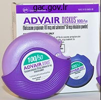
Cheap advair diskus 250mcg fast delivery
Echocardiography is normally the first examination carried out when the analysis is suspected, and will show regional wall movement abnormalities and thickening of the interventricular septum, with shiny echoes suggesting infiltration. Alternatively, the ventricles may seem thinned with international dysfunction and aneurysm formation. Diastolic dysfunction could additionally be seen in the course of the preliminary interstitial inflammatory stage when systolic perform is still normal. Patients may also current with congestive coronary heart failure, cor pulmonale, supraventricular and ventricular arrhythmias, conduction disturbances, ventricular aneurysms, pericardial effusions, mitral valve abnormalities, and sudden cardiac death. Acute myocardial inflammation ensuing from sarcoid infiltration may be seen as areas of focal thickening with increased signal intensity on T2-weighted black blood photographs. Late changes include wall thinning and delayed hyperenhancement thought to reflect chronic scarring. These modifications may be difficult to distinguish from persistent infarction, though they have a tendency to be in a noncoronary distribution and will spare the subendocardium. Echocardiography is beneficial to assess operate and focal wall motion abnormalities in typical locations for sarcoidosis, however is relatively nonspecific. Imaging Techniques and Findings Radiography Plain radiographs present no information regarding cardiac sarcoidosis. The extent of delayed hyperenhancement correlated with illness duration, ventricular operate, mitral regurgitation, and presence of ventricular tachycardia. Nuclear Medicine Thallium 201 scintigraphy myocardial perfusion studies sometimes show segmental areas of decreased uptake within the ventricular myocardium that disappear or lower in dimension throughout stress or after intravenous dipyridamole administration. Gallium 67 scintigraphy has additionally been used to present cardiac and extracardiac disease, for follow-up of energetic illness, and as a guide for potential sites for biopsy. More lately, Tc 99m sestamibi has been used as a perfusion agent, with a reverse distribution similar to that described in thallium. According to the Japanese pointers, 8 of 21 sufferers were identified with cardiac sarcoidosis. Angiography Coronary angiography is usually normal in patients with cardiac sarcoidosis. Cardiac amyloidosis typically has a poor prognosis, with a median survival of 13 months for sufferers presenting with major amyloidosis. Oral chemotherapy, together with melphalan and prednisone, has proven limited benefits to sufferers with cardiac involvement. Stem cell transplantation has shown promising outcomes for remedy of primary amyloidosis; nevertheless, the mortality associated with transplantation is 5 instances larger in amyloidosis compared with different hematologic malignancies. Medical remedy for endomyocardial eosinophilic disease consists of anticoagulation, diuretics, and digitalis. Corticosteroids, hydroxyurea, cytotoxic medicine, and imatinib all have been employed, with variable outcomes. Histologically, these patients have interstitial fibrosis with elevated amounts of collagen, glycoprotein, triglycerides, and ldl cholesterol within the myocardial interstitium. Differential analysis between constriction and restriction in these patients is particularly difficult because radiation may also induce pericardial fibrosis and constriction. Endocardiectomy has been carried out in patients with eosinophilic endomyocardial illness, with relatively excessive operative mortality. On imaging, visualization of a thickened pericardium, typically with focal distortion of the ventricular contour and atrial enlargement, permits confident analysis of constrictive pericarditis. Etiologies embody amyloidosis, eosinophilic endomyocardial illness, siderotic cardiomyopathy, sarcoidosis, radiation, storage illnesses, diabetes, and idiopathic. Amyloid coronary heart illness: new frontiers and insights in pathophysiology, diagnosis, and management. Frequency and distribution of senile cardiovascular amyloid: a clinicopathologic correlation. Prognostic significance of Doppler measures of diastolic function in cardiac amyloidosis: a Doppler echocardiography study. Echocardiographic findings in systemic amyloidosis: spectrum of cardiac involvement and relation to survival. Prognostic significance of ultrasound myocardial tissue characterization in patients with cardiac amyloidosis. Detection of left ventricular systolic dysfunction in cardiac amyloidosis with pressure fee echocardiography.
B6 (Pyridoxine (Vitamin B6)). Advair Diskus.
- Preventing another stroke.
- Improving thinking and memory in people aged 65 and older, when used in combination with folic acid and vitamin B12.
- Preventing reblockage of blood vessels after angioplasty, boosting the immune system, muscle cramps, eye problems, kidney problems, night leg cramps, arthritis, allergies, asthma, attention deficit-hyperactivity disorder (ADHD), Lyme disease, and other conditions.
- What other names is Pyridoxine (vitamin B6) known by?
- Autism.
- Treatment and prevention of pyridoxine deficiency.
Source: http://www.rxlist.com/script/main/art.asp?articlekey=96897
Purchase advair diskus toronto
Eventually, the lesion development is sufficient to overload the compensatory dilation, and lumen encroachment occurs (not shown). Reactive oxygen species induce necrosis and apoptosis, resulting in a necrotic core. Inflammatory cells promote cytokine and development issue release that stimulates fibrous cap formation. Risk Factors the risk factors for atherosclerosis are similar across the multiple arterial beds affected, whatever the end-organ perfused. They fall into two classes: those which are modifiable and people beyond our management. Modifiable danger elements could be further broken down into these which would possibly be predominantly a results of life-style indiscretions and those which are primarily manifestations of medical disease that can be treated (Table 88-1). The atherosclerotic process occurs in a stepwise style over time, and those with advanced age are extra likely to have a better burden and larger complexity of illness. Data from the Framingham research show that 7% to 9% of people seventy five years of age or older have carotid stenoses of 50% or extra. However, with the rising number of female smokers and disproportionate prevalence and rate of increase in weight problems, these gender differences are narrowing. For occasion, black populations have a 38% larger incidence than do white populations of ischemic stroke and stroke mortality adjusted for risk elements. This is evident from studies of widespread carotid artery wall thickness and belly calcification, during which familial factors contribute 64% to 92% and 50% of the variation, respectively. The majority of isolated riskassociated genes to date modulate other recognized cardiovascular danger elements somewhat than the atherosclerotic course of itself. Genes that work independently of known comorbid circumstances are the subject of intense ongoing analysis. The proposed mediators of this increased danger include immune advanced deposition; increased fibrinogen, von Willebrand factor, and other procoagulants; larger lipoprotein levels from glucocorticoid remedy; and direct vascular damage with endothelial cell progenitor cell depletion. Modifiable Risk Factors Many of the identified modifiable risk elements have wellestablished interactions with the pathophysiologic processes of noncoronary atherosclerosis. The black population has a higher fee of atherosclerosis than the white population does. Smoking Diabetes Hypertension Hypercholesterolemia Hyperhomocysteinemia C-reactive protein 0. Lipoxygenase also will increase free radical production and subsequently reduces nitric oxide formation. Homocysteine decreases nitric oxide availability in addition to its direct toxicity to the endothelium and its prothrombotic effects. The Edinburgh Artery Study specifically addressed the differential odds ratios by measuring threat factors and analyzing the prevalence of those two conditions in 1592 subjects both with and with no history of tobacco use. Increased levels of C-reactive protein promote apoptosis and stimulate procoagulant tissue factors, leukocyte adhesion molecules, and inhibitors of fibrinolysis. The hyperglycemia, insulin resistance, and fatty acid production related to diabetes reduce the bioavailability of nitric oxide, decreasing vasodilation and permitting increased clean muscle cell proliferation and platelet activation. Finally, diabetes will increase procoagulant tissue issue and fibrinogen manufacturing, resulting in a hypercoagulable state. Triglyceride-rich lipoproteins stimulate clean muscle cell proliferation and extracellular matrix deposition. This risk factor complex leads to a low-grade inflammatory state with increased levels of C-reactive protein, tumor necrosis factor, and fibrinogen. Moreover, each element of the metabolic syndrome independently increases atherosclerotic danger. Adipose tissue worsens insulin sensitivity and causes a system-wide proinflammatory state. Persistent hyperglycemia from insulin resistance and the high coprevalence of diabetes mellitus result in advanced glycation end-products that trigger extra arterial irritation. Both physical inactivity and weight problems have been shown to increase C-reactive protein levels and to cause endothelial dysfunction. They additionally worsen many different illness states that independently enhance the danger of illness. However, novel contributors of risk, especially these estimating inflammation, similar to high-sensitivity C-reactive protein, lipoprotein(a), and homocysteine, are difficult these current paradigms.
Buy advair diskus in united states online
In these cases, it may be necessary to improve the time between injection and image acquisition to optimize the distinction between myocardial and background activity. When inside the myocyte, ammonia N thirteen is integrated into the glutamine pool and turns into metabolically trapped. This heterogeneous retention might lead to an obvious perfusion defect in the inferolateral wall, limiting its analysis. Ammonia N thirteen allows the acquisition of ungated and gated images of wonderful high quality. Gated ammonia N 13 imaging can provide correct assessments of regional and global cardiac operate. The relaxation and stress emission photographs ought to each be corrected with its personal devoted transmission scan due to identified adjustments in cardiac and pulmonary volumes during pharmacologic stress. Emission Scans For rubidium eighty two, approximately the identical dose (40 to 60 mCi) is injected for the remaining and stress myocardial perfusion research because of the brief physical half-life of rubidium 82 (76 seconds). Some laboratories perform stress imaging first as a outcome of a standard scan could avoid the need for rest imaging. Imaging begins 90 to a hundred and twenty seconds after rubidium eighty two injection, or three to 5 minutes after ammonia N thirteen injection, to allow for clearance of radioactivity from the lungs and blood pool; the scan period is roughly 5 minutes for rubidium 82 or 20 minutes for ammonia thirteen N. Imaging begins with the bolus (short infusion) of rubidium 82 or ammonia thirteen N and continues for 7 to 8 minutes or 20 minutes. The advantage of this method is that it permits quantification of myocardial blood circulate (in mL/min/g) by becoming regional tissue and blood timeactivity curves to a suitable kinetic model. Fluorine 18 has a bodily half-life of 109 minutes, allowing regional distribution to clinical sites from native cyclotrons. Although regular myocardium preferentially makes use of free fatty acids, ischemic myocardium switches to preferential glucose metabolism. Because the latter is a poor substrate for additional metabolism and is impermeable to the cell membrane, it turns into virtually trapped in the myocardium, permitting the imaging of viable cells. This is the best strategy because a single injection and knowledge acquisition allows multiple picture reconstructions. With this approach, picture acquisition begins with the bolus injection of the radionuclide and continues for 7 to 8 minutes for rubidium eighty two or 20 minutes for ammonia N thirteen. List mode imaging requires significant computer power to perform the multiple reconstructions, particularly for the three-dimensional image acquisition mode. Stress testing is mostly performed utilizing adenosine, dipyridamole, or dobutamine. Quality control measures, together with routine inspection of the transmission and emission data and the transmission-emission alignment, should always be enforced to obtain high-quality studies. The level of statistical noise within the emission and transmission images should be checked to see if sufficient counts have been acquired; that is crucial with rubidium eighty two imaging due to the short half-life of the isotope. The most common sources of count-poor emission research embody massive patient size, inadequate radionuclide dose or supply. B, the correction of the emission-transmission misalignment (right), and the resulting normal perfusion examine. Acquisition of emission images earlier than full clearance of radiotracer from the blood pool could doubtlessly degrade image high quality. The optimal prescan delay for acquisition of ammonia thirteen N images is about 3 minutes and for rubidium 82 images is approximately ninety seconds in healthy subjects. In these medical settings, increased prescan delay is normally required to improve picture high quality. Misregistration of transmission and emission pictures can result from respiratory or affected person movement, and may result in inaccurate clinical outcomes. The extent and direction of this misalignment determine whether or not artifacts are obvious within the attenuation-corrected photographs. The corresponding common optimistic and adverse predictive values are 94% (range 80% to 100%) and 73% (range 36% to 100%), and the general diagnostic accuracy is 90% (range 84% to 98%).

Buy advair diskus 500mcg free shipping
However, this practice can lead to loss of useful counts in patients with quick blood pool clearance or loss of picture quality in sufferers with poor myocardial perform or perfusion. Thus, permitting a flexible delay permits more correct comparison between the 2 picture sets. In this mode, the responding detectors and time of each event are stored in a very giant information file. This is another advance made possible by elevated computing energy and it permits a retrospective determination as to which counts must be mixed to type an image. The information assortment can start on the time of injection and later parsed to create optimum images. However, its flexibility leads to more constant highquality imaging at the price of including two steps within the picture processing protocol. Therefore, the best flexibility in the study is achieved with an inventory mode acquisition, followed by initial fast reconstruction of photographs at predefined times, then evaluation and a second reconstruction with optional timing. The primary distinction between the 2 sequences is that in sinogram mode acquisition, a second resting injection is required with electrocardiographic trigger if gated info is desired. The list mode acquisition shops the electrocardiographic triggers in the occasion listing, which might then be binned retrospectively. This acquisition characteristic reduces scan time and saves radiation dose to the patient. Note that an additional injection is required to acquire gated knowledge in contrast with the list mode protocol. For instance, pharmacologic stress with use of regadenoson is delivered in a 10-second bolus before the stress radiotracer infusion. The knowledge are histogrammed right into a specified variety of frames, and areas are drawn over the left myocardium and ventricular cavity. These regions are utilized identically to all frames and plotted over time to assemble a time-activity curve. The time that the blood pool focus drops to one-half the myocardial focus is recognized and set as the scan delay. The record mode information are then rehistogrammed by starting at the delay point to reconstruct static and gated pictures. This is the "optimal timing" advantage made potential by record mode knowledge collection. Highcontrast pictures can be obtained by ready for the blood pool to attain half of the myocardium exercise concentration. Therefore, this protocol consists of a resting perfusion research followed by a glucose metabolism examine with elec- trocardiographic monitoring. As previously talked about, the mobilization of glucose transporters is promoted by shifting the myocardium from utilizing fatty acids to glucose by administration of a glucose load. Plasma glucose ranges have to be continuously monitored to keep the myocardium in the preferential glucose state (<140 mg/dL). Patients with diabetes mellitus current a selected problem due to their incapability to reliably produce endogenous insulin and their reduced cell response to exogenous insulin stimulation. This might lead to troubles in stabilizing blood glucose levels throughout uptake after infusion and through imaging. The transaxial data are then reoriented alongside the short axis and imported into the user-desirable show software for interpretation. The following quality control procedures are commonplace for several producers and found in a quantity of tips, such as these of the American Society of Nuclear Cardiology. It will also set up the baseline performance of the digital camera, from which the person can document adjustments that occur over time. The procedures outlined are essential to maintaining high diagnostic accuracy and anticipating potential equipment failures before they compromise picture quality. The high quality management program ought to be adapted to the particular traits of the system and think about particular person manufacturer suggestions. These procedures should be thought of in developing a program in your institution.
Buy on line advair diskus
Blalock-Taussig, central, Potts, and Waterston shunts all have been used, however the most well-liked present palliative shunts are the modified Blalock-Taussig shunt and the central shunt. The modified Blalock-Taussig shunt makes use of a polytetrafluoroethylene tube graft to connect the brachiocephalic artery or subclavian artery to the ipsilateral department pulmonary artery. A central shunt also uses a polytetrafluoroethylene tube graft, however connects the ascending aorta to the department pulmonary artery confluence. Palliative shunts are outgrown as the youngster grows, but can be a bridge in time to allow adequate development earlier than corrective surgical procedure. Shunt problems embrace shunt occlusion, branch pulmonary artery stenosis, and branch pulmonary artery distortion. Symptomatic infants with pulmonary hypoplasia can still be handled with initial palliation. Major aorticopulmonary collateral arteries-number and placement After palliative procedures, further findings embody the following: 1. Pulmonary artery stents-integrity, patency, stenosis After corrective surgery, further findings include the following: 1. Despite the surgery, the pulmonary artery (black arrow) and department pulmonary arteries (yellow arrowheads) remained aneurysmal and brought on airway compression. I Tetralogy of Fallot is the commonest type of cyanotic congenital coronary heart disease, but not monolithic in appearance, presentation, or therapy. It is the most common congenital heart lesion associated with a proper aortic arch. Pulmonary insufficiency is the nexus of late complications in tetralogy of Fallot. Cardiovascular magnetic resonance within the follow-up of sufferers with corrected tetralogy of Fallot: a evaluate. Cardiac outflow tract: a evaluate of some embryogenetic elements of the conotruncal area of the guts. A evaluate of the options for therapy of major aortopulmonary collateral arteries in the setting of tetralogy of Fallot with pulmonary atresia. Frequency of aberrant subclavian artery, arch laterality and related intracardiac anomalies detected by echocardiography. Coronary arterial anatomy in tetralogy of Fallot: morphological and scientific correlations. Tricuspid valve magnetic resonance imaging part contrast velocity-encoded circulate quantification for comply with up of tetralogy of Fallot. Is early major repair for correction of tetralogy of Fallot comparable to surgery after 6 months of age The influence of pulmonary valve alternative after tetralogy of Fallot repair: a matched comparison. Remodeling of the proper ventricle after early pulmonary valve substitute in kids with repaired tetralogy of Fallot: assessment by cardiovascular magnetic resonance. Preoperative thresholds for pulmonary valve replacement in sufferers with corrected tetralogy of Fallot using cardiovascular magnetic resonance. Indications and timing of pulmonary valve alternative after tetralogy of Fallot repair. Chronic pulmonary valve insufficiency after repaired tetralogy of Fallot: diagnostics, reoperations and reconstruction possibilities. Right ventricular dysfunction and pulmonary valve replacement after correction of tetralogy of Fallot. Aortic root dilatation in tetralogy of Fallot long-term after repair-histology of the aorta in tetralogy of Fallot: proof of intrinsic aortopathy. Quantitative morphometric analysis of progressive infundibular obstruction in tetralogy of Fallot. Demonstration of coronary arteries and major cardiac vascular structures in congenital heart illness by cardiac multidetector angiography. Accurate quantification of pulmonary artery diameter in patients with cyanotic congenital heart illness utilizing multidetector-row computed tomography. Right ventricular diastolic operate in kids with pulmonary regurgitation after restore of tetralogy of Fallot: volumetric evaluation by magnetic resonance velocity mapping. For anatomic obstructive lesions, the ductus arteriosus is commonly patent, providing retrograde move into the pulmonary circulation.
Discount 500mcg advair diskus with visa
With regard to the elements of the tricuspid valve complicated, the annulus anchors the tricuspid valve to the proper trigone of the cardiac fibrous skeleton, providing agency support for the whole complicated. Native pulmonary arteries (short arrows) obtain blood move, reconstituted from a dominant single major arterial pulmonary collateral artery (B, lengthy arrow) off the proximal descending aorta, which then feeds a mediastinal collateral community (A, long arrow). A, B, Four-chamber steady-state free precession views obtained in diastole and systole, respectively. C, D, Four-chamber dynamic perfusion and 15-minute delayed imaging reveal no enhancement or hyperenhancement, respectively. Four-chamber cine steady-state free precession exhibits proper atrial enlargement in each diastole (A) and systole (B). B, Short-axis darkish blood imaging exhibits a dysplastic thickened pulmonary valve (arrow). C, Fibrosis is present in the valve apparatus, evidenced by delayed hyperenhancement (arrow). Cine steady-state free precession in systole (D) and diastole (E) demonstrates both stenosis (turbulent antegrade signal, arrow) and insufficiency (retrograde signal), respectively. Turbulent sign (arrows) at the pulmonary valve corresponds to stenosis (A) and insufficiency (B). E, Quantitative analysis from part contrast imaging yielded a pulmonary regurgitant fraction of roughly 25%. The three leaflets, labeled as anterior, posterior, and septal, are uneven in size, shape, and function. Using three-dimensional echocardiography, Anwar and colleagues1 have reported the valve area to be four. The anterior leaflet is the biggest and most mobile, with a semicircular form and an average width of 3. The septal leaflet has a semioval form, paralleling the interventricular septum, from anteroseptal margin to the posterior proper ventricle wall. It is typically smaller than anterior leaflet, but is reported to have a width up to 3. The posterior leaflet extends alongside the posterior margin of the annulus from the inferolateral margin of the proper ventricular free wall to the septum. It is variable in dimension and morphology, containing one or more (up to four) scallops, with a mean width up to 2. The equipment generates opposing tension and capabilities to prevent leaflet prolapse into the best atrium. There are three teams of papillary muscular tissues, labeled as anteroseptal, anteroposterior, and posteroseptal. There are five distinct types of tricuspid valve chordae tendineae, every with variable leaflet distribution and insertion. Direct and oblique assessment ought to be made for the presence of tricuspid stenosis and regurgitation. Real-time echocardiography will present a regurgitant color stream and increased velocity measurement. Assessment should also address the morphology and measurement of the pulmonary arteries, pulmonary valve, right ventricle (including the right ventricular outflow tract), right atrium, proper atrial appendage, superior vena cavae, inferior vena cavae, and central hepatic veins. Imaging Technique and Findings Radiography Radiography demonstrates enlargement of the proper side of the center. Severe tricuspid regurgitation can lead to profound cardiomegaly, manifesting as wall to wall coronary heart on the posteroanterior chest radiograph, during which the cardiac silhouette occupies the whole diameter of the thorax. Ultrasound Echocardiography is the mainstay of tricuspid valve imaging, allowing the identification of tricuspid regurgitation and evaluation of the severity and cause. Twodimensional echocardiography is the normal method of evaluation and may include shade Doppler flow mapping. Three-dimensional echocardiography might maintain some advantages over the two-dimensional technique because of the difficult three-dimensional construction of the proper ventricle. Because of the motion of the valve in the course of the cardiac cycle, direct measurement of valvular regurgitation is difficult.
Syndromes
- Difficulty walking that gets worse over time; by age 25-30 the person is usually unable to walk
- Major depression
- Flank pain, severe
- Muscle cramps
- Anakinra (interleukin-1 receptor agonist)
- What other symptoms came before the change in skin turgor (vomiting, diarrhea, others)?
- Talk to the person
- Lithium
- On the top of the middle head, just forward of center (anterior fontanelle)
Advair diskus 500mcg otc
The free Tc 99m pertechnetate can then be reconstituted with the aforementioned perfusion brokers to be used for myocardial perfusion imaging. Dose and Image Quality the major driving force behind the increased radiation publicity risk for thallium is its long half-life, particularly in contrast with technetium. As a result, dosing is limited to 2 to 4 mCi, a fraction of that feasible for technetiumbased agents (25 to 30 mCi), for a typical myocardial perfusion research. Count statistics for thallium subsequently show to be less sturdy, usually making imaging artifacts more outstanding and precise scan interpretation extra of a problem. Studies have persistently proven that image quality is healthier and interobserver reader variability is decrease with technetium-based agents compared with thallium. Extraction and Biodistribution Tc 99m sestamibi has an in vivo first-pass myocardial extraction of 55% to 68%, which is considerably less than that of 201Tl. Related to a lowered extraction price is a nonlinear cellular uptake notably above coronary move charges of two to 2. Cellular uptake of Tc 99m sestamibi, like that of 201Tl, is more linear at basal move charges and truly increases relative to absolute blood move at lower rates. Approximately 1% of the dose is taken up by the myocardium and stays within the blood pool 1 hour after injection. Unlike 201Tl, technetiumbased agents are more firmly trapped inside the myocardium and as a result decay before they trade with the blood pool, making redistribution insignificant. Thus, the power to assess for viability is implied by the differential retention of the tracer between ischemic and infarcted myocardium. Also on account of minimal redistribution, photographs may be acquired at a time distant from injection, allowing the potential use in sufferers with acute coronary syndromes. Technetium Tc 99m Labeled Myocardial Perfusion Agents Despite the scientific worth of 201Tl as a myocardial perfusion agent, its bodily characteristics are suboptimal for gamma digicam imaging. Since its introduction to clinical use within the Nineteen Seventies, intense analysis subsequently led to the discovery of other cardiac nuclear tracers, significantly technetium Tc 99m labeled agents. These brokers have many bodily properties which are superior to 201Tl as listed in Table 22-1, but the two of most interest are its best photon energy peak (140 keV) and short half-life (6 hours) that permits a 10-fold dose enhance as compared with 201 Tl. These two attributes end in picture quality superior to that of 201Tl-based cardiac imaging, particularly in obese sufferers. Sequestration of the tracer is similarly associated to the transmembrane potential of the myocardial mitochondrial membrane. Sequestration of the tracer appears to be localized to hydrophobic elements of myocardial cells, notably the cell membrane, with no specific affiliation to mitochondria. Extraction and Biodistribution the first-pass myocardial extraction fee for Tc 99m tetrofosmin is 54%, which is somewhat decrease than that of Tc 99m sestamibi, with a sluggish fee of myocardial clearance and redistribution. As a result of the lower extraction rate, uptake of the tracer plateaus at higher coronary flow rates, significantly above 2 mL/min/g, probably underestimates extra modest coronary stenosis. Ischemic myocardium retains the flexibility to switch Tc 99m tetrofosmin throughout the cell membrane, however no uptake is measurable in necrotic tissue. This linear uptake with circulate in the hyperemic range has the potential to improve sensitivity for detection of delicate ischemic changes in relation to the other Tc 99m based tracers. This compound is smaller than Tc 99m sestamibi and bigger than 201Tl and chemically distinct. Partitioning of the tracer in the lipid phase of the cell membrane seems to lead to a conformational change, with a lowered capability for cellular reuptake. Extraction and Biodistribution the neutral lipophilic traits of Tc 99m teboroxime result in an extraction fee that exceeds 90%. However, on account of the speedy myocardial washout of Tc 99m teboroxime, subsequent gamma imaging needs to happen quickly to keep away from underestimation of a possible defect. In animal fashions, defect normalization can occur as rapidly as 8 minutes by use of adenosine stress in dogs with and without coronary occlusions. This is in all probability going a result of a conformational change within the tracer on sequestration within the plasma lipid layer, impeding additional reuptake. This has led to little scientific utility of this perfusion tracer for viability assessment. Technetium permits improved picture high quality in stress imaging with decrease total radiation publicity. As new myocardial tracers are being developed, image quality might need to be balanced with radiation exposure. Pharmacokinetics of thallium-201 in normal individuals after routine myocardial scintigraphy.
Advair diskus 250mcg with visa
Gamma photons can interact with matter by the photoelectric impact (top), Compton scatter (middle), or pair manufacturing (bottom). Compton scatter happens when photons carrying an power within the range related to radionuclides that are utilized in medical imaging (60 to 500 keV) work together with matter with a excessive density of loosely certain electrons. In addition, one other charged particle interplay is recognized as bremsstrahlung, which involves the interaction of charged particles (electrons) with the robust forces in the nucleus, resulting in photon emission. Because most medical imaging includes photon (gamma and x-ray) detection, our discussion focuses on the three main ways photons interact with matter: photoelectric impact, Compton scatter, and pair manufacturing. The photoelectric impact is the photon-matter interplay liable for the production of a photoelectron in scintillation crystals (used in gamma digicam detectors- see later). Three primary ancillary radiations can happen as a consequence of the photoelectric effect. The other radiation happens with the manufacturing of a characteristic x-ray through the initial photon-orbital electron interaction. The photon releases a part of its power to the interacting electron, proportional to the incident angle of interplay (0 to 90 degrees) between the photon and electron. The two kinds of radiation merchandise during a Compton scatter interplay include the scattered photon and the interacting electron, termed the recoil electron. The subsequent path of journey of the scattered photon and recoil electron are altered during this interaction, producing "scatter" of the photon from its original path. The angle of the photon after Compton scatter is decided by the energy of the incident photon, with lower energy incident photons extra prone to have a larger angle of deflection after this interaction. This scatter of photons from their authentic angle of travel provides a significant difficulty for outlining from the place these photons originated during an imaging procedure. The ultimate sort of interaction between a photon and matter is known as pair manufacturing. The probability of a selected kind of interplay between a photon and matter is decided by the power of the photon and the Z variety of the material. The power of the interacting photon and the Z variety of the interacting component determine the probability of a selected kind of photon-matter interaction. The most probably effect of an imaging photon interaction inside the human physique earlier than its arrival at the detection digicam is certainly one of Compton scatter. The final concept within the interactions with photons with matter known as attenuation and refers to the share of photons that work together with a given thickness of matter. The Z number defines the linear attenuation coefficient (�) for the actual materials, with higher Z quantity elements rising the likelihood of photon attenuation. This section details the fundamental instrumentation that makes up gamma (Anger) cameras and highlights some newer improvements on this expertise. A photon emitted from a affected person must travel along a path that enables it to cross via the collimator holes the place it encounters the scintillation crystal. The photomultiplier tubes detect this mild and generate an electrical signal relative to the depth of the detected light. These electrical alerts are individually detected and allow for determination of the originating location of the photon by way of computerized electronics and algorithms, and are amplified and converted to a digital picture. This technique requires a method of detecting photons, defining the spatial origination of those photons, determining their power traits and number, and Collimators A collimator is a device that restricts the passage of photons into the scintillation crystal to choose for photons traveling alongside particular paths. Collimators are typically manufactured from lead and are composed of multiple holes of defined diameter and depth, separated by intervening septa. To reach the scintillation crystal, photons must move by way of considered one of these holes, touring parallel to the lengthy axis of the outlet. Other types of collimators include slant-hole, converging, diverging, and pin-hole. The choice of a specific collimator is dependent upon the object being imaged, the power of the imaging photon, and the specified relationship between image sensitivity and resolution, with sensitivity outlined as the proportion of emitted photons from a given source which are able to pass through the collimator and interact with the crystal. As part of the flexibility to prohibit or allow photon transmission, collimators also affect the spatial resolution of the digital camera. The spatial decision is often measured by imaging a point source of radioactivity, and is measured as the width of a plot of image depth (peak photon counts) versus distance and is expressed at one half of the maximum depth. For two totally different collimators with equivalent hole lengths, these with smaller diameter holes have decrease sensitivity, but are in a position to resolve photon sources which are carefully opposed. Slant gap Converging Diverging Scintillation Crystals When a photon passes via a collimator, it interacts with the scintillation crystal.
Advair diskus 500 mcg low cost
A third modality used for blood move imaging is contrast-enhanced harmonic imaging. In this modality, distinction agents, that are primarily gas-filled bubbles, are injected into the bloodstream. Contrast agents boost the echogenicity of the blood, and are used to enhance the sign coming from the blood in Doppler ultrasonography and B-flow imaging. Doppler Ultrasonography the Doppler impact, named after the Austrian mathematician and physicist Christian A. Doppler (1803-1853) who first hypothesized it, simply states that for a stationary observer, the apparent frequency of a wave emitted from a shifting source changes proportional to the relative velocity of the source with respect to the observer. In simple terms, relative velocity is the rate of change of the gap between the source and the observer. Although Doppler developed his hypothesis for gentle waves, he also famous that the identical hypothesis utilized to sound waves. The Doppler impact is noticed most easily with sound waves because, within the audible range, our ears can hear and detect modifications in frequency (pitch). Imagine (or somewhat remember) a car passing by whereas honking, or an ambulance, hearth truck, or police automotive with sirens blaring. The pitch of the siren is famous to change whereas passing by: the siren starts with a high pitch; when getting close, the pitch starts to drop; the siren attains the actual pitch right when passing by and continues to drop thereafter. Doppler frequency shift observed for a goal shifting toward the observer: the stable curve represents the frequency spectrum of the transmitted sign, whereas the dashed curve represents the frequency spectrum of the obtained sign. In ultrasonography, the Doppler impact is applied to identify tissue motion, blood move, and vessel constructions. The simplest utility of Doppler impact in ultrasonography is fetal heart fee monitoring. These units detect the motion of the fetal coronary heart and convert it into audible sound. More sophisticated Doppler devices that visualize blood flow and vessel buildings are actually integrated into most trendy ultrasound imaging methods. The back-scattering coefficient of the cells is a quadratic function of their relative size with respect to the wavelength. In B-mode imaging, blood is almost anechoic because of the very small back-scattering coefficient of blood cells. In Doppler imaging, by which the encircling tissue is stationary, the transferring blood cells generate the Doppler signal. Although white blood cells are larger, red blood cells are extra numerous in the blood and generate a lot of the Doppler flow signal. The Doppler frequency shift fD is formulated as: fD = 2 i f0 i v s,r i cos c where f0 is the frequency of the transmitted ultrasound wave, vs,r is the relative velocity of the transferring target with respect to the transducer, and c is the pace of sound in tissue (on common 1540 m/s); is the angle of the course of the transferring goal with respect to the transducer. The Doppler sign is proportional to the transmitted ultrasound frequency, so higher frequencies are most popular. Another purpose for preferring higher frequencies is the back-scattering coefficient of blood cells. As talked about beforehand, the back-scattering coefficient of cells is a quadratic operate of the size of the cells in relation to the wavelength. Because wavelength is inversely proportional to the frequency, using greater ultrasound frequencies (shorter wavelength) will increase the back-scattering coefficient significantly. Attenuation in tissue, which is an exponential function of frequency, finally limits the frequency and penetration depth, nevertheless. It is strongest when the blood flow is parallel to the ultrasound beam, and is zero when the blood move is at right angles to the ultrasound beam. The Doppler sign can be proportional to the rate of the blood cells and the variety of blood cells within the pattern volume interrogated by the ultrasound beam. Because of this, ultrasound systems now use pulsed wave ultrasound methods, which measure the section shift within the acquired signal using a mathematical course of referred to as autocorrelation. This technique is achieved by interrogating the identical pattern quantity a quantity of times (at least 3, typically 10 to 20 times), and correlating the echoes.
Buy advair diskus online now
Coronary artery obstruction after the arterial switch operation for transposition of the good arteries in newborns. Long-term outcome after the mustard restore for easy transposition of the nice arteries: 28-year follow-up. Twenty-five-year experience with rastelli restore for transposition of the nice arteries. Combined arterial change and Senning operation for congenitally corrected transposition of the good arteries: affected person selection and intermediate outcomes. Intention-to-treat analysis of pulmonary artery banding in conditions with a morphological proper ventricle within the systemic circulation with a view to anatomic biventricular repair. Outcomes of definitive surgical restore for congenitally corrected transposition of the nice arteries or double outlet proper ventricle with discordant atrioventricular connections: danger analyses in 189 sufferers. Myocardial density and composition: a basis for calculating intracellular metabolite concentrations. Chan Truncus arteriosus is an uncommon but probably lethal congenital heart illness that manifests through the neonatal period or early infancy. It is outlined by a common origin of the aorta and the pulmonary arteries, resulting from an incomplete embryologic septation and separation of the aorta and the pulmonary trunk. Since then, different classifications had been proposed by Collett and Edwards3 in 1949, by Van Praagh and Van Praagh4 in 1965, and by the Society of Thoracic Surgeons in 2000. Subsequently, Van Praagh and Van Praagh4 proposed a unique classification with 4 types, 1 to 4. Van Praagh and Van Praagh kind 1 is similar to Collett and Edwards kind I, describing a brief pulmonary trunk arising from the frequent arterial trunk. Van Praagh and Van Praagh kind 2 describes separate origins of the left and right branch pulmonary arteries arising from the common arterial trunk, regardless of the distance separating their origins. Van Praagh and Van Praagh kind 3 describes one branch pulmonary artery arising from the widespread arterial trunk and the opposite connected to a ductus arteriosus or an aorticopulmonary collateral artery. Van Praagh and Van Praagh sort four describes the coexistence of a standard arterial trunk and an interrupted or a severely hypoplastic aortic arch. In 2000, the Society of Thoracic Surgeons proposed a uniform reporting system with modifiers that better describe anatomic features useful for surgical outcome studies. Synonyms are widespread arterial trunk, truncus arteriosus communis, and customary aorticopulmonary trunk. Truncus arteriosus is defined by the anatomy: the aorta and the pulmonary arteries arise from a common trunk above a single truncal valve. In Collett and Edwards kind I, a common arterial trunk divides into an aorta and a short pulmonary trunk, which divides into the left and right branch pulmonary arteries. Of patients with truncus arteriosus, 34% to 41% harbor a chromosome 22q11 deletion. Today, this chromosomal deletion is readily detected with the fluorescence in situ hybridization method. Genetic screening is necessary because 22q11 deletion is inherited in an autosomal dominant trend from one mother or father in 6% to 28% of circumstances. Knowledge of this deletion also heightens scientific suspicions for related anomalies, including athymia, hypocalcemia, and nasopalatal malformation. Truncus arteriosus can be associated with trisomy 8 and chromosomal 10p deletion. Similar to the closing of a zipper, the fusion proceeds upward via the truncus arteriosus, creating two spiraling lumens that turn into the ascending aorta and the pulmonary trunk. From this junction, the fusion also travels downward by way of the conus cordis until it meets the ventricular septum, separating the right and left ventricles. Separation of the systemic and the pulmonary circulations is accomplished by 9 weeks of gestation. Beginning in the seventh week, specialized tissue swellings from the conotruncal junction evolve into the aortic and the pulmonary semilunar valves; these are additionally completed by 9 weeks. Persistent truncus arteriosus is the failure of growth or fusion of the conotruncal ridges. Second, the failure to form separate aortic and pulmonary valves depart a single truncal valve. All three defects lead to essential hemodynamic derangements and their medical manifestations.

