Astelin dosages: 10 ml
Astelin packs: 1 sprayer, 2 sprayer, 3 sprayer, 4 sprayer, 5 sprayer, 6 sprayer, 7 sprayer, 8 sprayer, 9 sprayer, 10 sprayer
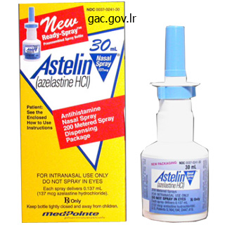
Generic astelin 10ml without prescription
Doing workouts that strengthen muscular tissues associated with the knee and carrying protecting padding can stop knee displacement. Patella the patella, or kneecap, is a flat sesamoid bone situated in a tendon that passes anteriorly over the knee (see fig. The patella, because of its position, controls the angle at which this tendon continues towards the tibia. Its proximal end is expanded into medial and lateral condyles, which have concave surfaces and articulate with the condyles of the femur (fig. A outstanding anterior crest extends downward from the tuberosity and attaches connective tissues in the leg. At its distal finish, the tibia expands to form a prominence on the medial side of the ankle referred to as the medial malleolus (mah-leo-lus), which is an attachment for ligaments. One of those bones, the talus (talus), can transfer freely the place it joins the tibia and fibula, forming the ankle. The largest of the tarsals, the calcaneus (kal-kane-us), or heel bone, is under the talus where it initiatives backward to form the bottom of the heel. The calcaneus helps assist body weight and offers an attachment, the calcaneal tuberosity, for muscles that move the foot. The instep or metatarsus (metah-tahrsus) consists of 5 elongated metatarsal bones, which articulate with the tarsus. The tarsals and metatarsals are certain by ligaments, forming the arches of the foot. A longitudinal arch extends from the heel to the toe, and a transverse arch stretches across the foot. Sometimes, however, the tissues that bind the metatarsals weaken, producing fallen arches, or flat feet. Fibula the fibula is an extended, slender bone located on the lateral facet of the tibia. Its ends are barely enlarged right into a proximal head and a distal lateral malleolus (fig. The lateral malleolus articulates with the ankle and protrudes on the lateral side. Each toe has three phalanges-a proximal, a center, and a distal phalanx-except the good toe, which has solely two as a result of it lacks the middle phalanx (fig. Most apparent is the incremental decrease in top that begins at about age thirty, with a loss of about 1/16 of an inch a 12 months. In the later years, compression fractures within the vertebrae could contribute considerably to loss of peak (fig. Overall, as calcium ranges fall and bone material gradually vanishes, the skeleton loses power, and the bones turn into brittle and more and more prone to fracture. However, the continued capability of fractures to heal reveals that the bone tissue is still alive and practical. Components of the skeletal system and particular person bones change to different degrees and at completely different rates over a lifetime. Bone matrix changes, with the ratio of mineral to protein growing, making bones extra brittle and prone to fracture. Trabecular bone, because of its spongy, much less compact nature, exhibits the changes of getting older first. It is also discovered within the higher a half of the femur, whereas the shaft is more compact bone. As remodeling continues throughout life, older osteons disappear as new ones are constructed subsequent to them. With age, the osteons may coalesce, additional weakening the overall constructions as gaps type. In the primary decade following menopause, ladies lose 15% to 20% of trabecular bone, which is two to thrice the speed of loss in males and in premenopausal women. During the identical time, compact bone loss is 10% to 15%, which is three to 4 occasions the speed of loss in males and premenopausal girls. By very old age, a lady may have only half the trabecular and compact bone mass as she did in her twenties, whereas a really aged man could have one-third much less bone mass.
Generic 10ml astelin free shipping
Nucleic acids the very giant and complex nucleic acids embody atoms of carbon, hydrogen, oxygen, nitrogen, and phosphorus, which form constructing blocks referred to as nucleotides (nukle-o-ti dz). Each nucleotide consists of a 5-carbon sugar (ribose or deoxyribose), a phosphate group, and considered one of several nitrogen-containing natural bases, known as nitrogenous bases (fig. Dna chains are held collectively by hydrogen bonds (dotted lines) and so they twist, forming a double helix. Many larger molecules have polar areas the place nitrogen or oxygen bond with hydrogen. Water solubility and lipid solubility are necessary elements in drug delivery and in movements of gear throughout the body. Because tissues and organs of various composition take in X rays in a unique way, the depth of X rays reaching the detector varies from place to place. Pet imaging makes use of radioactive isotopes that naturally emit positrons, that are atypical positively charged electrons, to detect biochemical exercise in a particular physique part. Useful isotopes in Pet imaging include carbon-11, nitrogen-13, oxygen-15, and fluorine-18. When one of these isotopes releases a positron, it interacts with a close-by negatively charged electron. Pet images of individuals with this disorder reveal intense exercise in two components of the mind that are quiet in the brains of unaffected individuals. Knowing the site of altered brain exercise might help researchers develop extra directed drug remedy. Starting materials are known as reactants; the resulting atoms or molecules are known as merchandise. Three types of chemical reactions are synthesis, in which large molecules build up from smaller ones; decomposition, in which molecules break down; and change reactions, during which components of two completely different molecules trade positions. The path of a reaction relies upon upon the proportion of reactants and merchandise and the power obtainable. Electrolytes that release hydrogen ions are acids, and those who launch hydroxide or other ions that react with hydrogen ions are bases. A tenfold distinction in hydrogen ion focus separates each whole number in the pH scale. An atom consists of electrons surrounding a nucleus, which has protons and neutrons. Electrons are negatively charged, protons positively charged, and neutrons uncharged. The atomic number of a component is equal to the variety of protons in each atom; the atomic weight is the same as the variety of protons plus the number of neutrons in each atom. Isotopes are atoms with the same atomic quantity however different atomic weights (due to differing numbers of neutrons). Electrons occupy house in areas known as electron shells that encircle an atomic nucleus. Atoms with completely crammed outer shells are inert, whereas atoms with incompletely stuffed outer shells gain, lose, or share electrons and thus turn out to be stable. Atoms that lose electrons become positively charged (cations); atoms that achieve electrons turn out to be negatively charged (anions). Ions with opposite expenses appeal to and be a part of by ionic bonds; atoms that share electrons join by covalent bonds. Electrolytes should be current in certain concentrations inside and out of doors of cells. Carbohydrates provide much of the vitality cells require and are built of straightforward sugar molecules. Lipids, such as fats, phospholipids, and steroids, supply vitality and are used to construct cell parts; their building blocks are molecules of glycerol and fatty acids. Proteins serve as structural materials, energy sources, hormones, cell surface receptors, antibodies, and enzymes that velocity chemical reactions with out being consumed.
Cheap 10ml astelin with amex
Interphase is divided into two hole phases (g1 and g2), when specific molecules and constructions duplicate, and a synthesis part (s), when Dna replicates. Mitosis can be thought-about in stages-prophase, metaphase, anaphase, and telophase. Interphase Once thought to be a time of rest, interphase is actually a really active period. During interphase, the cell grows and maintains its routine features as well as its contributions to the interior setting. If the cell is developmentally programmed to divide, it must amass essential biochemicals and duplicate a lot of its contents so that two cells can type from one. Mitosis Mitosis and cytokinesis constitute a form of cell division that happens in somatic (nonsex) cells and produces two daughter cells from an unique cell (fig. In distinction to mitosis is meiosis, a second type of cell division that happens solely in the cells that give rise to sex cells (sperm and eggs). In this manner, when a sperm fertilizes an egg, the total variety of 46 chromosomes is restored. During mitosis, the nuclear contents divide in an event referred to as karyokinesis, which means "nucleus motion. Mitosis should be very precise so that each new cell receives a complete copy of the genetic data. In prophase the chromatin fibers condense, making the person chromosomes visible beneath magnification. The centrioles of the centrosome replicate just earlier than the onset of mitosis, and through prophase, the two newly formed pairs of centrioles move to opposite sides of the cell (fig. Microtubules are assembled from tubulin proteins within the cytoplasm, and these structures affiliate with the centrioles and chromosomes. A spindle-shaped array of microtubules (spindle fibers) types between the centrioles as they transfer aside (fig. In metaphase spindle fibers connect to the centromeres so that a fiber accompanying one chromatid attaches to one centromere and a fiber accompanying the other sister chromatid attaches to its centromere (fig. The chromosomes move along the spindle fibers, and microtubules assist align them about midway between the centrioles. In anaphase the centromeres of the chromatids separate, and the sister chromatids at the moment are thought-about individual chromosomes. The separated chromosomes move in reverse directions, again as the result of microtubule activity. The spindle fibers shorten and pull their connected chromosomes toward the centrioles at reverse sides of the cell (fig. In telophase, the ultimate stage of mitosis, the chromosomes full their migration toward the centrioles. As the identical units of chromosomes strategy their respective centrioles, they begin to elongate and unwind from rodlike buildings to threadlike buildings. A nuclear envelope forms round every chromosome set, and nucleoli become visible throughout the newly shaped nuclei. Cytoplasmic Division Cytoplasmic division (cytokinesis) begins throughout anaphase when the cell membrane begins to constrict around the middle, which it continues to do by way of telophase. The musclelike contraction of a hoop of actin microfilaments pinches off two cells from one (fig. Centromeres separate, and sister chromatids move aside, with each chromatid now an individual chromosome; spindle fibers shorten and pull these new particular person chromosomes towards the centrioles. Chromosomes elongate and kind chromatin threads; nuclear membranes type round every chromosome set; nucleoli kind; microtubules break down. Metaphase Mitosis is usually known as mobile replica, as a result of it ends in two cells from one-the cell reproduces. This may be confusing, as a outcome of meiosis is the prelude to human sexual replica. Both mitosis and meiosis are types of cell division, with comparable steps but completely different outcomes, and occurring in various sorts of cells. Cleavage furrow (e) (b) Centromere Spindle fiber Prophase Chromosomes condense and turn into visible. Late Prophase Nuclear envelopes Telophase and Cytokinesis Nuclear envelopes start to reassemble round two daughter (d) nuclei. Chromosomes Sister chromatids (c) Mitosis Cytokinesis G1 phase Anaphase Sister chromatids separate and the ensuing chromosomes transfer to opposite poles of cell.
Purchase astelin 10ml with mastercard
Lipid-soluble substances, corresponding to oxygen, carbon dioxide, steroids, urea, ethanol, and common anesthetics, freely cross the cell membrane by simple diffusion. Facilitated diffusion contains provider molecules and channels that transport some substances into or out of cells, from a area of upper concentration to considered one of decrease focus. The larger spheres depict transported substances that cross through protein service molecules, and the small spheres symbolize ions, which cross the membrane by way of ion channels. This union of glucose and its provider molecule modifications the shape of the provider in a method that moves glucose to the inner surface of the membrane. Here, the glucose is launched, and the carrier molecule returns to its original shape and may pick up one other glucose molecule. Facilitated diffusion can move molecules solely down a concentration gradient and is restricted for a selected solute (a glucose provider can solely carry glucose, for example). The variety of provider molecules or channels limits the speed at which solutes can cross the membrane. If impermeant solute is current on each side of the membrane, water will move by osmosis to the facet with the larger impermeant solute concentration. Osmosis is sometimes summarized as "water follows salt," however water will transfer by osmosis towards any impermeant solute. Therefore, water moves from compartment B throughout the selectively permeable membrane and into compartment A by osmosis. This capacity of osmosis to generate sufficient pressure to carry a volume of water known as osmotic pressure. When the height of the column of water in A reaches a certain level, the downward force (hydrostatic pressure) will forestall any further net osmotic motion of water. The higher the concentration of impermeant solute particles (protein in this example) in a solution, the higher the osmotic strain of that answer. Water always tends to transfer by osmosis toward options of greater osmotic pressure. That is, water moves towards areas of trapped solute-whether in a lab train or within the body. Cell membranes are generally permeable to water, so water equilibrates by osmosis all through the body, and the concentration of water and solutes everywhere within the intracellular and extracellular fluids is basically the same. Solutions which have the next osmotic pressure than body fluids are known as hypertonic. If cells are put into a hypertonic answer, there shall be a net motion of water by osmosis out of the cells into the encompassing answer, and the cells shrink. Conversely, cells put into a hypotonic resolution, which has a lower osmotic pressure than body fluids, gain water by osmosis and swell. Although cell membranes are somewhat elastic, the cells may swell so much that they burst. Filtration Filtration (fil-trashun) is one other process that forces molecules by way of membranes by exerting pressure. One technique is to pour a combination of solids and water onto filter paper in a funnel (fig. The paper is a porous membrane via which the small water molecules pass, leaving the larger stable particles behind. An example of filtration is making espresso by the drip method, using a filter paper cone or basket. In the body, tissue fluid forms by filtration when water and dissolved substances are forced out via the thin, porous partitions of blood capillaries, however larger particles such as plasma protein molecules remain inside (fig. Blood strain Blood flow Larger molecules Smaller molecules by heart action, which is bigger inside the vessel than outdoors it. However, the large proteins left inside capillaries oppose filtration by drawing water into blood vessels by osmosis, stopping the formation of extra tissue fluid, which causes a condition known as edema. Although a heart beating is an active physique process, filtration is taken into account passive as a result of it could happen from the strain of gravity alone. It is slightly like the difference between making ready espresso by forcing water via espresso grounds using a French press (active) or by the drip method (passive). Active Transport When molecules or ions cross through cell membranes by diffusion or facilitated diffusion, their web motion is from regions of upper concentration to regions of lower focus.
Astelin 10ml lowest price
Thus, a thin layer of cytotrophoblastic cells called the trophoblastic shell is formed around the syncytiotrophoblast. The trophoblastic shell is interrupted solely at sites the place maternal vessels communicate with the intervillous areas. Chorionic villi constantly type out of the trophoblastic sprouts all through being pregnant. The chorionic villi can stay either free (floating villi) in the intervillous house or grow into the maternal side of the placenta (basal plate) to form primary stem villi or anchoring villi. Future progress of the placenta is completed by interstitial development of the trophoblastic shell. The diagrams illustrate the separation of the fetal and maternal blood vessels by the placental membrane, which is composed of the endothelium of the capillaries, mesenchyme, cytotrophoblast, and syncytiotrophoblast. The number of syncytial knots is elevated with gestational age of the placenta and can be used to evaluate villous maturity. Increased variety of syncytial knots can be related to some pathologic conditions, such as uteroplacental malperfusion. Several forms of cells are recognized within the connective tissue stroma of the villi: mesenchymal cells, reticular cells, fibroblasts, myofibroblasts, smooth muscle cells, and fetal placental antigen�presenting cells (placental macrophages), traditionally also recognized as Hofbauer cells (Plate one hundred, web page 892). Fetal placental antigen�presenting cells are the precise villous macrophages of fetal origin that take part within the placental innate immune reactions. In response to antigen, they proliferate and upregulate particular surface receptors that recognize and bind to a wide selection of pathogens. The vacuoles in these cells contain lipids, glycosaminoglycans, and glycoproteins. Primary villi represent the first stage of improvement during which the syncytiotrophoblast and cytotrophoblast form finger-like extensions into the maternal decidua. In secondary chorionic villi, the extra-embryonic connective tissue (mesenchyme) grows into the villi and is surrounded by a layer of cytotrophoblast. In tertiary chorionic villi, blood vessels and supportive cells differentiate within the mesenchymal core. In early being pregnant, villi are giant and edematous with few blood vessels surrounded by many cells of connective tissue. They are covered by a thick layer of syncytiotrophoblast and a steady layer of cytotrophoblast cells. In late (in term) pregnancy, the layer of cytotrophoblast seems to be discontinuous and nuclei of the syncytiotrophoblast combination to form irregularly dispersed projections called syncytial knots. More fetal blood vessels are discovered in the connective tissue core, which turns into less mobile and contains fewer placental macrophages. These villi emerge from the chorionic plate as giant stem villi and department into the more and more smaller villi. This higher magnification shows the straightforward cuboidal epithelium of the amnion and the underlying connective tissue. This higher magnification exhibits a cross-sectioned villus containing a quantity of bigger blood vessels and its skinny floor syncytiotrophoblast layer. Within the basal plate and the connective tissue stroma are clusters of cells, the decidual cells (arrows), which arose from connective tissue cells. Blood begins to circulate through the embryonic cardiovascular system and the villi at about 21 days. The intervillous areas provide the location of change of vitamins, metabolic merchandise and intermediates, and wastes between the maternal and fetal circulatory methods. During the primary 8 weeks, villi cowl the entire chorionic surface, but as progress continues, villi on the decidua capsularis begin to degenerate, producing a smooth, comparatively avascular surface known as the chorion laeve. This area of the chorion, which is the fetal element of the placenta, is identified as the chorion frondosum or villous chorion. The layer of the placenta from which the villi project is identified as the chorionic plate (Plate 99, web page 890). During the interval of fast progress of the chorion frondosum, at concerning the fourth to fifth month of gestation, the fetal a half of the placenta is split by the placental (decidual) septa into 15 to 25 areas called cotyledons. Cotyledons are seen as the bulging areas on the maternal aspect of the basal plate. Vessels within this a half of the endometrium provide blood to the intervillous areas.
Discount astelin on line
Mnemonic Devices Another method for remembering data is the mnemonic device. One kind of mnemonic gadget is a listing of words, forming a phrase, during which the rst letter of each word corresponds to the rst letter of each word that must be remembered. Another kind of mnemonic system is a word fashioned by the rst letters of the objects to be remembered. For example, ipmat represents the stages in the cell cycle: interphase, prophase, metaphase, anaphase, and telophase. Master a few new playing cards every day, and evaluate cards from previous days, and use them all once more on the end of the semester to prepare for the comprehensive nal exam. They could even come in handy later, such as in learning for exams for admission to medical college or graduate school. Divide your deck in half and ip half of the playing cards so that the answer quite than the query is exhibiting. Get used to identifying a construction or process from a description in addition to giving a description when supplied with a process or structure. This is extra like what might be anticipated of you in the actual world of the health-care professional. As essential as these are, you still have to master this materials in your path to turning into a health-care professional. Good time administration abilities are therefore essential in your examine of human anatomy and physiology. Daily repetition is useful, so scheduling several brief examine intervals every day can replace a last-minute crunch to cram for an exam. Thinking about these ideas for learning now can maximize research time all through the semester, and, hopefully, lead to tutorial success. A working information of the construction and function of the human body provides the foundation for all careers in the well being sciences. Working as a group and alternating leaders allows college students to verbalize the knowledge. Individual students can research and master one a half of the assigned materials, after which clarify it to the others in the group, which contains the information into the memory of the speaker. Be positive to use anatomical and physiological terms, in explanations and on a regular basis dialog, till they turn into a part of your working vocabulary, quite than intimidating jargon. Most essential of all-the group should keep on task, and never turn out to be a automobile for social interaction. Small examine teams reviewing and vocalizing materials can divide and conquer the educational task. Time administration expertise encourage scheduled learning, together with daily repetition as an alternative of cramming for exams. Before class Read the assigned textual content material previous to the corresponding class meeting. Photographs, line artwork, ow charts, and organizational tables help in mastery of the materials. Understanding physiology and the significance of anatomy, however, requires you to be ready to recall previous concepts. Critical pondering is all about linking previous concepts with current concepts underneath novel circumstances, in new methods. Check out McGraw-Hill on-line assets that may allow you to practice and assess your studying. McGraw-Hill Connect Interactive Questions Reinforce your information using assigned interactive questions. Learning anatomy and physiology requires familiarity with the language used to describe structures and features. Cells aggregate and work together to type tissues, which in turn layer and fold and intertwine to kind organs, which in turn join into organ systems. Mastering the principles of anatomy and physiology not solely will present you with a new appreciation on your day-to-day activities, skills, strengths, and well being, but will provide a basis so that you can help your future sufferers, for these of you going into health care. Just as we do at present, primitive individuals suffered aches and pains, injured themselves, bled, broke bones, developed diseases, and contracted infections.
Order genuine astelin online
The area of fusion surrounds the cell like a belt, and the junction closes the area between the cells. Tight junctions usually be part of cells that type sheetlike layers, similar to those who line the within of the digestive tract. The linings of tiny blood vessels within the brain include cells held tightly together (From Science to Technology 5. Another kind of intercellular junction, referred to as a desmosome, rivets or "spot welds" pores and skin cells, enabling them to type a reinforced structural unit. The cell membranes of sure different cells, corresponding to those in coronary heart muscle and muscle of the digestive tract, are interconnected by tubular channels called hole junctions. These channels link the cytoplasm of adjacent cells and allow ions, nutrients (such as sugars, amino acids, and nucleotides), and other small molecules to transfer between them (fig. Tissues may be distinguished from one another by variations in cell dimension, form, organization, and function. The tissues of the human physique embody 4 main types: epithelial, connective, muscle, and nervous. These tissues associate, assemble, and work together to kind organs that have even more specialized capabilities. This chapter examines epithelial and connective tissues intimately, and introduces muscle and nervous tissues. Chapter 9 discusses muscle tissue in more detail, and chapters 10, eleven, and 12 study nervous tissue. Micrographs are photographs of extremely skinny slices (sections) of prepared tissue specimens. Like a good line of police officers preserving out a crowd, the blood-brain barrier is a 400-mile community of capillaries within the mind whose cells are firmly connected by overlapping tight junctions. The blood-brain barrier shields mind tissue from toxins and biochemical fluctuations that could possibly be overwhelming. For many years researchers have tried to deliver medicine throughout the barrier by tagging compounds to substances that can cross, and injecting substances that quickly chill out the tight junctions. More recently, researchers have utilized nanotechnology to the issue of circumventing the blood-brain barrier. Nanoparticles that may cross the blood-brain barrier are manufactured from mixtures of oils and polymers, with a impartial or barely unfavorable charge. In one software, anesthetics or chemotherapeutics are loaded into phospholipid bubbles (liposomes). In one other application, insulin is delivered in inhaled nanoparticles 10 to 50 nanometers in diameter. Originally developed to present insulin to people with diabetes instead of injecting it, nanoparticle supply of insulin may be useful in sustaining memory in individuals who have mild cognitive impairment or Alzheimer illness, based on early findings in clinical trials. Epithelium covers the physique surface and organs, varieties the internal lining of body cavities, traces hole organs, and composes glands. It all the time has a free (apical) floor uncovered to the outside or internally to an open space. A thin, extracellular layer referred to as the basement membrane anchors epithelium to underlying connective tissue. Cancer cells secrete a substance that dissolves basement membranes, enabling the cells to invade tissue layers. Cancer cells additionally produce fewer adhesion proteins, or none in any respect, which permits them to spread into surrounding tissue. Tight junctions fuse cell membranes, desmosomes are "spot welds," and hole junctions form channels linking the cytoplasm of adjacent cells. Q Which intercellular junction is the most probably to permit substances to move from one cell to another However, vitamins diffuse to epithelium from underlying connective tissues, which have abundant blood vessels. Epithelial cells readily divide, so injuries heal quickly as new cells replace lost or broken ones. For example, pores and skin cells and the cells that line the stomach and intestines are continually broken and replaced. Epithelial tissues composed of thin, flattened cells are squamous; those with cubelike cells are cuboidal; and those with elongated cells are columnar.
Astelin 10 ml overnight delivery
The energy for this process is supplied by the mitochondria situated within the internal phase of those photoreceptors. It is In rods, after a interval of sleep, a burst of disc shedding happens as mild first enters the attention. The shedding of discs in cones additionally allows the receptors to remove superfluous membrane. Although not fully understood, the shedding process in cones also alters the dimensions of the discs in order that the conical form is maintained as discs are released from the distal end of the cone. The retinal pigment epithelial cells contain quite a few mitochondria and phagosomes. The arrow signifies the placement of the junctional advanced between two adjacent cells. This layer is thought to be a metabolic barrier that restricts the passage of enormous molecules into the inner layers of the retina. The region of the rod cytoplasm that contains the nucleus is separated from the inner segment by a tapering strategy of the cytoplasm. In cones, the nuclei are positioned near the outer segments, and no tapering is seen. The outer plexiform layer (5) is shaped by the processes of the photoreceptor cells and neurons. The outer plexiform layer is formed by the processes of retinal rods and cones and the processes of horizontal, interplexiform, amacrine, and bipolar cells. The processes enable the electrical coupling of photoreceptor cells to these specialized interneurons via synapses. A skinny course of extends from the region of the nucleus of every rod or cone to an inner expanded portion with several lateral processes. Normally, many photoreceptor cells converge onto one bipolar cell and kind interconnecting neural networks. The fovea is also unique in that the compactness of the inner neural layers of the retina causes the photoreceptor cells to be oriented obliquely. Horizontal cell dendritic processes Their processes make investments the other cells of the retina so utterly that they fill many of the extracellular area. Microvilli extending from their apical border lie between the photoreceptor cells of the rods and cones. In the peripheral regions of the retina, the axons of bipolar cells move to the inside plexiform layer where they synapse with several ganglion cells. Through these connections, the bipolar cells establish communication with a number of cells in each layer besides within the fovea, the place they might synapse only with a single ganglion cell to provide higher visual acuity on this region. Horizontal cells and their processes lengthen to the outer plexiform layer where they intermingle with processes of bipolar cells. The cells have synaptic connections with rod spherules, cone pedicles, and bipolar cells. Their processes branch extensively to present websites of synaptic connections with axonal endings of bipolar cells and dendrites of ganglion cells. Interplexiform cells and their processes have synapses in each internal and outer plexiform layers. These cells convey impulses from the inner plexiform to the outer plexiform layer. The inner plexiform layer consists of synaptic connections between axons of the bipolar neurons and dendrites of ganglion cells. It also accommodates synapses between intermingling processes of amacrine cells and bipolar neurons, ganglion cells, and interplexiform neurons. The ganglion cell layer (8) consists of the cell our bodies of enormous multipolar neurons. The web site where the axons converge to type the optic nerve is called the optic disc. From the middle of the optic nerve (clinically called the optic cup), central retinal vessels emerge.

