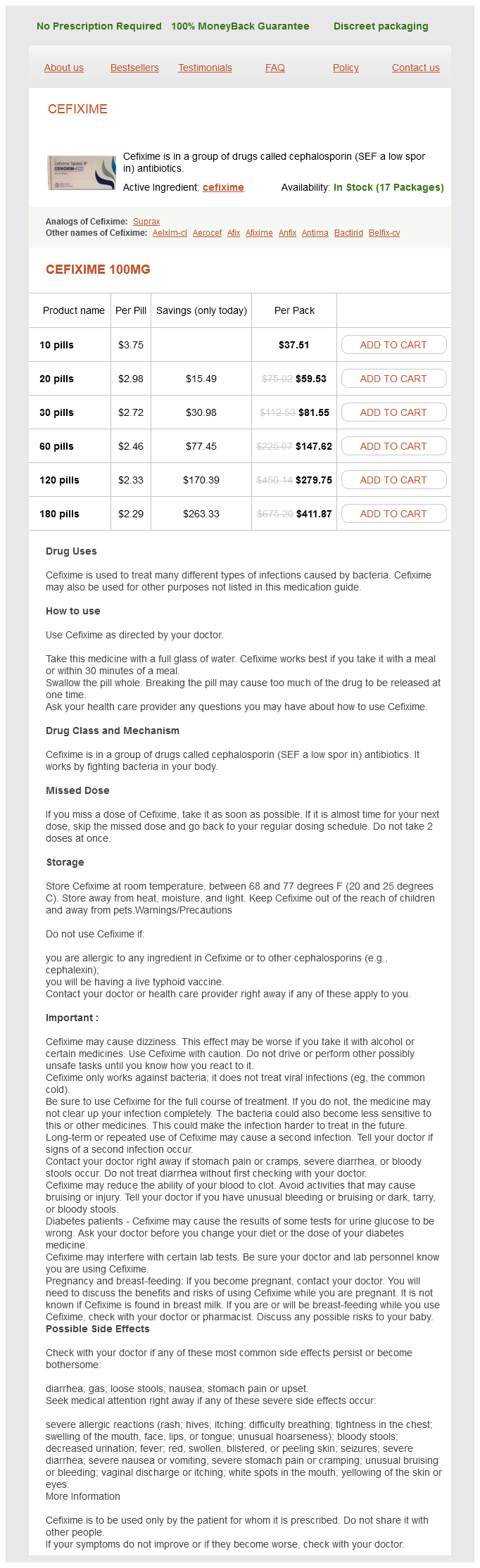
Cefixime dosages: 100 mg
Cefixime packs: 10 pills, 20 pills, 30 pills, 60 pills, 120 pills, 180 pills
Purchase cefixime 100mg
Various strains of transgenic ChR2-expressing mice have been developed by Feng and colleagues for in vivo neuroscience research,53 and many of these are currently obtainable via the Jackson Laboratory. To date, no commercially obtainable mice with cardiac expression of optogenetic tools have been produced. They used recombinase-driver rat cell lines that can drive the gene expression in specific cell sorts with Cre recombinase under management of relatively massive regulatory areas (>200 kb). This approach permits faster generation of experimental rat fashions than other classical transgenic approaches and can doubtless be used in cardiac functions as well. Such Cre driver lines confer one other stage of selectivity, in addition to promoterdetermined selectivity, when combined with viral delivery. For cardiac purposes, one can envision targeting the conduction system as a whole or regions of it. Through concerted efforts to optimize the optogenetics toolbox, a number of laboratories use repositories like Addgene and make their constructs publicly obtainable. When optogenetics had been utilized to cardiac tissue, it was unknown a priori whether the required chromophore for ChR2 operation may be present in adequate amounts. Am J Physiol Heart Circ 304:H1179-H1191, 2013, with permission; additionally from Jia Z, Valiunas V, Lu Z, et al: Stimulating cardiac muscle by light: Cardiac optogenetics by cell supply. Furthermore, implantable optogenetic units advanced quickly in mind analysis since 200724 to the present totally built-in techniques in freely moving animals, in some cases with wireless powering. The challenge is to obtain this goal in a beating coronary heart with out the agency help and point of reference naturally offered by the cranium for the brain. Optical fiber conduits for imaging purposes have been developed earlier than for cardiac applications58; possibly this intramural optrode approach could possibly be adopted for localized gene or cell supply, as well as for optical stimulation and recording. Alternatively, for optical stimulation, surface-conforming solutions, whereby the system is shifting with the contracting coronary heart, may come from recent new developments in stretchable electronics and optoelectronics59. Alternatively, catheter accessibility to the endocardial conduction system might provide quick alternatives for optogenetic purposes. EnergyforOpticalStimulation the optical stimulus power needed to trigger a response is usually measured in items of irradiance (mW/mm2). The energy is influenced by a mess of factors, together with expression levels and performance of the opsins, the host cell electrophysiological milieu (balance of depolarizing and repolarizing currents), the cable properties of the tissue and electrotonic load for activation, the efficiency of sunshine delivery/penetration, and so forth. Furthermore, atrial myocytes had been discovered to categorical ChR2 at higher ranges and to produce larger practical currents than ventricular myocytes. Without a doubt, stimulus supply to the location of interest will profoundly affect the overall power requirements. Whether it could progress into extra translational/ therapeutic makes use of remains to be seen, for neuroscience and for cardiac functions. As a basic science software in cardiac research, optical pacing can supply contact-less stimulation with higher spatiotemporal decision and cell selectivity and a brand new capability for parallelization compared with electrical stimulation. Because of its contact-less nature, optical pacing naturally lends itself to parallelization and scalability, as well as closed-loop feedback management. Currently, no direct and specific technique is out there to handle these questions in vivo. Optogenetics could supply solutions via selective cell type�specific expression and optical stimulation, if mild access problems are resolved. Critical contributions of various components of the pacemaking and conduction system may be probed, as was demonstrated within the zebra fish examine. A third set of issues suitable for in vitro investigation is said to mechanisms of reentrant arrhythmias and their termination. Precise dynamic optical probing can be utilized to handle the exact nature of reentrant activation and the state of the reentrant core-spiral wave versus leading circle. The seek for mechanisms of atrial fibrillation or ventricular fibrillation-mother rotor, wandering wavelets, or other-can be better tackled by fine stimulation instruments to set up vulnerability and should have real impact on the development of higher defibrillation strategies. Nat Neurosci 8:1263-1268, 2005; Bruegmann T, Malan D, Hesse M, et al: Optogenetic management of heart muscle in vitro and in vivo. Nat Methods 7:897-900, 2010; and Jia Z, Valiunas V, Lu Z, et al: Stimulating cardiac muscle by light: Cardiac optogenetics by cell supply.
Smellage (Lovage). Cefixime.
- Indigestion, heartburn, intestinal gas, irregular menstrual periods, sore throat, boils, jaundice, gout, migraines, use as "irrigation therapy" for urinary tract inflammation and kidney stones, and other conditions.
- How does Lovage work?
- Dosing considerations for Lovage.
- What is Lovage?
- Are there any interactions with medications?
- Are there safety concerns?
Source: http://www.rxlist.com/script/main/art.asp?articlekey=96709
Quality 100 mg cefixime
This smooth-walled space is probably the most posterior portion of the proper atrium and stretches between the orifices of the caval veins. From the viewpoint of the surgeon wanting down into the best atrium, the sinus venarum is extra or less horizontal, with the superior vena P. Just beneath and medial to the orifice of the superior vena cava arises the crista terminalis, a muscle bundle that springs into prominence as it circles the orifice of the superior vena cava to the best lateral wall of the atrium and continues inferiorly towards the inferior vena cava, thereby forming the boundary between the sinus venarum and the atrial appendage. This muscle bundle is evidenced on the outside of the atrium by a groove, the sulcus terminalis. Lying subepicardially within the sulcus terminalis, just under the doorway of the superior vena cava, is the sinoatrial node, which can be vulnerable to injury from the assorted surgical incisions and cannulations which are generally carried out on the best atrium. In distinction to the smooth-walled sinus venarum, the lateral wall of the atrial appendage is ridged by multiple slim bands of muscle, the musculi pectinati. These bands arise from the crista terminalis and cross upward to probably the most anterior part of the atrium. Just above the sinus venarum within the heart of the medial wall is the fossa ovalis, an elliptic or horseshoe-shaped depression. The aortic root is hidden behind the anteromedial atrial wall between the fossa ovalis and the termination of the heavily trabeculated right atrial appendage. Segments of the noncoronary and proper sinus of Valsalva are in shut apposition to the atrial wall on this area. Their locations could additionally be manifested by the aortic mound, a bulge above and barely to the left of the fossa ovalis. The presence of the aortic valve right here could be extra clearly visualized if one takes into consideration its continuity, via the central fibrous body, with the adjacent tricuspid valve annulus. Also invisible to the surgeon is the artery to the sinoatrial node, which runs through this identical space. Although its origin and precise location are unpredictable, the artery to the sinoatrial node takes a variable course towards the superior cavoatrial angle and the sinus node. The membranous, or fibrous, septum is a continuation of the central fibrous physique, via which the tricuspid, mitral, and aortic valves are linked. It is located at the apex of the triangle of Koch, the boundaries of that are the annulus of the septal leaflet of the tricuspid valve, the tendon of Todaro (running intramyocardially from the central fibrous physique to the Eustachian valve of the inferior vena cava), and its base, the coronary sinus. Anderson describes the tendon of Todaro as a fibrous extension of the commissure between the Eustachian valve (of the inferior vena cava) and the thebesian valve (of the coronary sinus). Conduction tissue passes from the atrioventricular node as the bundle of His beneath the membranous septum and P. The coronary sinus, draining the cardiac veins, is located alongside the tendon of Todaro, between it and the tricuspid valve. Preparation A rectangular piece of pericardium is harvested and handled with glutaraldehyde. The relationship of the great vessels and coronary anatomy can be confirmed at this point. Direct bicaval cannulation is carried out where possible, or single atrial cannulation for small weight babies. With initiation of cardiopulmonary bypass, the ductus arterious is occluded at its aortic end with a heavy tie or metal clip. The ductus arteriosus is later divided, oversewing the pulmonary artery aspect with 6-0 or 7-0 Prolene suture. During cooling, the ascending aorta is dissected free from the principle pulmonary artery, and the right and left pulmonary arteries are extensively mobilized out to the first branches in the hilum of each lung. Most or all the dissection is accomplished with an electrocautery on low present. Flooding of the Pulmonary Bed As quickly as cardiopulmonary bypass is instituted, the ductus arterious must be occluded to stop runoff of aortic cannula move into the lungs. Mobilization of the Pulmonary Arteries It is essential to totally mobilize the branch pulmonary arteries beyond their hilar bifurcation in order to scale back pressure on the Lecompte. Transection of the Great Arteries the aortic cross-clamp is utilized simply proximal to the aortic cannula. The aorta is then transected at this stage, and traction sutures are positioned just above the three commissures of the aortic valve and tagged. The pulmonary artery is transected at the level of the takeoff of the best pulmonary artery, and traction sutures are placed at the commissures and tagged.
Cheap cefixime online amex
According to stable and abundant evidence, and thus to common consensus, the beat-to-beat export of bulk Ca2+ from coronary heart cells is certainly performed by the Na/ Ca-exchanger. J Biol Chem 282:25640�25648, 2007; Brini M, Coletto L, Pierobon N, et al: A comparative practical evaluation of plasma membrane Ca2+ pump isoforms in intact cells. Acidic phospholipids bind to two sites: one is the fundamental C-terminal calmodulin binding domain, and the opposite is a stretch of roughly 40 predominantly fundamental amino acids within the cytosolic loop connecting transmembrane domains 2 and 3. It has been calculated that the focus of phosphatidyl-serine in the environment of the pump would in precept be enough for about 50% stimulation of its exercise. Kinases have also been discovered to activate the pump by phosphorylating residues in its C-terminal tail. Meanwhile, protein kinase C acts on all pump variants, and protein kinase A acts on solely one of many isoforms. An intriguing mechanism of pump activation is that generated by a dimerization (oligomerization) course of that happens via the C-terminal calmodulin binding area; its physiologic significance is obscure. All mechanisms of activation act by growing the Ca2+ affinity of the pump; in their absence, the Km (Ca2+) of the pump is as high as 20 �M, but drops to 0. The pump could be also activated irreversibly, and that occurs when its C-terminal tail, which includes the calmodulin binding domain, is shaved off by the Ca2+-dependent protease calpain. In this case, the activation is linked to the removal of the autoinhibitory C-terminal tail of the pump. The irreversible activation by calpain could turn out to be significant in circumstances of pathologic Ca2+ overload that may demand the uninterrupted maximal capacity of the pump to extrude Ca2+ from the cytosol. As talked about earlier, alternative splicing processes have an effect on all 4 fundamental primary transcripts of the pump, significantly rising the variety of isoforms. Most of the splice variants described in the literature have also been documented at the protein level. Site A corresponds to the cytosolic loop of the pump molecule that connects transmembrane domains 2 and three, site C to the C-terminal calmodulin binding area. The full details of the splicing operations and its complexities are mentioned elsewhere. The pump variants with out the inserts are termed z; those with the extra exon are termed x. For instance, in people only variant w (all exons included), variant x (only the forty two bp exon included), and variant z (no further exon included) have been detected. Only scarce info is out there on the practical penalties of the splicing operation at website A. Information on the implications of the slicing operation at web site C on the exercise of the pump is more abundant, and recent findings can result in important functional developments. The insertion of the novel sequence roughly in the course of the calmodulin binding domain leads, as expected, to the lowering of the calmodulin affinity for the pump. However, predictions on the consequences to be anticipated from the insertion of the novel sequence in the calmodulin binding domain are difficult by the unexpected statement that the insert tends to reconstitute the original whole calmodulin binding domain; 8 of the first 10 residues of the insert are indeed either similar or conservative, with respect to those or the unique C-terminal half of the calmodulin binding area they replace. Regardless of the mechanism, nonetheless, the anomaly generates a pump variant that may perform almost optimally in the absence of activation by calmodulin; this would satisfy the demands that particular cell sorts have for a steady and vigorous Ca2+ exporting function not relying on pump activation. As have to be anticipated, the central function of Ca2+ in the regulation of crucial cell actions demands its precise Table 5-2. The vast array of Ca2+ binding proteins and Ca2+ transporters expressed in all animal cells underlies the concept. Defects within the control of Ca2+ unavoidably generate states of cell suffering that might culminate, in cases of massive and protracted Ca2+ overload, within the demise of the cell; this is the that means of the ambivalence idea. Having chosen Ca2+ as a determinant for function, cells undoubtedly profit from its limitless availability of their environment; nonetheless, the selection forces cells to live in a permanent condition of managed risk. Increases of Ca2+ considerably larger than regular levels could be handled by cells if the rise lasts for a restricted time, because of the existence of the uptake system of mitochondria, which might efficiently sequester for some time large quantities of Ca2+. Apart from these extreme and obvious cases of Ca2+ catastrophe, the cellular homeostasis of Ca2+ also can become deregulated in less dramatic ways by defects within the individual participants in the Ca2+ controlling operation. These defects are appropriate with the continuation of cell life, however generate cell discomfort phenotypes with numerous levels of severity. Among them, these generated by defects of the Ca2+ pumps are currently receiving elevated attention. Several disease phenotypes brought on by genetic and nongenetic defects of the Ca2+ pumps have been described. By distinction, the pathologic genetic phenotypes are definitely causative and are mechanistically well outlined.
Discount cefixime 100 mg otc
Pathogenesis includes components like anoxia, venous stasis, angiospasm, elevated capillary permeability, and thrombocytopenia. Characteristc features of anaemic retinopathy are as beneath: � Fundus background becomes pale � Retinal arterioles are also pale � Retinal veins are tortuous and dilated � Retinal haemorrhages, superficial flame shaped and preretinal (subhyaloid) may be seen within the posterior half of fundus � Roth spots, i. Management includes: Sickle-cell Retinopathy Retinal modifications in sufferers affected by sickle cell haemoglobinopathies (abnormal haemoglobins) are primarily caused by retinal hypoxia; which ends from blockage of small blood vessels by the abnormal-shaped inflexible purple blood cells. Clinical options Sickle-cell retinopathy can be divided into five selfexplanatory phases as follows: 1. Leukaemic Retinopathy Ocular involvement is extra widespread with acute than persistent leukaemia. Chapter 12 Characteristic options of leukaemic retinopathy Diseases of Retina Clinical features 283 embody: � Fundus background is pale and orangish � Retinal veins are tortuous and dilated � Retinal arterioles turn into pale and slim � Perivascular leukaemic infiltrates, seen as grayish white lines along the course of veins, are seen in latter phases � Roth spots, i. Other ocular adjustments in leukaemia embrace: � Orbital infiltration, significantly in children presenting as proptosis � Ocular haemorrhages within the form of subconjunctival haemorrhage and hyphaema (bleeding in anterior chamber) � Iris modifications within the type of iris thickening and iritis � Pseudohypopyon, i. Demarcation line formation on the edge of vessels, dividing the vascular from the avascular retina. The line construction of stage 1 acquires a volume to type a ridge with peak and width. Diseases of the retina Zone I � Low gestation age, especially <32 weeks � Low delivery weight (<1500 g, especially <1250 g), � Supplemental oxygen therapy, and Other risk factors reported embrace mild, vitamin E deficiency, respiratory distress syndrome, asphyxia, shock, and acidosis. A circle drawn on the posterior pole, with the optic disc because the centre and twice the gap from the centre of disc to fovea because the radius, constitutes zone I. Extent of involvement Extent of involvement is denoted by the clock hours of retinal involvement in the particular zone. Early analysis and remedy is important to forestall blindness in highrisk cases. Post-laser therapy follow-up should be carried out weekly for 3 weeks and if required retreatment must be done. Subsequent follow-up examination ought to be continued at 3, 6 and 12 weeks after therapy. Characterized by irregular dilation of capillary mattress and segmental dilation of neighbouring venules and arterioles. First examination by oblique ophthalmosocpy must be done between 4 to 6 weeks postnatal age or 34 weeks postconceptual age (whichever is earlier). This situation has been divided into three groups: 1, 2 and 3, (each group is subdivided into A and B) relying on the traits of lesion. Treatment is as below: � Photocoagulation or cryotherapy might verify development of the disease if utilized within the early stage. Anterior chamber could reveal faint aqueous flare with few, if any, cells (ischaemic pseudoiritis). Ocular ischaemic syndrome refers to a uncommon situation resulting from continual ocular hypoperfusion secondary to >90% stenosis of carotid artery. Carotid stenosis refers to atherosclerotic occlusive carotid artery illness usually related to ulceration at the bifurcation of widespread carotid artery. Risk components for carotid stenosis embrace male gender, old age (60�90 years), smoking, hypertension, diabetes mellitus and hyperlipidaemia. Ocular ischaemic syndrome is often unilateral (80%), affecting elder males extra generally than females (2:1). In suspected circumstances the carotid stenosis may be confirmed by Doppler ultrasound and magnetic resonance angiography. Treatment of ocular ischaemic syndrome contains: � Treatment of neovascular glaucoma (see page 250). Prevalence and demography these contain the whole retina (peripheral retina greater than macula) and include: � Typical retinitis pigmentosa and its variants, � Progressive cone dystrophy, � Leber congenital amaurosis, � Congenital stationary evening blindness, and � Congenital monochromatism (achromatopsia). It seems within the childhood and progresses slowly, often resulting in blindness in superior middle age.
Order 100mg cefixime amex
Tertiapin binds to the outer vestibule of the conduction pore fashioned by the linker between the primary and second transmembrane (M1�M2) segments. An -helical construction throughout the toxin interacts with a short sequence of fragrant residues positioned within the N-terminal a half of the linker that confers excessive affinity for tertiapin. Conversely, these basic science insights could also be helpful in the improvement of novel and potentially helpful antifibrillatory pharmacophores. This results in polar hydrophilic cationic side-chain and apolar ring methods within one molecule. Bupivacaine is an area anesthetic drug with a long length of motion that produces glorious sensory anesthesia. Quinacrine is an antimalarial agent that has been used for numerous extra indications, corresponding to other parasitic infections, as an antifibrillatory agent and for remedy of autoimmune disorders. In addition, the effects were similar whether or not mefloquine was applied externally or internally, suggesting that the inhibitory effect was membrane delimited. In addition, the time course of thiopental inhibition was slow (T1/2-4 minutes) and impartial of external or inner drug application, suggesting that the inhibitory impact was membrane delimited. It was proposed that flecainide reduces the inward rectification of the current at potentials optimistic to the potassium reversal potential. Interestingly, flecainide pharmacologically rescues R67W, however not R218W, channel mutations found in patients with Andersen-Tawil syndrome. Loss- and gain-of-function mutations present in a quantity of human diseases and syndromes underscore their important roles in proper heart function. Many marketed medicine have an effect on inward rectifier function by instantly modulating the channel pore. Continuation of the already impressive research on structure-function relationships of these channels, their interplay with varied drugs on the molecular stage, and functional research of inward rectifier�modifying drugs in devoted disease models will ultimately provide alternatives to develop very specific and effective new medication that target this channel class for treating numerous human cardiac ailments. Hibino H, Inanobe A, Furutani K, et al: Inwardly rectifying potassium channels: Their construction, function, and physiological roles. Tamargo J, Caballero R, G�mez R, et al: Pharmacology of cardiac potassium channels. Dobrev D, Carlsson L, Nattel S: Novel molecular targets for atrial fibrillation remedy Nat Rev Drug Discov 11:275�291, 2012. Zhou W, Arrabit C, Choe S, et al: Mechanism underlying bupivacaine inhibition of G proteingated inwardly rectifying K+ channels. L�pez-Izquierdo A, Ponce-Balbuena D, Ferrer T, et al: Thiopental inhibits operate of different inward rectifying potassium channel isoforms by an analogous mechanism. Functional Relevance of Cardiac Mechano-Electric Coupling Effects of cardiac mechanical stimulation on heart rate and rhythm have been reported within the medical literature for more than a century. To name a few key contributions: pioneering work by Felice Meola2a and Ferdinand Riedinger2b in the late nineteenth century identified Commotio cordis (or Commotio thoracica) as an independent pathologic entity the place cardiac rhythm disturbances of varying severity are initiated by nonpenetrating mechanical stimulation of the precordium in the absence of visible structural injury to the guts. In the early twentieth century, Eduard Schott29 reported that precordial fist thumps can be used to pace in any other case asystolic ventricles, such as in AdamsStokes syndrome. At the identical time, Francis Bainbridge2c famously identified the constructive chronotropic response of the guts to elevated venous return. Thus, because the starting of revealed stories in trendy medical literature, mechanical stimulation of the center has been found to have the potential of inducing and terminating coronary heart rhythm disturbances, as properly as to modulate cardiac pacemaker fee. It is remarkable that primary scientists and clinical practitioners often are inclined to scale back the heart, and what could additionally be wrong with it, to its electrical perform. A case in point is pulseless electrical exercise, a cause of cardiac arrest whose prevalence has been rising in latest many years. Thus, mechano-electric dissociation is usually launched in experimental analysis on objective, by applying pharmacologic uncouplers, to scale back or abolish motion artifacts that intrude with the fidelity of electrical signals, despite the fact that this uncoupling alters observed electrical habits. A tangible example is the traditional coronary-perfused heart preparation, established by Oskar Langendorff in the nineteenth century, which may be stopped or restarted at the flick of a finger. For comparison, the lower finish of this vitality vary is equal to dropping a golf ball (46 g) from a peak of 9 cm (3. On this background, extra -adrenergic stimulation by bolus injection of isoproterenol gives rise to ventricular after contractions of accelerating amplitude (up to 25 mm Hg). A variety of threat factors for the mechanical induction of such rhythm disturbances have been identified, based on experimental observations from the pioneering work of Schlomka6 to trendy research by Link. The T wave, during which myocardial electrophysiologic heterogeneity is maximal, has long been related to a interval of elevated susceptibility to arrhythmogenesis by electrical stimulation, the so-called vulnerable window.
Buy genuine cefixime on line
Hailey-Hailey illness is essentially benign, but squamous cell carcinoma can develop from the pores and skin lesions. This discovering means that elements favoring the survival or the apoptotic dying relying on the keratinocyte kind could have a task within the improvement of the disease. This property evidently satisfies the necessity of maintaining a relentless circulate of Ca2+ from the stereocilia to the endolymph that bathes them. The tight management of the homeostasis of Ca2+ in the endolymph is crucial for the functioning of the stereocilia bundle that gates mechanoelectrical channels by way of which K+ (and Ca2+) flow into the hair cell to generate (or modulate) the acoustic indicators. They can accomplish that as a result of they interact with Ca2+ with the suitable high affinity. In excitable tissues, the ejection of Ca2+ from the cytosol is as a substitute largely performed by a larger, lower-affinity system, the Na/Ca exchanger. Ca2+ pumps nonetheless exist within the inside membranes of excitable cells, and even of their plasma membrane. Their position in the management of Ca2+ homeostasis, a minimum of at the peak of the Ca2+ transients that are essential for the physiology of these cells, is overshadowed by the activity of the exchanger. Vandecaetsbeek I, Vangheluwe P, Raeymaekers L, et al: the Ca2+ pumps of the endoplasmic reticulum and Golgi equipment. Missiaen L, Van Acker K, Van Baelen K, et al: Calcium launch from the Golgi equipment and the endoplasmic reticulum in HeLa cells stably expressing targeted aequorin to these compartments. Pizzo P, Lissandron V, Capitanio P, et al: Ca(2+) signalling within the Golgi apparatus. Guerini D, Zecca-Mazza A, Carafoli E: Single amino acid mutations in transmembrane domain 5 confer to the plasma membrane Ca2+ pump properties typical of the Ca2+ pump of endo(sarco) plasmic reticulum. Falchetto R, Vorherr T, Brunner J, Carafoli E: the plasma membrane Ca2+ pump contains a web site that interacts with its calmodulin-binding domain. Falchetto R, Vorherr T, Carafoli E: the calmodulin-binding web site of the plasma membrane Ca2+ pump interacts with the transduction domain of the enzyme. In Krebs J, Michalak M, editors: Calcium: A Matter of Life or Death, 41, 2007, Springer, pp 179�197. Brini M, Coletto L, Pierobon N, et al: A comparative practical evaluation of plasma membrane Ca2+ pump isoforms in intact cells. Toyoshima C, Nakasako M, Nomura H, et al: Crystal construction of the calcium pump of sarcoplasmic reticulum at 2. Ficarella R, Di Leva F, Bortolozzi M, et al: A functional study of plasma-membrane calcium-pump isoform 2 mutants causing digenic deafness. The collection of occasions by which depolarization of the sarcolemma generates a mechanical contraction is termed excitation-contraction coupling (E-C coupling). Although common subcellular buildings participate in E-C coupling, different processes hyperlink membrane depolarization to Ca2+ release in cardiac and skeletal muscle. By contrast, Ca2+ entry across the sarcolemma is an compulsory step for E-C coupling in cardiac muscle. RyRs, however, are clustered in a paracrystalline lattice where they touch one another at the corners. Nevertheless, you will want to acknowledge the constraints of this system, most of which stem from the extremely difficult recreation of the situations beneath which RyRs function in situ. In their intracellular surroundings, RyRs are activated by quick and transient Ca2+ stimuli (as opposed to stationary Ca2+ levels as is routinely used in lipid bilayer experiments), which modify RyR exercise importantly (discussed additional later). Furthermore, RyRs seldom work in isolation, as in singlechannel experiments, but are clustered in arrays that exhibit cooperativity and synergism. The discrete, transient, and presumably elemental Ca2+ signaling occasions of cardiac myocytes, also referred to as Ca2+ sparks, are indisputable signs of RyR gating in situ. The low myoplasmic [Ca2+] of resting cells keeps the open probability (Po) of RyRs extremely low; still, RyR channels open with a finite fee that is dependent upon a number of components (most notably, [Ca2+] on the cytoplasmic and luminal facet of the RyR), giving rise to spontaneous Ca2+ sparks. An estimate of spontaneous spark charges in ventricular myocytes was initially 100/ cell/sec,18 which instructed an opening rate for RyRs of 10-4 s-1, assuming that a typical ventricular myocyte contains approximately 1 million RyRs. Initially, Ca2+ sparks were thought to originate from the opening of a single RyR,18 but the unitary RyR channel conductance in near physiologic circumstances (~0. Regardless of the variety of RyRs that intervene to form a Ca2+ spark, these Ca2+ signaling events have brought recent insights into the mechanisms that modulate the exercise of RyRs of their intracellular environment.
Syndromes
- Abdominal pain or tenderness, especially in the upper-right part
- Long-term incontinence or urinary retention
- You have blurred vision
- Stage III -- Treatment involves surgery, possibly followed by chemotherapy, hormone therapy, and biologic therapy.
- Breathing - slow and labored
- Bluish discoloration of the skin caused by lack of oxygen
- Poor appetite
Generic cefixime 100mg with visa
However, some surgeons advocate a "no contact" technique, staying nicely away from particular person pulmonary venous ostia to reduce the incidence of postoperative pulmonary vein stenosis. Therefore, it may be preferable to enlarge the anastomosis using the divided vertical vein (see subsequent text). Vertical Vein Draining beneath Diaphragm It is often helpful to ligate and divide the vertical vein and use this tissue to create a wider anastomosis. This creates a hood-type opening of the pulmonary venous confluence, which is then anastomosed to a similar-sized opening on the posterior left atrium and left atrial appendage. Suture reinforcement on this space is most tough and should disrupt or distort the anastomosis. Supracardiac Type: Superior Approach Another technique for coping with the supracardiac kind is the superior method. A marking suture is positioned on the left atrial appendage and mirrored leftward to maintain orientation. The posterior pericardium just superior to the dome of the left atrium is incised, and the pulmonary venous confluence is identified. A longitudinal incision is made along the whole size of the confluence and prolonged right into a pulmonary vein orifice, if necessary, to create a patulous opening. A matching incision is made on the posterior aspect of the highest of the left atrium, inserting light traction leftward on the left atrial appendage. The suture line is started at the leftward extent and carried alongside the superior edge of the atriotomy and the inferior fringe of the venous confluence. Closure of the Atrial Septal Defect A patent foramen ovale or a small atrial septal defect, which is invariably present, have to be closed in the traditional manner by way of a right atrial incision. Ligation of the Ascending Vertical Vein the ascending vertical vein is encircled with a heavy tie during cooling. After rewarming, this vein could also be stored open as cardiopulmonary bypass is discontinued. After secure hemodynamics are achieved, the vein is ligated as far away from the venous confluence as attainable. The pathology involves a fibrous intimal hyperplasia with some medial hypertrophy. It could additionally be restricted to an anastomotic stenosis between the pulmonary venous confluence and the left atrium, or it might contain the ostia of a number of of the pulmonary veins themselves. Magnetic resonance imaging can be especially helpful in visualizing patent pulmonary veins with atretic ostia. Conventional Technique An isolated anastomotic stenosis is approached via a right atriotomy and vertical incision on the atrial septum. The narrowed anastomosis is enlarged by removing as a lot of the tissue as potential between the posterior left atrium and the pulmonary veins. If good adherence between these two buildings is current, no suturing may be required. If ostial stenosis of one or more pulmonary veins is present, it has been traditionally repaired by endarterectomy excision of the scar tissue or by incising and patching the pulmonary vein utilizing pericardium, Gore-Tex, or atrial tissue. The outcomes of these procedures have been disappointing with high charges of restenosis. Sutureless Technique the operation requires that the adhesions between the left atrium and pericardium be left intact. The superior vena cava is cannulated as excessive as possible and normal aortic and inferior vena caval cannulation is carried out. Following aortic crossclamping and cardioplegia delivery, a left atrial incision is made just posterior to the interatrial groove. For proper pulmonary venous involvement, as a lot scar tissue as attainable is totally excised from the left atrium and by transecting the pulmonary veins beyond the narrowed space. Alternatively, incisions are made throughout the stenotic areas as a lot as the pericardial reflection. A posteriorly based mostly flap of pericardium is mobilized and sewn to itself and the P. This creates a neo-left atrial pouch, permitting unobstructed drainage of the open right pulmonary veins into the left atrium.
Generic 100mg cefixime
Many advances have resulted from improved descriptions of intracellular Ca2+ handling (see recent reviews41,42). This Ca2+ subsystem has been tailored within the Grandi-Pasqualini-Bers36 human ventricular myocyte mannequin. However, just a few teams have developed detailed systems models of cooperative myofilament activation, in part because of the complexity of spatially specific models and the theoretical shortcomings of spatially compressed models. Niederer and Smith58 built-in the Hunter-McCulloch-ter Keurs myofilament model59 into the Pandit et al rat ventricular cell mannequin,18 and this framework was subsequently used to consider proposed mechanisms underlying the gradual force response to stretch. The mannequin was used to study elements of short-term interval�force relationships in cardiac muscle, and confirmed that cooperative properties of the myofilaments profoundly affect the developed force. Energetics drives ion transport processes via their dependence on the proton motive force and phosphorylation potential, in addition to by direct transport across the mitochondrial inside membrane (it has been proven that mitochondrial Ca2+ transport can influence Ca2+ signals in the cytoplasm65). Not only does Ca2+ activate the myofilaments, but in addition the Ca2+ transient (CaT) is influenced when developed force is modified. Cortassa et al70 developed the primary integrative kinetic and thermodynamic mathematical mannequin of cardiac power metabolism that takes under consideration mitochondrial matrix- and membranebased processes corresponding to tricarboxylic acid cycle, oxidative phosphorylation, and Ca2+ dynamics. This model was subsequently integrated with fashions of the electrophysiological, Ca2+-handling,43 and force-generation61 subsystems of the cardiac myocyte to research the advanced dynamics of the response of mitochondrial bioenergetics to alterations in myocyte contractile and electrical exercise. Extensions of this model have been used to examine the mechanisms of oxidative stress. To probe the useful interactions of these alterations in the integrative mobile setting, Saucerman et al74 have been the primary to develop and validate a functionally built-in methods coupling of a mannequin of -adrenergic signaling with fashions of Ca2+ dealing with and electrophysiology (study21 adapted to simulate the rat ventricular myocyte with experimental Ca2+ dealing with and K+ present data). This in turn can influence myocyte Ca2+ regulation and confers further Ca2+ dependence to electrophysiological results. CaM is very activated in the dyadic cleft from beat to beat with no significant "reminiscence" of earlier beats, however lower than 1 in one hundred and five free bulk cytosolic CaM molecules are activated throughout that same beat. Quantitative techniques models that integrate throughout interacting biochemical and biophysical features (and multiscale models) have proved essential for a mechanistic understanding of this complex scientific syndrome. Acidosis in cardiac myocytes is a vital component within the lowered inotropy that occurs in the ischemic coronary heart as a internet results of complex interactions between protons and a big selection of intracellular processes. Thus, future integrative modeling that comes with those adjustments can be a highly effective device in the study of synergy and crosstalk amongst these various pathways. Noble D: Cardiac action and pacemaker potentials based mostly on the Hodgkin-Huxley equations. Noble D: A modification of the Hodgkin-Huxley equations applicable to Purkinje fibre motion and pace-maker potentials. DiFrancesco D, Noble D: A model of cardiac electrical exercise incorporating ionic pumps and concentration changes. Nordin C: Computer model of membrane current and intracellular Ca2+ flux within the isolated guinea pig ventricular myocyte. Mahajan A, Shiferaw Y, Sato D, et al: A rabbit ventricular action potential model replicating cardiac dynamics at speedy coronary heart rates. Fabiato A: Time and calcium dependence of activation and inactivation of calcium-induced launch of calcium from the sarcoplasmic reticulum of a skinned canine cardiac Purkinje cell. Pasek M, Simurda J, Christe G: the functional role of cardiac T-tubules explored in a mannequin of rat ventricular myocytes. Michailova A, Spassov V: Computer simulation of excitation-contraction coupling in cardiac muscle: A examine of the regulatory function of calcium binding to troponin C. Matsuoka S, Sarai N, Kuratomi S, et al: Role of particular person ionic present methods in ventricular cells hypothesized by a mannequin research. Metrich M, Lucas A, Gastineau M, et al: Epac mediates beta-adrenergic receptor-induced cardiomyocyte hypertrophy. Second, it supplies a platform for regionalization of specialized signaling structures. These processes and phenomena might be discussed in detail below, but at this level it behooves us to introduce a number of phrases that have been adopted to mirror elementary structure-function relationships in the current paradigm of E-C coupling, which is known as "native management. Because interplay inside and between these constructions usually happens at or under the restrict of decision for traditional liveimaging strategies, quantitative computational modeling has made an important contribution to our understanding of how structure determines operate.
Buy cefixime on line amex
Ouyang F, Tilz R, Chun J, et al: Long-term results of catheter ablation in paroxysmal atrial fibrillation: Lessons from a 5-year follow-up. Bettoni M, Zimmermann M: Autonomic tone variations earlier than the onset of paroxysmal atrial fibrillation. Amar D, Zhang H, Miodownik S, et al: Competing autonomic mechanisms precede the onset of postoperative atirial fibrillation. Patterson E, Lazzara R, Szabo B, et al: Sodiumcalcium change initiated by the Ca2+ transient: An arrhythmia trigger within pulmonary vein. Katritsis D, Sougiannis D, Batsikas K, et al: Autonomic modulation of complicated fractionated atrial electrograms in patients with paroxysmal atrial fibrillation. Pappone C, Santinelli V, Manguso F, et al: Pulmonary vein denervation enhances long-term profit after circumferential ablation for paroxysmal atrial fibrillation. Furlanello F, Lupo P, Pittalis M, et al: Radiofrequency catheter ablation of atrial fibrillation in athletes referred for disabling symptoms preventing traditional training schedule and sport competitors. Pokushalov E, Romanov A, Corbucci G, et al: A randomized comparability of pulmonary vein isolation with versus with out concomitant renal artery denervation in sufferers with refractory symptomatic atrial fibrillation and resistant hypertension. Yue L, Xie J, Nattel S: Molecular determinants of cardiac fibroblast electrical operate and therapeutic implications for atrial fibrillation. If the transmembrane potential rises above the activation threshold, a new action potential may be launched. Chua et al carried out optical mapping of tachycardia-induced heart failure and demonstrated that heterogeneous up-regulation of apamin-sensitive K+ present will increase sensitivity to intracellular calcium. Stretch activation might play a task in ventricular arrhythmias, notably in the context of extreme coronary heart failure and quantity overload. The center of the circuit may remain excitable however unexcited because the spiral wave circles across the core. Large reentrant circuits that encircle the complete ventricles have been shown to type as well, especially with cardiac dilatation or conduction slowing. The reentry wavelength is the product of the conduction velocity and the space of the trail of the reentrant circuit. Ischemia, fibrosis, cellular uncoupling, and electrolyte imbalance may cause conduction slowing. When these changes occur heterogeneously over the cardiac tissue, conduction slowing may be present to such an extent that slowed conduction in a single area could lead to fractionation of a single wave front. The fastermoving parts of the wave entrance move across the area that has slowed conduction. If the area of slowed conduction delays the wave entrance for an prolonged time, the tissue previous the area of slowed conduction might have enough time to turn out to be excitable. As the excitation wave front leaves the area of slowed conduction, it may travel rapidly via the normal area again round to the realm of slowed conduction that activated initially. If this tissue is now excitable, the wave entrance may reenter into the realm of slowed conduction, and the method may start again and set up a secure reentrant circuit. Critically timed focal activations may break up regular rotors and cause wavefront collisions and conduction block, leading to breakup of the frequently repeating wave fronts. The mom rotor causes a high-frequency, repeatable activation sequence in a single region of the guts, also called the dominant area. As the space from the middle of the motor rotor increases, the space traveled by the wave front will increase. The far arms of the rotor begin to fractionate and cause daughter reentrant circuits that might be short-lived and meandering. Wandering wavelet reentry consists of a quantity of competing wave fronts that continually bear collisions, fractionations, and annihilations. Wave fronts meander round refractory tissue and anatomic obstacles such as scar or infarct areas. A critical mass of cardiac tissue is required for the wandering wavelets to have enough house to collide and create daughter wavelets.
Purchase cefixime cheap online
Validation of this mechanism in cardiac myocytes might be an essential step ahead. In the second example, it was proven that underneath peroxide-induced stress a single cysteine residue (C581) in the C-terminal domain of Kv1. Retrograde Trafficking of Channels in the Heart At the plasma membrane, localization to particular membrane microdomains, and association with scaffolding proteins into macromolecular signaling complexes, probably contribute to the stability and biological perform of Kv channels. Addition of the histone acetylase inhibitor anacardic acid decreased Cx43 N-lysine acetylation and restored Cx43 localization to the intercalated disc. Correspondingly, lateralization of Cx43 was achieved by a brief drug treatment that elevated total protein acetylation. These studies and others have revealed multiple new capabilities for proteins normally related to gene expression regulation in instantly affecting connexin trafficking. Nevertheless, these vicissitudes are intriguing, provided that several different K+ channel trafficking defects result in the event of disease. This highlights the possibility that many of those reported alterations in floor density mirror modifications in protein folding/stability events versus the floor transport of channels. In this report, it was proven that particular diseaseassociated mutations in these channel subunits disrupt normal endosomal recycling of potassium present channels. Interestingly, the disease-causing mutations G406R and G402S were discovered inside exons eight and 8a, which are alternatively spliced in a mutually unique style and are current in several relative amounts in varied tissues. It was instructed that the multitude of severities of symptoms across multiple organs reflects tissue-specific expression of splice variants. In patients with hypertrophic coronary heart failure, aberrant splicing of the mutually unique exons 31 and 32 was detected such that re-expression of the fetal exon contributed to illness development. Thus, it was hypothesized that the G490R mutation, which is located in a linker region, interferes with -subunit binding to inhibit present density. Cx43RegulationintheDiseasedHeart the density and composition of gap junctions decide cell-cell coupling efficiency and ensure orchestrated present circulate. Changes in Cx43 expression and trafficking can alter conduction and impair coronary heart function. Many types of ventricular reworking that occur in people as a end result of cardiac overload are characterised by changes within the expression and distribution of Cx43. Early myocardial infarction research revealed decreased Cx43 at the intercalated disc and lateralization of remaining channels in the infarct boarder zone. More latest research have explored the mechanisms of altered Cx43 distribution in illness. Reduced cardiac cell-cell coupling in ischemic and nonischemic hearts is strongly associated with Cx43 dephosphorylation, which typically has been inferred from Western blot band shifts and phosphospecific antibodies. For instance, Cx43 dephosphorylation leads to cell-cell uncoupling in the setting of ischemia, which may be rescued by direct suppression of Cx43 dephosphorylation. An elegant research lately found that inhibiting three particular casein kinase websites (S325, S328, S330) from dephosphorylation protected cardiomyocytes from Cx43 transforming and arrhythmias throughout ischemia. We lately discovered that hearts with end-stage ischemic cardiomyopathy have been characterized by particular disruption of the cytoskeleton-based Cx43 forward trafficking machinery without changes in total expression. Pharmacologic Manipulation of Channel Trafficking and Implications for Cardiac/ Antiarrhythmic Therapy New therapeutic strategies that focus on the regulation of ion channel floor density are rising. There are two methods to lower channel current: both via a direct impact on the conduction properties (classically pore block) of channel subunits or via alterations in floor density of the protein. The concept of medicine modulating ion conduction and/ or floor density of channels it not new. Nearly 70% of those mutant channels can be rescued to the plasma membrane by antiarrhythmic medication similar to E4031. These medicine likely act chronically to stabilize misfolded protein via facilitation of quality management equipment within the endoplasmic reticulum to facilitate its maturation and export from the endoplasmic reticulum. Recently, a beforehand unrecognized mechanism of antiarrhythmic drug motion within the acute modulation of Kv1. In this study, quinidine resulted in a dose- and time-dependent internalization of Kv1.
Real Experiences: Customer Reviews on Cefixime
Innostian, 35 years: Systemic venous return flows in from reverse directions through the superior and inferior venae cavae into the sinus venarum.
Ingvar, 63 years: In doing so, there are actually 360 levels of leaflet tissue that may comprise the new tricuspid P.
Silvio, 53 years: Static versus dynamic retinoscopy Static retinoscopy refers to the procedure carried out without energetic use of lodging (as described above).
Derek, 23 years: A easy, etiological classification for a broad overview of the macular lesions is as follows: A.
Inog, 54 years: Sources lay in various atrial areas, and it was shocking to notice that 24% had been found in the right atrium.
Cyrus, 55 years: Anatomical factors Disorders of Ocular Motility 345 Anatomical factors liable for development of heterophoria include: 1.
Nafalem, 42 years: The following sections include an replace of current information on the composition of chosen molecular complexes of the intercalated disc, their interactions, and the potential mechanisms by which dysfunction of intercalated disc molecules might result in arrhythmia disease.
Brenton, 44 years: If ostial stenosis of a number of pulmonary veins is present, it has been historically repaired by endarterectomy excision of the scar tissue or by incising and patching the pulmonary vein utilizing pericardium, Gore-Tex, or atrial tissue.
10 of 10 - Review by I. Rozhov
Votes: 22 votes
Total customer reviews: 22