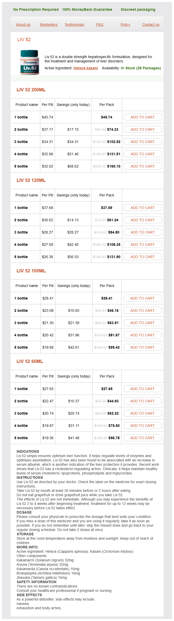
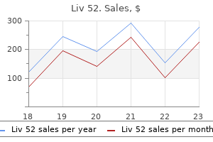
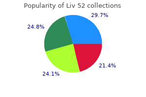
Liv 52 dosages: 200 ml, 120 ml, 100 ml, 60 ml
Liv 52 packs: 1 bottle, 2 bottle, 3 bottle, 4 bottle, 5 bottle

Order generic liv 52 from india
The greater occipital nerve pierces the fascia just below the superior nuchal ridge, along with the occipital artery. It supplies the medial portion of the posterior scalp as far anterior as the vertex. The lesser occipital nerve arises from the ventral main rami of the second and third cervical nerves. The lesser occipital nerve passes superiorly along the posterior border of the sternocleidomastoid muscle and divides into cutaneous branches that innervate the lateral portion of the posterior scalp and the cranial surface of the pinna of the ear. Less generally, repetitive microtrauma from working with the neck hyperextended. Tension-type headache, which is much more common, occasionally mimics the ache of occipital neuralgia. SignS and SympTomS A affected person suffering from occipital neuralgia experiences neuritic ache within the distribution of the greater and lesser occipital nerves when the nerves are palpated at the stage of the nuchal ridge. Some sufferers can elicit ache with rotation or lateral bending of the cervical backbone. Testing is aimed primarily at figuring out an occult pathologic course of or other diseases that will mimic occipital neuralgia (see "Differential Diagnosis"). Screening laboratory checks consisting of a whole blood rely, erythrocyte sedimentation rate, and automated blood chemistry must be performed if the diagnosis of occipital neuralgia is in question. Neural blockade of the higher and lesser occipital nerves can help confirm the analysis and distinguish occipital neuralgia from tension-type headache. The higher and lesser occipital nerves can simply be blocked on the nuchal ridge. More typically, patients with headaches involving the occipital area are suffering from tension-type headache. Axial computed tomography scan after intravenous distinction demonstrates a cystic-appearing, hypodense mass with irregular, rimlike distinction enhancement (arrow) within the medial facet of the left temporal lobe. To perform neural blockade of the greater and lesser occipital nerves, the affected person is positioned in a sitting position with the cervical spine flexed and the forehead on a padded bedside table. For remedy of occipital neuralgia or other painful situations involving the larger and lesser occipital nerves, a complete of eighty mg methylprednisolone is added to the native anesthetic with the primary block, and 40 mg of depot steroid is added with subsequent blocks. After the skin is prepared with antiseptic resolution, a 1�-inch, 22-gauge needle is inserted just medial to the artery and is superior perpendicularly until the needle approaches the periosteum of the underlying occipital bone. Paresthesias could additionally be elicited, and the affected person must be warned of this chance. The lesser occipital nerve and several superficial branches of the higher occipital nerve are then blocked by directing the needle laterally and slightly inferiorly. This vascularity, coupled with the shut proximity to arteries of each the greater and lesser occipital nerves, signifies that the clinician must fastidiously calculate the whole dose of local anesthetic that can be safely given, especially if bilateral nerve blocks are being performed. This vascularity and the proximity to the arterial supply give rise to an elevated incidence of postblock ecchymosis and hematoma formation. These complications may be decreased if guide stress is applied to the area of the block instantly after injection. Application of chilly packs for 20 minutes after the block also can lower the quantity of ache and bleeding. Care should be taken to keep away from inadvertent needle placement into the foramen magnum, as a result of the subarachnoid administration of native anesthetic in this area results in instant complete spinal anesthesia. As with different headache syndromes, the clinician must be positive that the analysis is right and that the patient has no coexistent intracranial illness or illness of the cervical backbone that could be erroneously attributed to occipital neuralgia. Clinical Pearls the most common purpose that larger and lesser occipital nerve blocks fail to relieve headache ache is that the affected person has been misdiagnosed. Vallejo R, Benyamin R, Kramer J: Neuromodulation of the occipital nerve in ache management, Tech Reg Anesth Pain Manag 10(1):12�15, 2006. In Atlas of interventional pain administration, ed 3, Philadelphia, 2009, Saunders, pp 24�28. Also often known as idiopathic intracranial hypertension, pseudotumor cerebri is seen most frequently in overweight ladies between the ages of 20 and 45 years. If epidemiologic research look solely at overweight ladies, the incidence will increase to roughly 20 circumstances per one hundred,000 patients. Predisposing elements embrace ingestion of various medications together with tetracycline, vitamin A, corticosteroids, and nalidixic acid (Table 8-1). In many sufferers, nonetheless, the exact explanation for pseudotumor cerebri stays unknown.
Discount liv 52 200 ml overnight delivery
It is characterised by tenderness and burning ache within the plantar floor of the forefoot, with painful paresthesias within the two affected toes. This ache syndrome is believed to be brought on by perineural fibrosis of the interdigital nerves. Radionuclide bone scanning could also be useful to determine stress fractures of the metatarsal or sesamoid bones that may be missed on plain radiographs. Coronal (short axis) T2-weighted magnetic resonance image through the forefoot demonstrates a hypointense lesion situated between the third and fourth metatarsal heads (arrows). A whole of three mL non-epinephrine-containing native anesthetic and forty mg methylprednisolone is drawn up in a 12-mL sterile syringe. The affected interdigital space is identified, the dorsal floor of the foot at this point is marked with a sterile marker, and the skin is ready with antiseptic answer. While the clinician is slowly injecting, the needle is advanced from the dorsal surface of the foot toward the palmar surface. Because the plantar digital nerve is situated on the dorsal facet of the flexor retinaculum, the needle should be advanced virtually to the palmar surface of the foot. The needle is eliminated, and stress is utilized to the injection website to keep away from hematoma formation. Because of the confined area of the delicate tissues surrounding the metatarsals and digits, mechanical compression of the blood supply after injection is a chance. The illness could also be recognized as a result of the characteristic radiographic findings of collapse of the second and, less commonly, third metatarsal head or heads. Like the scaphoid, the second metatarsal joint is extraordinarily vulnerable to this illness because of the tenuous blood supply of the articular cartilage. This blood provide is definitely disrupted, and this often leaves the proximal portion of the bone without nutrition and results in osteonecrosis. Investigators consider that the relative immobility of the second and third metatarsals, combined with the intense load transmission, makes these bones significantly susceptible to the event of avascular necrosis. The ache is deep and aching, and the affected person typically complains of increased pain on weight bearing and a limp when strolling. The affected person may or could not have a transparent historical past of foot trauma that may be identified because the inciting incident for the disease. Subtle swelling over the affected joint or joints may be appreciated on careful physical examination. B, Early collapse of the dorsal central portion of the metatarsal with flattening of the articular floor. C, Further flattening of the metatarsal head with continued collapse of the central portion of the articular surface with medial and lateral projections; the plantar articular cartilage remains intact. D, Loose our bodies form from fractures of lateral projections and separation of a central articular fragment. E, End-stage degenerative arthrosis with marked flattening of the metatarsal head and joint area narrowing. Administration of gadolinium adopted by postcontrast imaging may assist delineate the adequacy of blood supply; contrast enhancement of the metatarsal joint is an effective prognostic sign. Ultimately, surgical repair within the type of total joint arthroplasty is the remedy of alternative. Injection of the joint with local anesthetic and steroid is a comparatively safe method if the clinician is attentive to detail and specifically makes use of small amounts of local anesthetic and avoids high injection pressures that may further damage the joint. Approximately 25% of sufferers will complain of a transient enhance in pain after this injection method, and sufferers should be warned of this risk. SignS and SympTomS the pain of plantar fasciitis is most severe when first strolling after a period of non�weight bearing and is made worse by prolonged standing or strolling. Patients can also have tenderness alongside the plantar fascia because it moves anteriorly. Pain is increased by dorsiflexing the toes, which pulls the plantar fascia taut, and then palpating along the fascia from the heel to the forefoot. Although characteristic radiographic adjustments are lacking in plantar fasciitis, radionuclide bone scanning might show elevated uptake the place the plantar fascia attaches to the medial calcaneal tuberosity; it can also rule out stress fractures not seen on plain radiographs. This sagittal quick tau inversion recovery magnetic resonance picture demonstrates discontinuity of the plantar fascia, with extensive edema of the flexor digitorum brevis muscle (arrowhead). This sagittal T2-weighted magnetic resonance picture demonstrates a big gentle tissue mass in the plantar side of the foot. The mass is homogeneous and exhibits a thick capsule, simulating a fluid assortment.
Purchase cheap liv 52 line
CompliCaTionS and piTfallS Careful remark for the event of decrease extremity compartment syndrome through the early part of this condition is important if bleeding is important, especially in anticoagulated sufferers. Given the overlap of symptoms of tennis leg with deep venous thrombosis, the clinician must have a high index of suspicion for the event of deep venous thrombosis, especially during the rest part of recovery or if anticoagulants have been discontinued. As the harm to the musculotendinous unit heals, scar formation can happen and may result in continual pain and functional incapacity. If this happens, surgical excision and reconstruction of the musculotendinous unit could also be required. A excessive index of suspicion for the insidious onset of lower extremity compartment syndrome or deep venous thrombosis is necessary, to keep away from disaster. Kwak H-S, Lee K-B, Han Y-M: Ruptures of the medial head of the gastrocnemius ("tennis leg"): scientific end result and compression impact, Clin Imaging 30(1):48�53, 2006. Pai V, Pai V: Acute compartment syndrome after rupture of the medial head of gastrocnemius in a toddler, J Foot Ankle Surg 46(4):288�290, 2007. TreaTmenT Initial treatment of the pain and practical incapacity associated with tennis leg contains rest, elevation, use of elastic compressive wraps, and utility of ice to the affected extremity to reduce swelling and ache. The ankle joint is susceptible to the event of arthritis from varied situations which have the flexibility to damage the joint cartilage. Less widespread causes embody the collagen vascular diseases, infection, villonodular synovitis, and Lyme disease. Collagen vascular disease typically manifests as polyarthropathy quite than as monarthropathy limited to the ankle joint, though ankle ache secondary to collagen vascular illness responds exceedingly properly to the therapy modalities described right here. Magnetic resonance imaging of the ankle is indicated within the case of trauma, if the diagnosis is in query, or if an occult mass or tumor is suspected. Bursitis of the ankle and entrapment neuropathies such as tarsal tunnel syndrome may confuse the diagnosis; both these conditions could coexist with arthritis of the ankle. Primary and metastatic tumors of the distal tibia and fibula and backbone, in addition to occult fractures, may manifest in a way just like arthritis of the ankle. TreaTmenT Initial therapy of the pain and functional disability related to arthritis of the ankle includes a mixture of nonsteroidal antiinflammatory drugs or cyclooxygenase-2 inhibitors and bodily remedy. To perform intraarticular injection of the ankle, the affected person is placed within the supine place, and the skin overlying the ankle joint is prepared with antiseptic answer. With continued disuse, muscle wasting may occur, and a frozen ankle secondary to adhesive capsulitis might develop. The needle is fastidiously advanced via the pores and skin, subcutaneous tissues, and joint capsule and into the joint. Physical modalities, together with local heat and gentle rangeof-motion exercises, must be introduced several days after the patient undergoes injection. At this level, a triangular indentation indicating the joint space is definitely palpable. The injection technique described is extremely effective in treating the ache of arthritis of the ankle joint. In Atlas of pain management injection strategies, ed 2, Philadelphia, 2007, Saunders, pp 497�500. The midtarsal joints are prone to the event of arthritis from varied circumstances which have the ability to harm the joint cartilage. Collagen vascular illness usually manifests as polyarthropathy rather than as monarthropathy restricted to the midtarsal joint, though midtarsal ache secondary to collagen vascular disease responds exceedingly properly to the therapy modalities described right here. SignS and SympTomS Most patients present with ache localized to the dorsum of the foot. Some patients complain of a grating or popping sensation with use of the joints, and crepitus could also be present on physical examination. In addition to pain, patients with arthritis of the midtarsal joint often expertise a gradual decrease in useful ability because of decreased midtarsal range of motion that makes simple on a regular basis duties similar to walking and climbing stairs fairly difficult. Magnetic resonance imaging of the midtarsal joint is indicated if aseptic necrosis, an occult mass, or a tumor is suspected. Primary and metastatic tumors of the foot can also manifest in a manner similar to arthritis of the midtarsal joint. TreaTmenT Initial treatment of the ache and useful incapacity related to arthritis of the midtarsal joint includes a combination of nonsteroidal antiinflammatory medicine or cyclooxygenase-2 inhibitors and physical therapy. The major complication of intraarticular injection of the midtarsal joint is an infection, although this must be exceedingly uncommon if strict aseptic approach is followed.
Purchase liv 52 online pills
Immunohistochemical expression of gastric mucin and p53 in minimal deviation adenocarcinoma of the uterine cervix. Gastric-type endocervical adenocarcinoma: An aggressive tumor with unusual metastatic AdenocArcinomAs of the cervix And their Precursors And neuroendocrine tumors � 157. Invasive endocervical adenocarcinoma: A new pattern-based classification system with necessary clinical significance. Role of lymphovascular invasion in sample C invasive endocervical adenocarcinoma. Analysis of 29 instances with emphasis on minimally invasive cervical tumors and the ability of metastases to simulate main ovarian neoplasms. Pattern classification of endocervical adenocarcinoma: Reproducibility and evaluate of standards. Adenocarcinoma of the uterine cervix with choriocarcinomatous and hepatoid differentiation: Report of a case. Colloid carcinoma of the intestinal type within the uterine cervix: Mucin immunohistochemistry. Endocervical adenocarcinoma with morphologic options of both usual and gastric types: Clinicopathologic and immunohistochemical analyses and high-risk detection by in situ hybridization. The prognostic significance of histologic type in early stage cervical cancer � A multi-institutional study. Absence of human papillomavirus an infection in minimal deviation adenocarcinoma and lobular endocervical glandular hyperplasia. Endocervical adenocarcinomas with prominent endometrial or endomyometrial involvement simulating major endometrial carcinomas. Mucinous tumors of the ovary associated with mucinous adenocarcinomas of the cervix: A clinicopathologic evaluation of sixteen circumstances. Well-differentiated villoglandular adenocarcinoma of the uterine cervix: Oncogene/tumor suppressor gene alterations and human papillomavirus genotyping. Well differentiated villoglandular adenocarcinoma of uterine cervix: A clinicopathological study of 24 cases. Adenocarcinoma of the uterine cervix with predominantly villoglandular papillary development pattern. Villoglandular adenocarcinoma of the cervix: Clarity is required on the histological definition for this difficult analysis. Villoglandular papillary adenocarcinoma of the uterine cervix: A clinicopathological analysis of 13 instances. Clinicopathologic features of villoglandular papillary adenocarcinoma of the uterine cervix. Primary signet-ring cell carcinoma of the cervix: Case report and evaluate of the literature. Colloid carcinoma of the uterine cervix: A case report with respect to immunohistochemical analyses. Signet-ring cells in squamous cell carcinoma of the cervix and in non-neoplastic ectocervical epithelium. Adenocarcinoma of the uterine cervix of intestinal kind containing numerous Paneth cells. Pseudomyxoma-type invasion in gastrointestinal adenocarcinomas of endometrium and cervix: A report of 2 instances. Histopathologic subtyping of cervical adenocarcinoma reveals increasing incidence rates of endometrioid tumors in all age groups. Minimal-deviation endometrioid adenocarcinoma of the cervix: A case report with ultrastructural evaluation demonstrating abnormal ciliation of the tumor cells. Endometrial endometrioid adenocarcinoma of the uterine corpus involving the cervix: Some instances probably symbolize impartial primaries. Minimal deviation endometrioid adenocarcinoma of cervix: A clinicopathological and immunohistochemical examine of two instances. Minimal-deviation endometrioid adenocarcinoma of the uterine cervix: A report of 5 cases of a particular neoplasm that may be misinterpreted as benign.
Generic liv 52 100ml fast delivery
It was hoped that it might exchange the barbiturates as a sedative and due to this fact prevent the frequent deaths caused from unintended and intentional barbiturate overdosage. Thalidomide given to girls during being pregnant produced delivery defects, most notably phocomelia, an arrested growth of the limbs of the affected newborn. The more lucky have been born with only disfigurations of the nostril, eyes, and ears. The most severely aftlicted died of malformation of the heart or gastrointestinal tract. This drug catastrophe spurred the Congress to strengthen the existing legal guidelines regarding new drugs. Without dissent, on October 10, 1962, the KefauverHartis Drug Amendments to the Food, Drug, and Cosmetic Act of 1938 were passed by each houses of Congress. The Comprehensive Drug Abuse Prevention and Control Act of 1970 established 5 "schedulesn for the classification and control of drug substances which are subject to abuse. These schedules present for reducing ranges of control, from schedule I to schedule V. The medication in the five schedules may be described as follows: � Schedule I: Drugs with no accepted medical use, or different substances with a excessive potential for abuse. In this class are morphine, cocaine, methamphetamine, amobarbital, and different such drugs. In this category are specified quantities of codeine, hydrocodone, and related agents. In this class are specified quantities of difenoxin, diazepam, oxazepam, and related brokers. Included in this category are specified portions of dihydrocodeine, diphenoxylate, and comparable brokers. The pregnancy classes A, B, C, D, and X are faraway from all human prescription drug and biological product labeling. The requirement nows that the labeling features a abstract of the dangers of utilizing a drug during pregnancy and lactation, a discussion of the data supporting that summary, and relevant data to help health care providers make prescribing selections and counsel girls about the use of medication during pregnancy and lactation. The labeling additionally contains relevant information about pregnancy testing, contraception, and infertility for health care suppliers prescribing for females and males of reproductive potential. It creates a consistent format for offering information about the dangers and benefits of prescription drug and/or biological product use during being pregnant and lactation and by females and males of reproductive potential. Medication Exposures During Pregnancy and Lactation Every lady in the general inhabitants has a 3% to 5% risk of having a child with a delivery defect or psychological retardation. Two essential components to consider when assessing the teratogenic potential of a medicine are the stage of being pregnant at which the exposure occurred and the quantity of treatment taken. It is crucial to consider every publicity on a case-by-case basis in order to give an accurate risk assessment. All overseas drug manufacturing and distributing firms whose merchandise are imported into the United States are also included on this regulation. Exempt from the registration and itemizing necessities are hospitals, clinics, and the assorted well being practitioners who compound pharmaceutical preparations to be used in their respective establishments and practices. Also exempt are analysis and educating establishments during which drug merchandise are ready for functions aside from sale. Under this method, the first four numbers, the labeler code of the 10-character code, determine the manufacturer or distributor. The final six numbers identify the drug formulation and the commerce bundle dimension and type. The section that identifies the drug formulation is the product code, and the segment that identifies the trade package deal dimension and kind is the package code. The producer or distributor determines the ratio of use of the last six digits for the two codes, as a 3:3 digit product code to bundle code configuration. Only one such sort of configuration could also be chosen for use by a manufacturer or distributor, who then assigns a code number to each product to be included in the drug listing. Even when a drug producer discontinues the manufacture and distribution of a product, the quantity may not be used again. It also prohibits resale by well being care establishments of Orphan Drug Ad of 1983 Drugs intended for the treannent of "rare diseases and conditions" may be designated orphan drugs to help promote analysis on rare ailments. The legislation supplies tax credit and designated years of promoting exclusivity as incentives.
Buy liv 52 100 ml with amex
These tumors are distinguished from pseudoendometrioid metastases of other origins by scientific findings and from Krukenberg tumors with a glandular component by the absence or rarity of signet-ring cells. The massive to small gland sample is in marked contrast to the looks of a Krukenberg tumor. As with many other glandular metastases, marked maturation with a cystic pattern has resulted. Typical heterogenous morphology with small glands in a desmoplastic stroma (left) abruptly interfacing with a glandular sample with a garland-like cribriform sample and conspicuous dirty necrosis. The web site of the primary tumors in one research was 77% within the rectosigmoid, 9% ascending colon, 9% cecum, 5% descending colon, and barely the transverse colon. Metastatic colonic carcinomas are among the commonest ovarian tumors, excluding these within the sex cord�stromal class, that are associated with estrogenic or androgenic manifestations on account of functioning stroma. The ovarian tumors, which are bilateral in ~60% of cases, might kind nonspecific stable plenty, but are more typically solid and cystic, typically being predominantly cystic. Sectioning sometimes reveals friable or mushy yellow, red, or grey tissue and cysts with necrotic, mucinous, clear, or bloody contents. Multiple thin-walled cysts with mucinous contents may not often simulate a mucinous cystic neoplasm. On low energy the ovarian parenchyma is usually effaced but in smaller tumors it may separate nodules of tumor that often have a prominent desmoplastic stroma. Classic garland-like association of cribriform glands at the periphery of a giant gland whose lumen is crammed with necrotic debris. Typical colloid morphology, which is rarely a feature of main mucinous carcinoma of the ovary. Left: Metastatic clear cell colonic adenocarcinoma mimics a primary ovarian clear cell adenocarcinoma. Right: A hanging micropapillary sample doubtlessly mimics a serous adenocarcinoma. Endometrioid adenocarcinoma, which is within the differential diagnosis, is often negative for both markers. Mucin-containing goblet cells may be scattered among mucin-free cells, however are sometimes absent. Cysts lined by well-differentiated mucin-rich or flattened nonspecific epithelium are current in occasional circumstances. Occasionally the appearance is that of a colloid carcinoma, or, when the first intestinal tumor (sometimes small intestinal) is of the rare clear cell type, an endometrioid carcinoma of secretory sort or clear cell carcinoma is simulated. The stroma could additionally be desmoplastic, edematous, or mucoid, and incorporates luteinized cells in 30% of instances. Metastatic tuMors to the ovary � 565 Differential prognosis Primary endometrioid adenocarcinoma. Features favoring or establishing a prognosis of metastatic colonic carcinoma embody: � A recognized major intestinal most cancers, bilaterality, multiple nodules, soiled necrosis (particularly when in depth and associated with a quantity of of the other options listed here), segmental necrosis, and higher-grade nuclei and mitotic activity than in endometrioid carcinomas with related degrees of glandular differentiation. However, metastatic intestinal carcinomas may focally have a deceptive appearance that will simulate a benign or borderline mucinous tumor. The presence of bilaterality, outstanding dirty necrosis, and the immunoprofile noted above all favor or establish the latter prognosis. The ovarian spread is usually associated with pseudomyxoma peritonei (see Chapter 20). The appendix may exhibit a mucocele or exhibit no gross abnormality; generally solely the presence of appendiceal serosal mucin signifies an appendiceal lesion. Bilateral mucinous cystic neoplasms are seen with jelly-like materials being evident on the proper. The sectioned floor shows the everyday jelly-like appearance of the cyst contents in these instances with a spotlight of more stable yellow neoplastic tissue. The ovarian parenchyma is largely replaced by swimming pools of mucin with scant epithelium. Pools of mucin dissect through an otherwise unaltered ovarian stroma, so-called pseudomyxoma ovarii. Typical tall columnar cells are seen and have extensively retracted from the adjoining stroma. This feature is typical of metastatic low-grade appendiceal mucinous tumors but may be seen with teratomaassociated primary tumors.
Order liv 52 canada
Blood and urine cultures ought to be instantly obtained in all patients thought to be affected by retropharyngeal abscess to enable quick implementation of antibiotic therapy while the workup is in progress. Gram-negative and anaerobic antibiotic protection also needs to be started empirically instantly after blood and urine tradition samples are taken. Antibiotic therapy may be tailor-made to the culture and sensitivity stories as they turn into obtainable. Antibiotics alone are not often profitable in therapy of retropharyngeal abscess until the prognosis is made very early in the course of the disease; surgical drainage of the abscess is required to impact full restoration. Careful attention to airway administration is obligatory in patients suspected of affected by retropharyngeal abscess, and early endotracheal intubation in a managed setting is most well-liked to waiting till respiratory compromise is already present. The insidious onset of airway compromise related to retropharyngeal abscess can lull the clinician into a sense of false security, and failure to recognize the spread of infection into the central nervous system can lead to everlasting neurologic harm. If retropharyngeal abscess is suspected, the algorithm listed in Table 20-2 should be followed. Clinical Pearls Delay in analysis places the patient and clinician at large danger for a poor outcome. The clinician ought to assume that all patients who present with sore throat, fever, neck pain, painful and troublesome swallowing, and posterior pharyngeal swelling are affected by retropharyngeal abscess until proved otherwise and will deal with these sufferers accordingly. TreaTmenT the rapid initiation of therapy of retropharyngeal abscess is necessary if the patient is to avoid important morbidity and mortality. The remedy of retropharyngeal abscess is aimed toward two targets: (1) therapy of the infection with antibiotics and (2) drainage of the abscess to relieve compression on adjacent constructions including the airway. Wever K, Armstrong A: Retropharyngeal and mediastinal abscesses: postanesthetic complication of gynecologic surgery, Obstet Gynecol 94(5):857, 1999. Brook I: Microbiology and administration of peritonsillar, retropharyngeal, and parapharyngeal abscesses, J Oral Maxillofac Surg 62(12):1545�1550, 2004. Frequently, the patient presents with midline ache after extended activity requiring hyperextension of the neck, corresponding to portray a ceiling, or following prolonged use of a pc monitor with too excessive a focus. Decreased vary of movement is invariably present, and ache will increase with extension of the decrease cervical and higher thoracic backbone. Testing is aimed primarily at figuring out an occult pathologic process or different diseases that may mimic cervicothoracic bursitis (see "Differential Diagnosis"). Screening laboratory exams consisting of a whole blood count, erythrocyte sedimentation price, antinuclear antibody testing, and automated blood chemistry must be carried out to rule out occult inflammatory arthritis, an infection, and tumor. Pain syndromes that may mimic cervicothoracic bursitis embody cervical pressure, cervical fibromyositis, inflammatory arthritis, and disorders of the cervical spinal twine, roots, plexus, and nerves. Congenital abnormalities corresponding to Arnold-Chiari malformation and Klippel-Feil syndrome may also manifest equally to cervicothoracic bursitis. If these remedies fail to present speedy relief, injection of local anesthetic and steroid into the world between the interspinous ligament and the ligamentum flavum is a reasonable subsequent step. For symptomatic reduction, cervical epidural block, blockade of the medial branch of the dorsal ramus, or intra-articular injection of the facet joint with native anesthetic and steroid may also be considered. The proximity to the vertebral artery, mixed with the vascular nature of this area, renders the potential for intravascular injection high, and the injection of even a small amount of native anesthetic into the vertebral artery may end up in seizures. Many patients also complain of a transient improve in headache and cervicalgia after injection of the cervical facet joints. T1-weighted (A) and T2-weighted (B) sagittal magnetic resonance imaging of the cervical backbone that demonstrates lack of segmentation of the C4 and C5 vertebrae (arrows). Injection of local anesthetic and steroid is extremely efficient in the treatment of cervicothoracic bursitis pain that fails to respond to more conservative measures. In Atlas of pain administration injection techniques, ed 2, Philadelphia, 2009, Saunders, pp 52�56. Yung e, Asavasopon S, Joseph J, et al: Screening for head, neck, and shoulder pathology in patients with upper extremity indicators and symptoms, J Hand Ther 23(2):173�186, 2010. Brachial plexopathy has many causes, but a few of the extra widespread ones embrace compression of the plexus by cervical ribs or irregular muscle tissue. SignS and SympTomS Patients affected by brachial plexopathy complain of pain radiating to the supraclavicular area and upper extremity. The pain is neuritic and may take on a deep, boring quality because the plexus is invaded by tumor. Movement of the neck and shoulder exacerbates the pain, so patients often try to keep away from such motion.
Cheap liv 52 amex
The clinician could really feel some resistance to injection due to fibrosis of the surrounding tissue. If important resistance is encountered, the needle is probably in the tendon or nodule and should be withdrawn until the injection can proceed without important resistance. In Atlas of ache administration injection techniques, ed 2, Philadelphia, 2007, Saunders, pp 275�277. The major complications associated with injection are associated to trauma to an infected or previously broken tendon. The joints are additionally subject to invasion by tumor from main malignant tumors, including thymoma, or from metastatic disease. Laboratory evaluation for collagen vascular illness is indicated in patients affected by costosternal joint pain if other joints are involved. If trauma has occurred, costosternal syndrome could coexist with fractured ribs or fractures of the sternum itself, which could be missed on plain radiographs and will require radionuclide bone scanning for correct identification. Neuropathic pain involving the chest wall can also be confused or coexist with costosternal syndrome. Diseases of the constructions of the mediastinum are possible and may be difficult to diagnose. SignS and SympTomS Physical examination reveals that the patient vigorously makes an attempt to splint the joints by keeping the shoulders stiffly in a impartial place. The costosternal joints and adjoining intercostal muscular tissues may be tender to palpation. Use of an elastic rib belt might provide symptomatic aid and protect the costosternal joints from extra trauma. Radiograph of a sternum from a cadaver with rheumatoid arthritis exhibits giant erosions of the articular floor of each the manubrium (M) and the physique of the sternum (S). Subtle irregularities of the second and third sternocostal joints are evident, most prominently in the sternal facet of the left third sternocostal joint (arrowheads). The skin overlying the affected costosternal joints is ready with antiseptic solution. The costosternal joints are recognized; they want to be simply palpable as a slight bulging at the level the place the rib attaches to the sternum. After the needle is in proximity to the joint, 1 mL of solution is gently injected. Physical modalities, together with local heat and gentle range-ofmotion workouts, ought to be introduced a quantity of days after the affected person undergoes injection for costosternal joint ache. CompliCaTionS and piTfallS Because many pathologic processes can mimic the pain of costosternal syndrome, the clinician must carefully rule out underlying cardiac illness and ailments of the lung and structures of the 60 � Costosternal Syndrome 193 mediastinum. The major complication of the injection method is pneumothorax if the needle is positioned too laterally or deeply and invades the pleural space. The danger of this complication can be significantly decreased with strict consideration to accurate needle placement. The joint is also topic to invasion by tumor from main malignant tumors, including thymoma, or from metastatic disease. The CliniCal Syndrome the manubrium articulates with the body of the sternum by method of the manubriosternal joint on the angle of Louis. The manubriosternal joint is a fibrocartilaginous joint or synchondrosis, which lacks a real joint cavity. With severe trauma, SignS and SympTomS Physical examination reveals that the patient vigorously makes an attempt to splint the joint by keeping the shoulders stiffly in a impartial place. It is commonly traumatized throughout acceleration-deceleration accidents and blunt trauma to the chest. Laboratory analysis for collagen vascular disease is indicated in patients suffering from manubriosternal joint ache if other joints are involved. Pathologic processes that inflame the pleura, corresponding to pulmonary embolus, an infection, and Bornholm illness, may also confuse the prognosis and complicate treatment.
Real Experiences: Customer Reviews on Liv 52
Ford, 38 years: Secondary changes much like those seen in typical leiomyomas may be grossly apparent On microscopic examination, endothelium-coated plugs of benign clean muscle occupy the lumina of myometrial veins exterior of leiomyomas.
Yespas, 53 years: Tears of the medial meniscus are categorised by their orientation and shape (Table 98-2).
Kasim, 55 years: Deep Nabothian cysts of the endocervix: A attainable source of confusion with minimal-deviation adenocarcinoma (adenoma malignum).
Yugul, 33 years: Metastatic carcinoma, nevertheless, could rarely coexist with decidual cells in the identical node.
Giacomo, 46 years: These tumors were disproportionately grade 3 and related to myometrial and lymphovascular invasion.
9 of 10 - Review by G. Akascha
Votes: 70 votes
Total customer reviews: 70