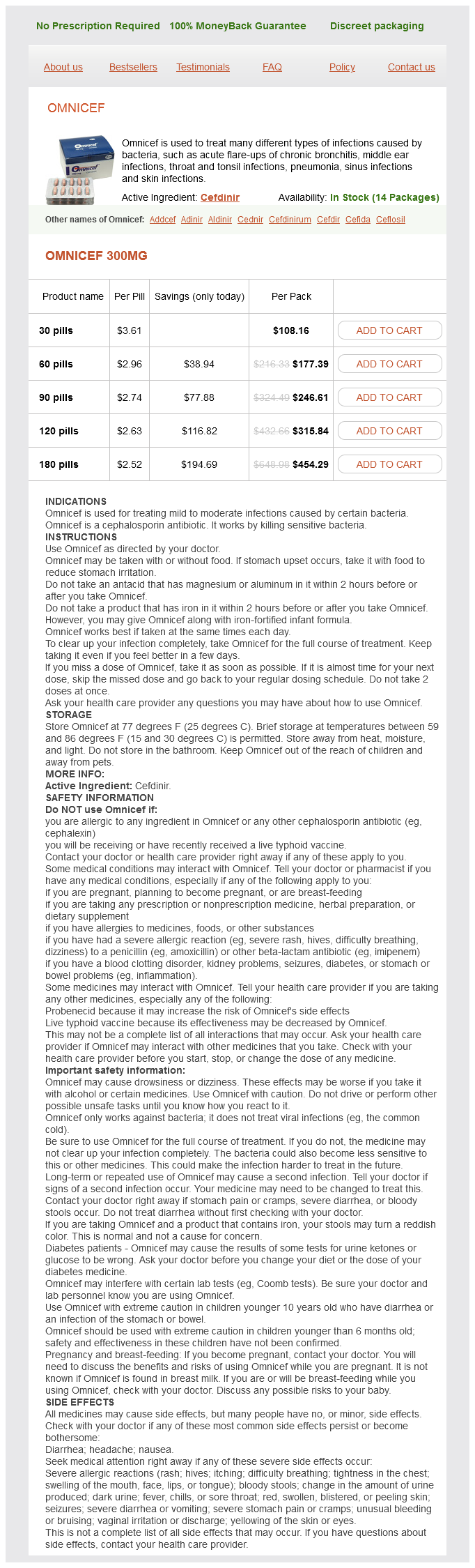
Omnicef dosages: 300 mg
Omnicef packs: 30 pills, 60 pills, 90 pills, 120 pills, 180 pills
Buy discount omnicef 300mg online
Closest to the epiphysis is the resting zone (also termed the reserve or prehypertrophic zone) of chondrocytes, beneath which lies the zone of proliferation with its flat, compact chondrocytes stacked in neat rows. As they move towards the metaphysis, these chondrocytes enter the zone of hypertrophy, the place each chondrocyte is giant, roughly spherical, and has an apparent area across the cell. The cartilage matrix becomes calcified on the lower reaches of this zone, resulting in death of the entrapped chondrocytes. Capillaries from the bone marrow invade upward between the spicules of calcified cartilage matrix, and osteoblasts combination alongside these vessels and lay down a thin layer of osteoid on this calcified cartilage scaffold. This area is rich in osteoclasts, which act to lower the variety of calcified cartilage spicules and the overall variety of trabeculae in the main spongiosa. The woven bone within the main spongiosa is roofed by lamellar bone as the secondary and tertiary spongiosa (mature trabeculae) are formed. This progressive modeling of cartilage to bone supplies the engine by which the bone grows in length. This adjustment occurs in the "cut back" zone within the metaphysis in young growing animals, the place osteoclasts are plentiful beneath the periosteum and osteoblasts are very active on the endocortical floor. Increases in bone width with age occur from growth at the periosteum as osteoblasts deposit osteoid on the outer floor of the existing cortex. Intramembranous Ossification Flat bones [like these of the calvarium (skull) and the scapula] and foci of woven bone are formed by intramembranous ossification. In this process, bone is laid down immediately in the mesenchymal collagenous matrix quite than by transmutation of a preformed cartilage model. At such sites, osteoblasts are derived from mesenchymal stem cells, and organized at random. Woven bone is normally present in few places within the skeleton: on the surface of calcified cartilage because of normal bone modeling, immediately subjacent to the deepest zone of articular cartilage (in small amounts), and at websites of tendon and ligament insertion. Molecular Regulation of Bone and Cartilage Development Bone and cartilage differentiation, growth, and mineralization are influenced by a selection of hormones, which are systemically out there molecules, in addition to development components, which are produced locally (Table 23. For example, Indian hedgehog (Ihh) is a master regulator of bone improvement, serving to control endochondral ossification by coordinating chondrocyte proliferation and differentiation in the development plate, and osteoblast differentiation within the primary spongiosa. All these ligands have regulatory exercise in the development plate and are important for regular improvement of the cartilage facilities that presage endochondral ossification. During skeletal development and repair, cell proliferation and differentiation in cartilage and bone are coordinated by cell�cell signaling. For instance, canonical Wnt signaling (via beta-catenin T-cell factor) is a key regulator of skeletogenesis by accelerating endochondral ossification and suppressing chondrocyte formation, resulting in shortening of the expansion plate and increased calcification of the hypertrophic zone. In explicit, Wnt/beta-catenin signaling works in concert with Ihh signaling within the development plate, with Ihh stimulating reserve zone chondrocytes to enter proliferation while Wnt acts downstream to promote osteoblast maturation. Similar controlling mechanisms of osteoblast proliferation and differentiation happen in adult mesenchymal progenitor cells during fracture restore, so initiation of bone formation in fracture restore initially requires Ihh signaling and later Wnt signaling in differentiated osteoblasts. Every part will comprise both old and new bone, and thus offers a glimpse on the history of the bone. The elements that mediate the set level for this balance embrace physical forces. Bone transforming is split into two major categories: targeted remodeling in response to harm or adjustments in biomechanical loading, and stochastic reworking in response to homeostatic mineral requirements. First, cartilage cores persist in trabecular and cortical bone for months or longer in rats. Third, the method of forming and shaping the diaphysis occurs by periosteal bone resorption and endosteal intramembranous bone formation on secondary spongiosa within the "reduce" zone in the metaphysis. Biomechanics Physical forces acting on the skeleton represent a significant extrinsic affect on postnatal bone improvement and maintenance. Cartilage thickness in a mature joint can also be affected by local stress and the surroundings created by physical exercise. In distinction, the hydroxyapatite mineral crystals within the collagen matrix impart compressive energy. Within the osseous matrix, osteocytes are thought to act in part as a community of mechanical sensory cells. The healthy skeleton is ready to frequently reply to mechanical stimuli by initiating or inhibiting bone reworking to keep bone structure for typical strains inside a standard physiological vary, a capability known as the mechanostat.
Diseases
- Basaran Yilmaz syndrome
- Richards Rundle syndrome
- Acrofacial dysostosis atypical postaxial
- Pure red cell aplasia
- Hyperglycinemia, isolated nonketotic type 2
- Pertussis
- Hypothalamic hamartoblastoma syndrome
- Paroxysmal dystonic choreoathetosis
- Stickler syndrome, type 2
- Uhl anomaly
Purchase 300 mg omnicef fast delivery
Major mechanisms of enormous intestinal diarrhea embody hypersecretion, large-bowel malabsorption, and/or direct colonic mucosal damage. Small intestinal malabsorption allows fermentable vitamins to enter the colon, where bacteria generate osmotically energetic merchandise that maintain water in the lumen. This course of, and the extent of the ensuing diarrhea, is finest demonstrated with cholera toxin. This process leads to severe diarrhea and demise, with little morphological proof of mucosal damage. Malabsorptive diarrheas related to loss of enterocytes, crypt cells or both, typically accompanied by Gastritis and gastric ulceration Esophageal, abdomen, and intestinal most cancers, and/or acute gastritis Ulcerative, hemorrhagic, and necrotic gastroenteritis and/or colitis Ulcerative, hemorrhagic, and necrotic gastroenteritis Table adapted from Handbook of Toxicologic Pathology, second ed. These lesions maybe site-specific, nonetheless, offering a clue to the sort of toxicant involved. By inflicting distension, these compounds elicit contractions that occur in either direction alongside the bowel. In a few circumstances, for example, organophosphate-insecticide toxicosis, diarrhea is through extended stimulation of muscarinic receptors due to inhibition of acetylcholinesterase at the synapse. The consequences of diarrhea are systemic in nature, and include dehydration, acidosis, and electrolyte alterations. Intracellular hydrogen ion concentrations increase and potassium concentrations lower. This electrolyte imbalance results in improper maintenance of intracellular pH ranges, and reduces the exercise of a number of enzyme systems. Reduced intracellular potassium is the results of a failure in mobile electrolyte transport. Vomiting maybe stimulated by direct mucosal irritation or by stimulation of the vomiting middle in the central nervous system. Vomiting may happen if the esophageal or pyloric lumen is obstructed by a scar or neoplasm. Bilateral stomach vagotomy and bilateral splanchnic nerve transection completely inhibit emesis induced by these anticancer medicine. Agonists of dopamine D2 receptors corresponding to apomorphine, L-dopa, and bromocryptine act in the chemoreceptor set off zone of the brainstem area postrema. Phenothiazine medication with vital dopamine D2 antagonists properties, similar to chlorpromazine and promethazine, block the emetic actions of dopamine D2 agonists. Emetine, the precept ingredient of ipecac, and opiates, such as morphine, act nonspecifically on the chemoreceptor trigger zone to provoke an emetic response. The space postrema is unprotected by a whole blood�brain barrier, thus permitting chemicals in blood to penetrate with relative freedom into this mind space. Polyps and neoplastic lots induced by carcinogenic agents as properly as partly or fully circumferential scars may end in physical obstruction. This activity represents contraction of each longitudinal and circular muscle layers. Neuronal networks concerned in peristalsis are advanced and incompletely understood; nonetheless, cholinergic excitation mechanisms play a major position. These compounds cause intestinal distension and elicit contractions that occur in both path alongside the bowel. Endogenous opioid peptides perhaps involved in segmentation motility, since morphine locks the gut right into a continuous segmentation sample of motility. In morphine-dependent rats, diarrhea, which is the other of the acute effects of morphine, is a major withdrawal event. Extrinsic nervous input affecting motility contains each stimulatory and inhibitory nerve fibers. Both vagal and sacral innervation to the massive intestine is particularly lively throughout defecation. Tonically energetic inhibitory neurons can account for a low responsiveness of circular muscular tissues to myogenic pacemakers. A model toxicity of inhibitory nerve enter that results in suppression of both cholinergic and serotoninergic synaptic transmission is norepinephrine overdose.
Buy discount omnicef online
Anatomic (structural) indices-Appropriate for mature and developing individuals A. Lesions (especially regional destruction of the parenchyma or particular neuronal populations) 3. Some modalities for assessing neurotoxicants and their results are carried out throughout life, like neurological and behavioral evaluations in addition to electrophysiological testing. Others are often confined to the postmortem setting, corresponding to macroscopic and microscopic evaluation (routine neuropathology). A few can be used in life as well as after death, like noninvasive imaging and assessments of nerve conduction velocities. This part offers an outline of typical anatomic pathology methods as well as a brief introduction to different key methods by which toxicologic neuropathology experience may be essential. Damage to a neuronal inhabitants often is accompanied by a rise in the variety of reactive glia within the quick neighborhood of the stricken cells. These reactions take place to render the glial cells more proficient at filling defects, supporting other Morphologic Evaluation Basic Patterns for Neurotoxicant-Induced Lesions Gross observations may reveal toxicant-induced alterations in neural structure at the time of necropsy. Generally irregular absolute mind weights or mind dimensions are judged to be biologically vital markers of neurotoxic damage. Histopathologic lesions are a common manifestation of neurotoxicant exposure, and certainly microscopic evaluation is often the most effective means for identifying many neurotoxicants. Acute cell death (neuronal necrosis) often presents as shrunken neurons with hypereosinophilic cytoplasm and dark, condensed, or fragmented nuclei. The difficulty in detecting small numbers of useless neurons or disintegrating axons microscopically in the "vast pink wasteland" of a big mind part has driven the event of special strategies designed to preferentially label toxicantinjured cells. Upper: Severe nigropallidal encephalomalacia (arrows) presents as nicely demarcated, bilaterally symmetrical zones of pallor (cream colored with a thin brown rim, where the adjoining grey matter is mild brown) in the affected basal nuclei of this horse that had consumed yellow star thistle (Centaurea solstitialis) for an extended period. Processing situations: Formalin fixation by immersion; paraffin embedding; H&E staining. Relative to adjoining cells with normal options, necrotic neurons (circles delineate consultant examples) have shrunken, spiky profiles; hypereosinophilic cytoplasm; dark, condensed or fragmented nuclei; and are sometimes bordered by clear retraction spaces. Cerebral cortex (upper) and cerebellum (lower) of a rat exposed to an unspecified neurotoxicant. As with injured neurons, reactive glia may be detected more simply if visualized using a special method to reveal the responding cells. Adult exposures to neurotoxic agents sometimes yield necrotic neurons, axonal or myelin degeneration, neuropil vacuolation, and/or reactive gliosis. The distribution of lesions among numerous neural cell populations often is determined by cell type�specific architectural or practical elements. For occasion the rich synaptic beds in the cerebral and cerebellar cortices as nicely as the hippocampus make these three areas common targets for neurotoxicants. The cell type� specific elements that direct neurotoxic results to a specific space also assist to explain species variations in responsiveness to numerous agents. The amino cupric silver process requires frozen tissue, while the Fluoro-Jade methodology could be carried out on routinely processed. These methods are more and more utilized in assessing the nervous system in regulatory studies. However, neural analysis in the majority of regulatory-type nonclinical studies is completed both as one facet of a basic screen for toxicity to all organ techniques. The differing aims of these research warrant a smaller listing of neural tissues for general studies relative to the bigger battery of nervous system domains used for research devoted mainly to an in depth neuropathology evaluation. When identified in advance, the timing of necropsies ought to be organized to occur when toxicant-induced neural lesions are at their peak, maintaining in thoughts that many agents exhibit a quantity of peaks characterised by distinct sorts of lesions. This precept is well illustrated by the excitotoxic agent kainic acid, by which neuronal necrosis happens at 1�4 days following exposure but synaptic terminal disintegration develops at 4�14 days. In many cases, neurotoxicity research are designed to recapitulate particular necessities listed in regulatory tips. The small size of the formaldehyde molecule permits it to simply and shortly penetrate dense neural tissues, whereas the reactive aldehyde group promotes speedy tissue fixation. Nervous system samples fastened on this manner could additionally be processed routinely together with samples from different organs, thus reducing the cost and labor required to prepare tissue blocks for histological sectioning. This method permits the use of fixative mixtures that include methanol-free formaldehyde (often freshly prepared from paraformaldehyde powder) and/or glutaraldehyde. Left: Compared to the small, spherical to oval, isolated nuclei of resting microglia (arrow), activated microglia have elongated, serpentine- or spindle-shaped nuclei and commonly type small nodules at sites of parenchymal damage.
Purchase 300mg omnicef mastercard
The unitized structure of the lesion with distinct borders, notably the presence of a skinny capsule, helps distinguish the cholangioma. The organic potential, in addition to the pathogenesis of the varied bile duct proliferative lesions, has generated a reasonable amount of analysis interest and a fair larger diploma of speculation. Cholangiofibrosis, cholangiofibromas, and cholangiocarcinomas are typically believed to represent a continuous spectrum of lesions. Neoplasms of bile duct epithelium are extraordinarily uncommon as spontaneous lesions in the rodent, although bile duct proliferation is comparatively widespread. Endothelial Cell Neoplasia Hemangiomas are benign neoplasms formed by endothelial cells. They kind pretty nicely circumscribed but hardly ever encapsulated plenty of vascular channels lined by a single layer of well-differentiated endothelial cells. Hemangiomas might assume a capillary type composed of multiple smallcaliber vessels compressed right into a mass. By gross inspection, hemangiosarcomas might seem as single or multiple raised, dark red, solid to fluidfilled lots. There are a number of brokers related to an elevated danger of hemangiosarcoma in rodents, particularly mice. Iron overload, vinyl chloride, and exposure to the pyrrolizidine, riddelliine, can cause hemangiosarcomas in mice. The differential prognosis of hepatic endothelial neoplasms presents few problems. The distinction between benign and malignant endothelial neoplasms of the liver relies totally on the comparatively regular mobile look and the uniform single-cell pavementing of hepatocyte surfaces by the benign hemangioma. Hemangiomas must be distinguished from dilated vascular spaces corresponding to telangiectasia and peliosis hepatis. Kupffer Cell Sarcoma Kupffer cell sarcomas might kind pale masses within the liver that are seen by gross remark and will have an space of central necrosis. Kupffer cell sarcomas could come up as primary neoplasms from the resident Kupffer cell population. Typically, Kupffer cell sarcomas form single or a quantity of pale nodules inside the liver. Histologically, Kupffer cell sarcomas are composed of spherical to oval uniform cells with ample foamy eosinophilic cytoplasm. Enlarged multinucleate cells could additionally be scattered all through the mass, and could be frequent. The development sample can be invasive, and tumor cells might dissect alongside sinusoids or blood vessels. Gall Bladder the gall bladder represents an unusual consideration within the evaluation of toxicity. The lesions embrace edema, ulceration, transmural irritation, and granulation tissue formation. Inflammatory lesions of the gall bladder wall may also appear in association with conditions that cause vasculitis. In toxicology studies, treatment-related vasculitis must be readily differentiated from cholecystitis based upon the appearance of typical vascular lesions in gall bladder, and often in different tissues. Dogs are also susceptible to an uncommon lesion, cystic mucinous hyperplasia, of the gall bladder mucosa. In toxicology studies, it has been reported to result from long-term administration of progestagens corresponding to oral contraceptives and corticosteroids. The lesion is characterized by intensive hyperplasia of mucosal epithelium forming quite a few long intraluminal projections with cysts that may include various, sometime plentiful, accumulation of mucus. Cytoplasmic adjustments in gall bladder mucosal epithelium have been reported as a manifestation of toxicity in some research. In mice, accumulation of eosinophilic crystalline materials, also called hyalinosis, was observed in gall bladder and stomach mucosal epithelium following long-term administration of Penicillin V.
White Dragon Flower (Orris). Omnicef.
- How does Orris work?
- What is Orris?
- Dosing considerations for Orris.
- Are there safety concerns?
- Purifying blood, skin diseases, bronchitis, cancer, improving appetite and digestion, inflammation of the spleen, liver and kidney problems, vomiting, constipation, bad breath, teething pain, and other conditions.
Source: http://www.rxlist.com/script/main/art.asp?articlekey=96636
Order generic omnicef on line
The trend toward product registration in multiple geographic regions has led to efforts toward standardizing regulatory requirements around the globe. Wide consensus exists that nonclinical safety evaluation research help human scientific trials of equal length. Some exceptions exist to these rules, particularly when the risk�benefit evaluation reveals that exposure to the potential harmful results of a compound is less detrimental to the patient than the withdrawal of a drug providing obvious profit. In distinction, decreased tolerance for threat is current when coping with diseases thought of "minor" or cosmetic, or for which there are protected alternatives. Experimental Design for Discovery and Safety Studies Studies undertaken throughout product discovery and improvement generally must be designed prematurely. Specific sections of standard research protocols describe the take a look at article and control materials, the animal demographics, methods, and how knowledge high quality might be assured. The toxicologic pathologist shall be instrumental in making certain the accuracy of tissue and fluid sampling in addition to the analytical battery to be used. In addition, the pathologist ought to evaluate other portions of the research protocol. For example, acute research in nonhuman primates could additionally be designed during which the test topics are immature (younger than three. In basic, doses for animal research are chosen that are a multiple of the effective dose as predicted from in vitro and efficacy discovery research, where doses were escalated until intolerance was observed. From this start line, doses are decreased and given over a quantity of days (often various between 5 and 14 days, depending on compound availability). Typical pathology endpoints are organ weights; clinical chemistry and hematologic parameters (comparable to the check panels performed within the diagnostic pathology setting) in blood samples; and histopathologic modifications in specimens from all main organs in addition to demonstrably distinctive organ regions. Where attainable, baseline knowledge collection (prior to preliminary treatment) is obtained for noninvasive (clinical pathology values, whole body weight) or minimally invasive. The full constellation of pathology endpoints usually is examined at the end of the treatment interval (the "terminal necropsy") and likewise after a treatment-free interval (the "restoration necropsy"). The group sizes for scheduled necropsies rely upon the timing with respect to the ultimate treatment. For terminal necropsies, the numbers of animals for all groups (including the management cohort) normally are set at 5 males for rodents and a couple of males for nonrodents for discovery research, and 10 per intercourse for rodents and 4�5 per sex for nonrodents for regulatory research. For example; preliminary group sizes in 2-year carcinogenicity research in rodents normally are set at 50�65 per intercourse per remedy dose for a last per group count of 25 at termination. Toxicologic pathologists are essential members of the discovery and improvement program, significantly by way of their advocacy to keep away from major experimental errors. For occasion, pathologists can make sure that sampling practices will permit acquisition of each morphologic and medical pathology parameters, and avoid interference from other sampling on these parameters. Pathologists can also argue towards the frequent practice of returning animals from discovery research to the colony for reuse, since residual modifications from the prior experiment complicate the interpretation of modifications observed in later research. Toxicologic pathologists are divided into those that believe that the diagnoses for various variations of the situation ought to be consolidated (the "lumpers") and folks who advocate that every function must be represented individually in the textual content description and data tables (the "splitters"). When the pathologist supplies multiple diagnoses to a typical pathological response, the analysis may be obscured and the data tables significantly expanded, making interpretation tough. However, occasionally splitting the prognosis will make clear the interpretation of a response, and in those situations splitting is appropriate. The separation of those elements into particular person diagnoses might make the detection of one other renal effect difficult by masking a possible compound-related effect on the behavior of a background lesion. Toxicologic pathology analysis is undertaken to discriminate real therapy effects from incidental findings ("background pathology" or "normal abnormalities"). Examples embrace minimal elevations in serum actions of hepatocellular leakage enzymes [e. Of course, variability amongst pathologists in whether or not or not these adjustments are recorded could impact the historical control knowledge tables, that are often used to interpret the incidence and severity of these sort of adjustments if the findings in the concurrent control group are complicated, inconclusive, or skewed. The most essential function of the pathologist follows the medical mannequin, where all the obtainable info is used to achieve an accurate prognosis.
300mg omnicef sale
These sensory nerve fibers have the potential when hyperstimulated to provoke a cascade of proinflammatory events and transmit nociceptive information to the central nervous system. Movement of a swallowed bolus from the mouth to the stomach and into the intestinal tract is a propulsive occasion, caudally progressing in entrance of a contraction wave of the circular muscle layers in varied digestive tract segments. Effective gastric emptying requires coordinated propulsive contractions in the antrum that progress to the pyloric canal, as nicely as correctly timed leisure of the higher duodenum. Some medication and toxicants scale back the speed of gastric emptying by producing contractions of the duodenum that abolish the antralduodenal strain gradient required for efficient emptying. Contractions of the circular muscle occur more or less randomly however are somewhat fixed in timing and site by the electrical slow waves or electrical management exercise generated initially in interstitial cells (of Cajal), which serve as bioelectrical pacemakers for smooth muscle. Clusters of propulsive contractions associated with contractile rings migrate 5�30 cm caudally, thereby propelling content material toward the cecum. Toxic agents that induce excessive migrating clustered contractions would abnormally pace propulsion via the small gut, which can impact electrolyte, nutrient, and water absorption. Migrating motor complexes are bands of contractile activity that transfer caudally over the abdomen and small gut during fasting to sweep digested food remains out of the abdomen. These motor complexes are energetic during times of fasting and continue till one other meal is consumed. The central nervous system exerts some extent of control over the activity of those complexes, however the precise complexes are initiated within the enteric nervous system. The mucosa is exposed to the very best concentration of orally administered compounds and may modify these compounds prior to their entry into the blood. Compound solubility and transport mechanisms will result in contact with different enzymes. Xenobiotic metabolism additionally can be carried out by luminal microorganisms; furthermore, luminal organisms can have an result on mucosal enzyme activity. Factors affecting the metabolic activity of the intestinal microflora should be taken into account in research of the biotransformation of orally ingested xenobiotics. Marked differences exist in microbial composition and metabolism of the intestine flora of different species of animals, and environmental factors similar to medication (especially antibiotics), food plan, and xenobiotics can modify microbial metabolism, and thus the toxicity, of overseas compounds. Presystemic clearance can occur for some toxicants both throughout the enterocyte or inside the gut lumen itself. This gut-associated first-pass effect represents the irreversible extraction and/or biotransformation of toxicants passing through enterocytes on their way into the lacteals or portal venous blood. Metabolites produced by enterocyte biotransformation can enter the intestinal lumen, the portal venous system, lacteals, or just remain saved within the cell. Conjugated water-soluble compounds formed throughout transport into enterocytes are probably to be excreted relatively shortly into the intestinal lumen, and due to this fact are cleared from the body (Table 15. After oral administration, when the concentration of a xenobiotic within the enterocyte could be very excessive, intestinal biotransformation reactions will usually be capacity-limited. The colon is three- to fivefold more lively than the small intestine in sure enzymatic processes. This augmentation of enzyme activity is just like that which happens within the liver. As occurs in the liver, chronic intake of ethanol may even enhance the level of exercise for several intestinal enzyme pathways. Sulfation proceeds extra rapidly in the proximal than in the distal small gut and colon. Enzymes of enterocytes are absolutely competent to perform oxidative, reductive, hydrolysis, and conjugation reactions. Some enzyme activities, such as nitroreductase and dechlorinase, perhaps attributable to each mucosal enzymes and luminal microflora. Both glucuronidation and sulfation reactions improve solubility of xenobiotics and thus play a major role in intestinal first-pass clearance for varied xenobiotics. Intestinal presystemic elimination of a dopamine prodrug, N-(N-acetyl-Lmethionyl)-O,O-bis(ethoxycarbonyl)dopamine, signifies that catechol ester hydrolysis, amido hydrolysis, and catechol O-methylation can even occur in enterocytes.
Purchase 300 mg omnicef amex
Retinoids, like isotretionoin (13-cisretinoic acid), have been recognized to affect the meibomian glands, inflicting blepharoconjunctivitis in humans and delicate conjunctival erythema and crusting of the eyelid margins in New Zealand white rabbits. Toxaphene administered orally to cynomolgus monkeys has been reported to produce meibomian gland irritation, enlargement, or each. The pathogenesis involves infected diverticula (arising from the glandular ducts) in both the dorsal (superior) and ventral (inferior) eyelids. The pharmacologic effect is suspected to be related to a failure to secrete glandular contents. Continued production of secretory material leads to glandular rupture and a considerable lipogranulomatous reaction. The mechanism is unknown however might involve eyelash lengthening in the course of the anagen phase of the lash development cycle and stimulation of melanogenesis. Ptosis (drooping eyelids, especially affecting the dorsal lid) and eyelid retraction are relatively frequent reactions to systemic or domestically administered xenobiotics. Other less common medicine associated with ptosis are tetraethylammonium, a ganglion blocker agent; guanethidine, a sympatholytic drug used to reduce lid retraction; penicillamine, a metal-chelator; the sedatives primidone, barbiturates and alcohol; and the chemotherapic agent vincristine. No common mechanism of action has been defined to clarify the toxic response to these brokers. On the opposite finish of the spectrum, stimulants similar to phenylephrine, amphetamine, and cocaine are related to higher eyelid retraction. Blepharitis (inflammation of the eyelid) and conjunctivitis (inflammation of the inside lid lining) are frequent reactions to quite a few topical ocular medicine. Drug preservatives similar to thimerosal and benzalkonium chloride in addition to antiviral agents like vidarabine, trifluridine, and idoxuridine are reported to trigger ocular irritation. Most generally used ophthalmic preparations current the potential for incidentally causing blepharitis and conjunctivitis. Mutations within the desmoglein 3 (Dsg3) gene had been reported to be the primary issue underlying ulcerative blepharitis in Dsg3bal and Dsg3bal-Pas mice (balding and balding Pasteur, respectively). These animals develop a pemphigus-like reaction with formation of suprabasilar vesicles affecting the mucocutaneous junctions. Such changes are identical to these noticed in humans and animals with pemphigus vulgaris. Among the medicine that do target these organs are phenazopyridine, sulfadiazine, and salicylazosulfapyridine. These medication are reported to cause marked necrosis of the lacrimal gland, with evolution to fibrosis through an unknown mechanism. Due to their close proximity, the cornea and conjunctiva are exposed concurrently. One of the most popular routes for clinical administration of ocular therapeutics is through subconjunctival injections. Subconjunctival administration of medicine associated with biodegradable drug implants designed for sustainable drug delivery is also frequent practice in nonclinical toxicology studies. These polymers degrade via hydrolysis of their ester bonds into lactic acids, glycolic acids, and caproic acid-and eventually into water and carbon dioxide. In most instances, these subconjunctival implants cause minimal to no tissue reaction, and when one does develop native conjunctival hyperemia and edema are the commonest responses. When subconjunctival injections are part of the study design, the conjunctiva ought to be evaluated microscopically, especially within the space of the injection. The conjunctiva and cornea are at nice threat from accidental publicity to caustic chemicals corresponding to sturdy acids and bases, which may easily erode the epithelium to expose the underlying tissues. In delicate acid burns the preliminary medical alterations are turbidity and dulling of the conjunctival surface with tissue hyperemia and probably petechial hemorrhages. There is loss of conjunctival epithelium adopted in delicate cases by fast regeneration (5�10 days). Acid and alkali burns (described in additional detail later) could cause neurotrophic keratitis, a type of corneal degeneration ensuing from altered corneal sensory enervation. Surfactants can disrupt the tear movie, sensitizing the corneal surface to an infection or deep ulceration. Among them, benzalkonium chloride, which is used to increase the penetration of certain medicine to the eye, could cause conjunctival hyperemia, corneal opacification, corneal edema, and infrequently epithelial degeneration and necrosis. The most typical acids which might be involved in ophthalmic injuries are sulfuric and sulfurous acids, chromic acid, nitric acid, hydrochloric acid, sulfur dioxide, and silver nitrate; the commonest bases are hydroxides of sodium, calcium, magnesium, ammonium, and potassium.
Buy cheap omnicef on line
Mineralocorticoids have results on ion transport by epithelial cells, particularly renal cells, resulting within the conservation of sodium (chloride and water) and lack of potassium. In the distal convoluted tubule of the mammalian nephron, a cation trade exists that promotes the resorption of sodium from the glomerular filtrate and the secretion of potassium into the lumen. Glucocorticoid hormones enhance glucose manufacturing with a concomitant breakdown of proteins for purposes of gluconeogenesis. Glucocorticoids additionally suppress inflammation along with attenuation of fibroplasia and immunological responses. The suppression of the immunological responses is essentially associated to the stabilization of lysosomal membranes of phagocytic cells, inhibition of a selection of lymphoid cell capabilities, and lysis of lymphocytes. The enhance in blood glucose is an important physiological response in antagonistic situations, however an important physiological effect of the glucocorticoids in stressful circumstances is to quench the inflammatory response to prevent it creating to the point the place it overwhelms the animal. In some situations, scientific indicators of hypoadrenocorticism may be noticed in affiliation with decrease urinary and plasma corticosteroid ranges. In Vitro Assessment In vitro studies are extraordinarily useful in determining the precise mobile consequences of xenobiotic exposure on steroidogenesis. In many situations, the outcomes of those in vitro assessments are helpful in correlating the development of adrenocortical degeneration to an inhibited pathway of steroidogenesis. This is a human adrenocortical carcinoma cell line that retains full adrenocortical steroidogenic enzyme functionality and might secrete aldosterone, cortisol, and androgens, estrogens, and progestogens and their precursors in response to acceptable problem. Rodent cell traces subsequently have little use in establishing mechanisms of toxicity, a minimal of when it comes to human relevance. Morphologic Evaluation Following the death of a test animal, morphological evaluation commences with macroscopic remark of the adrenal glands to detect modifications in dimension, color, and/or appearance. Subsequent histological examination of adrenal tissue (including each cortex and medulla) on midsagittal sections stained with hematoxylin and eosin (H&E) is carried out routinely. Microscopic examine may be supplemented by the use of particular stains and methods to affirm or identify a selected pigment, cell sort, or enzyme. Histomorphometric analysis can be utilized to assess refined variations in cell dimension, width of the totally different cortical zones, and cortical:medullary ratio. Likewise, the age of the test animal, to a lesser diploma, may be a factor within the development of chemically induced adrenocortical lesions. For instance, adrenal cortical necrosis was induced in rats at 50 days of age but not at 25 days by the administration of seven,12-dimethylbenz[a]anthracene. Adrenal hypertrophy could also be a results of stress or indeed might result from functional impairment of the adrenal cortex and decreased capacity to secrete glucocorticoids. Atrophy of the thymus and different lymphoid tissues is a helpful surrogate marker for adrenocortical competence, as this atrophy is induced by excess glucocorticoid secretion; similarly, the stress leukogram is helpful proof. Stress is a natural adaptive response designed to better equip the animal to Use of Animals as Models the adrenal cortex of animals is susceptible to develop proliferative and degenerative lesions, the etiology of which can be either spontaneous in nature or induced experimentally. Therefore, chemical testing using various domestic or laboratory animals is a sound means of assessing the toxic potential for people uncovered to varied xenobiotic chemicals. Species, Age, and Sex Variables the right alternative of species of check animal is crucial. This remark suggests that differences in metabolism play a role in the growth of adrenal cortical toxicity and in the inhibition of steroidogenesis. Elevation of the latter is diagnostic and proof of adrenocortical functional competence. By distinction, toxicity to the adrenal cortex could also be apparent as a end result of marked and infrequently irreversible histopathological lesions (degeneration, necrosis, and fibrosis). However, in circumstances of pharmacotoxicological inhibition of steroidogenic enzymes there may be no histopathological lesions however grossly impaired glucocorticoid manufacturing, clearly a toxicological concern. Prolonged use of exogenous glucocorticoids can mimic a syndrome of extra adrenal cortical function. More important from a toxicologic point of view are the degenerative effects of chemicals on the adrenal cortex that result in major adrenal cortical hypofunction.
Order genuine omnicef on-line
This treatment must be averted in children (except in these with hyperuricemia secondary to cancer and chemotherapy). Drug interplay of allopurinol with alcohol, caffeine, and thiazide diuretics may enhance the uric acid stage; it might additionally enhance the chance of skin rash if used with ampicillin and amoxicillin. Allopurinol reduces endogenous uric acid by selectively inhibiting the action of xanthine oxidase, the enzyme liable for changing xanthine derivatives to uric acid (the finish product of purine catabolism). Allopurinol has no analgesic, anti-inflammatory, or uricosuric (increasing excretion of uric acid) actions. Allopurinol is used to management main hyperuricemia that accompanies extreme gout and to prevent possibilities of acute gouty attack. Advise sufferers to drink enough fluid (at least 3,000 mL, or three quarts, per day) to produce a urine output of a minimum of 2,000 mL, or 2 quarts, per day. Instruct patients to report diminishing urine output, cloudy urine, an uncommon shade or odor of urine, pain or discomfort on urination, and the onset of itching or rash. Tell patients to stop taking allopurinol if a skin rash seems even after 5 or more weeks of remedy. Patients ought to reduce exposure to ultraviolet gentle or sunlight and never drive or engage in probably hazardous actions till their response to this drug is known. Allopurinol might cause drowsiness, headache, dizziness, nausea and vomiting, diarrhea, and abdominal ache. Allopurinol can be contraindicated Uricosuric Agents Probenecid and sulfinpyrazone are uricosuric medicine which may be used to lower the quantity of urate in patients with more and more frequent gouty attacks. Probenecid is extra more doubtless to trigger allergic dermatitis, however a rash might seem after the use of either compound. Another adverse effect of utilizing probenecid is nephrotic syndrome (a scientific state characterised by edema, varied irregular substances present within the urine, decreased plasma albumin, and normally increased blood cholesterol). These agents work by competitively inhibiting the renal tubular reabsorption of uric acid, thereby selling its excretion and lowering serum urate ranges. Uricosuric remedy must be initiated if a quantity of acute attacks of gouty arthritis have occurred or when plasma levels of uric acid in patients with gout are so excessive that tissue damage is nearly inevitable. Uricosuric therapy is contraindicated in sufferers with blood dyscrasias and uric acid kidney stones. Safety throughout Chapter thirty-six Drugs Used to Treat Musculoskeletal Conditions 641 pregnancy, lactation, and in children younger than 2 years of age has not been established. These brokers should be used cautiously in sufferers with a historical past of peptic ulcer. There is an increased danger of nitrofurantoin toxicity if used with probenecid or sulfinpyrazone. Advise sufferers to drink fluids liberally (approximately 3,000 mL per day) to maintain day by day urine output of at least 2,000 mL or extra. Physicians may advise the restriction of high-purine foods throughout early remedy till uric acid levels stabilize. Foods excessive in purine embody organ meats (sweetbreads, liver, kidneys), meat extracts, meat soups, and gravy. What drug is specifically used for gout and what are the commonest antagonistic results of this agent Muscle Spasms and Pain Muscular spasms and pain are often related to traumatic injuries and spasticity (an inability of opposing muscle groups to transfer in a coordinated manner) from such chronic debilitating issues as cerebral palsy, stroke, or head and spinal twine accidents. Muscle spasms can be brought on by an overmedication of antipsychotic medication, epilepsy, and hypocalcemia. The transmission of impulses from motor nerves to muscle cells happens across spaces known as neuromuscular junctions. Whereas a single, prolonged contraction known as 642 Unit foUr Effects of Drugs on Specific Systems a tonic spasm, a quantity of, rapidly repeated contractions are generally identified as clonic spasms. Most muscle spasms and strains are self-limited and reply to relaxation, physical therapy, and the short-term use of aspirin and other analgesics. Nonpharmacologic treatments embrace the immobilization of the affected muscle, the application of heat or chilly, ultrasonography, hydrotherapy, and massage. Local anesthesia may impact the relief of restricted muscle teams, and the native anesthetic block of efferent somatic motor outflow is typically used to relieve localized skeletal muscle spasms. Spasticity results from increased muscle tone brought on by hyperexcitable neurons or an absence of inhibition in the spinal twine (or at the skeletal muscles).
Real Experiences: Customer Reviews on Omnicef
Kamak, 42 years: The effectiveness of those bone biomarkers in shortterm and long-term pharmacological research has been demonstrated in the rat, dog, and monkey. Homocysteine seems to induce a sequence of alterations, which includes platelet adhesion, easy muscle cell proliferation, formation of foam cells, and ultimately lack of the endothelial layer at the site of atherogenic lesions. Dramatic age-related adjustments in reticulocyte counts can occur in pigs (increase at 3�7 days, lower at approximately three weeks, improve at 8 weeks, and decrease at approximately three months of age). In addition, the small intestine capabilities to biotransform compounds, leading to bioactivation or detoxing (Table 15.
Jack, 62 years: Thus, scientists resist distilling info down to easy "yes" or "no" solutions, preferring as a substitute to current possibilities and theories while recounting all of the proof in support and towards them. The mechanism is unknown but could contain eyelash lengthening during the anagen phase of the lash development cycle and stimulation of melanogenesis. Additional carcinogenicity fashions have been developed to research particular cancers, including urinary bladder, pancreatic, gastric, and thyroid cancer fashions in rats, a fish liver neoplasm model, and rat and mouse colon cancer models. Gross alterations in color, form, dimension, and consistency should be sought by viewing the uncut floor and minimize sections of the muscle tissue.
Lisk, 23 years: Additionally, zinc-deficient rats treated with carcinogens could develop multiple neoplasms of the esophageal mucosa. The contribution of prescription drugs and chemical compounds to society is finest understood by evaluating the mortality fee of populations earlier than and after the introduction of particular products. The relationship of C cells to follicular cells in cases of diffuse hyperplasia is similar to that famous in juvenile thyroid glands. The New Generations of Targeted Cancer Therapies Cancer stays the scourge of modern medication and efforts to control this complicated disease proceed to be a serious focus of biotechnology and pharmaceutical firms globally.
Tizgar, 52 years: In fact, these are sections through the tubulus rectus which is the place the seminiferous tubule empties into the rete testis. Progression of irritation and subsequent fibrosis past the thin pancreatic extracellular capsule causes perilobular fats necrosis because of the diffusion of activated hydrolytic enzymes launched from dead acinar cells and the extraordinary neutrophilic response that follows. Abrasions may be incidental in rodents, often associated to caging conditions or environmental irritants. The variety of generations of nonrespiratory bronchioles in rodents and rabbits is much like that in humans.
9 of 10 - Review by T. Keldron
Votes: 274 votes
Total customer reviews: 274