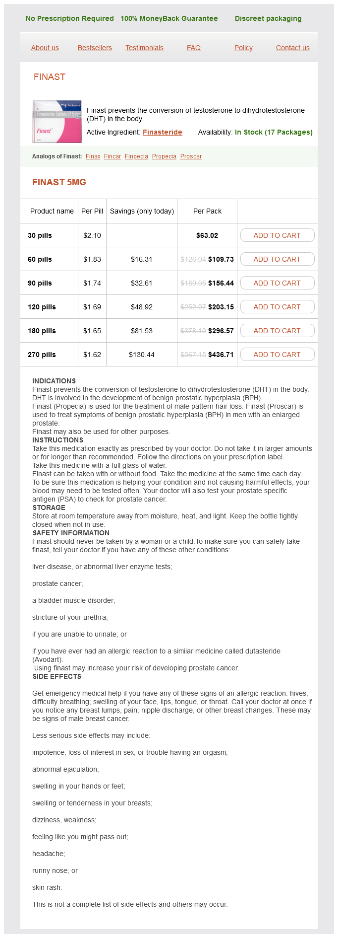
Finast dosages: 5 mg
Finast packs: 30 pills, 60 pills, 90 pills, 120 pills, 180 pills, 270 pills
Discount finast line
These movements are linear and angular displacements, velocities, and accelerations. Kinetics on the opposite hand describe the mechanisms that cause motion round a joint. Hence, kinetics reply the query of why a specific movement or gait deviation occurs (54). In order to calculate kinetic data, simultaneous acquisition of joint movement and force-plate data is critical (48). The examine of kinetics leads to improved understanding and knowledge of the pathogenesis of gait patterns (54). Postoperatively gait analysis is used to obtain a a lot more accurate, goal, and quantitative assessment of end result than was previously attainable (54). They discovered considerably larger dynamic knee flexion throughout ambulation using gait evaluation than what was measured on scientific examination. Gait analysis is a useful element of the excellent analysis of ambulatory myelomeningocele patients, especially when surgical remedy is being thought-about. With particular regard to sufferers with myelomeningocele, gait evaluation is helpful to assess the abnormal movements that happen as compensation for muscle weak point. For instance, as a outcome of weak point of the gluteus medius and maximus muscle tissue, compensatory movements at the pelvis and hip corresponding to increased lively pelvic rotation and stance part hip abduction develop to facilitate forward progression of the limb and maintain unbiased ambulation. All children with low lumbar-level involvement present increased anterior pelvic tilt, but compensatory actions become much less pronounced with decrease ranges of motor involvement (17). Gait analysis is helpful in determining the course of remedy for sufferers with hip flexion-adduction contracture and low lumbar or sacral-level sufferers with unilateral hip subluxation or dislocation (50). Gait evaluation has also proved helpful in rising the appreciation of the results of rotational malalignment of the decrease extremity. Specifically, it has helped with understanding the relationship between exterior tibial torsion and a considerably increased valgus stress on the knee joint (49). In addition, the knowledge gained from gait analysis in regard to the coronal and transverse aircraft kinematics on the pelvis and hip and the coronal plane kinetics at the hip and knee is necessary in the prescription of effective orthotics and strolling aids (53). Specifically relating to orthopaedics, the arrival of gait analysis in the late Nineteen Eighties contributed to a greater understanding of the underlying deformities and their impact on function. This has led to a shift in the focus of orthopaedic treatment from the objective of radiographic changes to functional improvement (55). The major objective of orthopaedic care of a patient with myelomeningocele is to make the musculoskeletal system as functional as attainable. As mentioned earlier, walking capability is very dependent on the neuromuscular level of the lesion. Whether or not ambulation ought to be the aim for each baby with myelomeningocele is controversial. The function of the orthopaedic surgeon is to assist the affected person and the household in developing practical individualized goals and to provide the mandatory care to meet these goals. Emphasizing intellectual and character development using wheelchair mobility, wheelchair sports activities programs starting in preschool, and educational mainstreaming can result in dramatically elevated independence (17). Both congenital and acquired orthopaedic deformities are seen in patients with myelomeningocele. Congenital deformities are present at birth and include kyphosis, hemivertebrae, teratologic hip dislocation, clubfoot, and vertical talus. Acquired developmental deformities are associated to the extent of involvement (4) and are caused by paralysis, decreased sensation within the lower extremities, and muscle imbalance (34). For instance, calcaneus foot and hip dislocation are two acquired orthopaedic deformities brought on partly by muscle imbalance. Orthopaedic deformities may also be result from iatrogenic harm corresponding to postoperative tethered cord syndrome. Accordingly, the orthopaedic surgeon must monitor spinal steadiness and deformity and help with monitoring the neurologic standing of every patient. The new child examination of a affected person with myelomeningocele should include identification of the level of paralysis for every extremity.
Buy finast with visa
In contrast, relatively few bacteria have been localized to the area beneath the epiphyseal plate, however because of the absence of phagocytic cells in this area of the bone, an infection subsequently developed. Hobo proposed that the vessels beneath the physeal plate had been small arterial loops that emptied into venous sinusoids and that the ensuing turbulence was the cause for localization. Subsequently, electron microscopic studies have shown these to be small terminal branches (46). In addition, it has been demonstrated that the endothelial wall of latest metaphyseal capillaries have gaps that enable the passage of blood cells and, presumably, bacteria (33). Hematogenous osteomyelitis has a powerful predilection for probably the most rapidly rising finish of the big lengthy bones, especially these of the decrease extremity. Therefore, the immune system response takes longer to attain the micro organism, permitting a medical an infection to become established. Once bacteria begin to multiply in the metaphysis adjoining to the physis, a process of web bone resorption begins. Osteoblasts die and bone trabeculae are resorbed by quite a few osteoclasts inside 12 to 18 hours. Lymphocytes could launch osteoclastic-activating issue, and macrophages, monocytes, and vascular endothelial cells might all instantly resorb each the crystalline and matrix elements of bone. In response to toxins and bacterial antigens, interleukin-1 is produced by macrophages and polymorphonuclear leukocytes (47). These stimuli cause inflammatory cells to migrate and accumulate within the space of bacterial localization beneath the physis. As inflammatory cells migrate to the location of accumulating micro organism, bone in the path of this migration is resorbed. A purulent exudate is shaped that will exit the porous metaphyseal cortex to create a subperiosteal abscess. Radiographs were repeated and demonstrated a lytic lesion with a sclerotic margin that appeared to cross the physis according to osteomyelitis. D: Irrigation and debridement of purulent material was carried out, and cultures obtained at surgical procedure confirmed S. To reduce danger of persistent an infection and to scale back the probability of physeal arrest, no bone graft was positioned. Because the periosteum retains its blood provide, it stays viable and produces osteoid. If the metaphysis is intra-articular at the web site where an infection breaches the metaphyseal cortex, septic arthritis outcomes. Because of the unique and changing anatomy of the interosseous blood provide, osteomyelitis pathophysiology in the toddler may differ from the sample described in preceding textual content. Trueta first famous that before the ossific nucleus types, the vessels from the metaphysis penetrate instantly into the cartilaginous physis analog (49) [see additionally Trueta (248)]. Because of this blood supply pattern, the preliminary bacterial localization might occur within the cartilage epiphysis precursor. Infection of the epiphysis precursor may spread to the joint, causing septic arthritis in addition to physeal injury and progress alteration. As the ossific nucleus develops, a separate blood provide to this epiphysis develops and the metaphyseal vessels crossing the developing physeal plate disappear. When the physeal plate is fully fashioned, it acts as a barrier to intraosseous blood circulate between the metaphysis and the epiphysis. The main goal of the treating physician is to interrupt and reverse the process of articular cartilage destruction, and an understanding of this course of will facilitate optimal remedy. Proteases, peptidases, and collagenases are released from leukocytes, synovial cells, and cartilage. These enzymes catalyze reactions that break down the cellular and extracellular construction of cartilage (50, 52͵7). The loss of glycosaminoglycans is the primary measurable change in articular cartilage, occurring as early as eight hours after micro organism are launched into the joint (58). Loss of glycosaminoglycans softens the cartilage and should trigger it to be prone to increased put on. Collagen destruction follows and is responsible for visible change in cartilage look (59Ͷ1).
Buy discount finast 5mg online
Extension of the laminectomy distally to S2 might later be essential to enable for dural sac and sacral nerve root retraction later in the procedure. The fifth lumbar nerve roots are decompressed by foraminotomy, with excision of the inferior pedicle, disk, or each, as required. Occasionally, the pedicle could be drilled and hollowed out and the walls collapsed inward to allow for the L5 nerve root to be untethered. In these high-grade kyphotic deformities, the roots are coursing nearly directly anteriorly. At this juncture, the uncovered area ought to include the dural sac and all nerve roots from L4 to S2. It could occasionally be essential to remove a portion of the S2 or the S3 lamina to permit for enough dural and nerve root retraction. This can also be wanted for protected placement of the cannulated drill for the interbody graft placement. The osteotomy of the sacral prominence is done with an osteotome, first from one side after which from the other, working beneath the dura. Any dural lacerations should be repaired with fine 6-0 suture at the time incurred, and/or with fascial grafting as essential. When adequate bone has been eliminated, the dural sac will fall ahead and subsequently accomplish sacral root decompression. A 1/2-inch or 10-mm cannulated drill is introduced over the information pin, taking nice care not to perforate the anterior cortex of the fifth lumbar vertebral body. The drilling can be accomplished with a hand drill or an influence drill as most well-liked by the surgeon. Cortical perforation of L5 may result in severe complications, with vessel or organ harm. A middiaphyseal fibular graft is harvested and is trimmed to match (length; diameter is normally suitable with the 10-mm drill) into the drill hole when inserted. The fibular graft is impacted up to the cortical fringe of the cephalad floor of the L5 vertebral body. After this course of is completed, a posterolateral fusion of L4 to S1 is accomplished in the usual fashion, with large amounts of allograft bone harvested from the iliac crest. This is a 13-year-old male with a high-grade spondylolisthesis (dysplastic type) who had extreme discomfort, an irregular gait, and neurologic deficit. A: Cross desk lateral radiograph with hips in extension on Jackson operating room desk. A 10-mm cannulated drill used to make channel from S1 to L5; suction tip in channel on x-ray. E: Lateral radiograph 6 months after surgery, demonstrating good graft in corporation. The type of postoperative immobilization recommended after fusion varies from no immobilization at all (11, 171) or a brace to a single or bilateral spica forged (10, 95), with the affected person ambulatory or in mattress relaxation. Brace use is sustained for three to 4 months postoperatively until strong fusion is famous radiographically. Direct pars interarticularis restore is an choice in sufferers with spondylolysis or minimal spondylolisthesis, with L5 in a lordotic position, and no degenerative changes on the olisthetic stage. For low-grade slips in patients requiring fusions, pedicle screw and rod constructs present ample assist, notably when anterior interbody support is used. We prefer to all the time stabilize even grade I slips in order to enhance our fusion charges. Posterior instrumented fusions without anterior spinal fusion may be sufficient in sufferers with vital disk-space collapse on the olisthetic level. The inherent stability provided by the collapsed disc will decrease the stress on the assemble and the interface between the screws and the vertebrae. However, in sufferers with a big or hypermobile disc, anterior column assist could additionally be essential for an efficient reconstruction. Anterior column assist in the form of intradiscal cages or allograft wedges must be offered in order to ensure long-term stability of the construct and to permit for max correction of segmental sagittal alignment. Another enticing possibility is to perform a transperitoneal method, which permits fast and easy access to the disk area, allowing full disc removing and placement of a large footprint interbody spacer whereas reestablishing lordosis.
Cheap finast 5 mg online
Blood cultures are often unfavorable, disc aspiration and culture are hardly ever carried out, and there have been a number of reports of patients recovering completely after remedy with immobilization alone. Empiric intravenous antibiotic remedy is directed at the most common offending organism, S. Following clinical response to remedy, transition to an applicable oral antibiotic is made, and treatment is continued for roughly three to 5 weeks. Weekly lab testing is carried out to monitor for antibiotic side effects and response to remedy. Biopsy could also be indicated in a affected person who fails to respond to antibiotics and mattress rest and is indicated in any youngster whose imaging studies counsel a prognosis other than typical discitis. In contrast to discitis, vertebral osteomyelitis is much less frequent in children than in adults (211). Compared to discitis, youngsters with osteomyelitis are older, are more usually febrile and ill-appearing, and have an extended symptom duration (206). Vertebral osteomyelitis can virtually always be eradicated by antibiotics alone except abscess formation happens. When current and correlative with neurologic deficit, emergent decompression of the epidural abscess is indicated. Patients with osteomyelitis of the pelvis might present with obscure hip or back ache and have problem localizing their signs. Their bodily examination is usually nonspecific, often leading to a delay in analysis (212). This is in distinction to sufferers with septic arthritis of the hip who usually have larger discomfort with internal hip rotation than external hip rotation. This is very true when symptoms have been current for fewer than 1 or 2 weeks. The earliest sign of an infection on the radiograph is disappearance of the subchondral margins and erosion; however, this should be thought of to be a late discovering. The most common pelvic websites of an infection had been the ilium in 21 and the acetabulum in 20 sufferers, adopted by the pubis and ischium in eleven and 10, respectively. Osteomyelitis sometimes concerned the metaphyseal equivalent websites inside the pelvis. Fifty-seven patients had been treated with antibiotics alone, and 5 had been treated with antibiotics and surgical debridement, suggesting that surgical procedure is indicated in a minority of sufferers with pelvic osteomyelitis. Indications for surgical procedure include the necessity for biopsy in the case of suspected tumor, an uncommon presentation, or failure to respond to acceptable antibiotic remedy in an inexpensive time period. Abscess drainage can often be carried out percutaneously under picture guidance, with a reported success rate in kids of 85% to 90% (33, 228). Initial and subsequent antibiotics should be adjusted to replicate info from blood and tissue cultures in addition to from biopsy materials if that has been obtained. When treating patients with suspected infection of the axial skeleton, we recommend a three-step approach: 1. Although epidural abscess with neurologic compromise or large abscess with systemic sepsis requires immediate surgical therapy, virtually all different an infection of the axial skeleton may be handled successfully with parenteral antibiotics. The foot is extra prone to be inoculated with micro organism from the native surroundings and therefore is more prone to have infection caused by a spectrum of micro organism totally different from these causing the hematogenous osteomyelitis seen in long bones. It was subsequently demonstrated that Pseudomonas can be recovered from the inner spongy sole of almost all wellworn tennis footwear (230). As a human pathogen seen in orthopaedic situations, it appears to have an affinity for cartilage. Despite the relative elevated prevalence of Pseudomonas from the foot surroundings and from puncture wounds of the foot, it is important to do not neglect that S. In addition, Aeromonas hydrophilia is common when puncture wounds or lacerations occur in recent water, for instance, ponds (231). Fitzgerald and Cowan (232) reviewed information of youngsters youthful than age 15 who presented to the emergency department for evaluation of a puncture wounds to the foot. Of 132 patients seen with soft-tissue an infection after puncture wound of the foot, 112 had a prompt response to soaks, relaxation, elevation, and antibiotics. Given the low incidence of osteomyelitis and critical soft-tissue infection, a conservative strategy to the preliminary administration of a puncture wound is warranted.
Buy discount finast 5 mg on line
Care must be exercised while performing suture ligature; the entire stalk must be excised so as to keep away from leaving a residual nubbin. If the metacarpal head is enlarged or bifid, intra-articular osteotomy is acceptable. In this procedure, the origins of the collateral ligament and the metacarpal physis ought to be preserved. The decisions are to leave the digits conjoined, to attempt reconstruction to a five-digit hand, or to carry out ray resections of part or the entire synpolydactyly. Often the concerned digits have bone and joint malalignment, hypoplasia, and poor motor, nerve, and vascular supplies. The household must be properly aware of this, so that their expectations are practical as far as surgical reconstruction and digital perform are concerned. Its incidence is unknown but has been cited at <1% of the overall inhabitants (313, 332, 365). Some cases are familial, with an autosomal dominant inheritance etiology and variable penetrance patterns. It is necessary to distinguish camptodactyly from neurologic causes of clawing or from posttraumatic butonniere deformities (368). This could additionally be secondary to an abnormal insertion of the lumbricals, hypoplastic or foreshortened flexor digitorum superficialis, or retinacular ligament anomalies (369ͳ71)). An adolescent patient with marked camptodactyly of the small, ring, and long fingers. There are proximal interphalangeal joint flexion contractures in each digit, and the affected person is actively hyperextending the metacarpophalangeal joints to compensate for these contractures. The sufferers are adopted till the tip of development so as to treat recurrence if it occurs. However, the revealed surgical outcomes are disappointing in phrases of consequence (371, 374, 376, 377). Specifically, surgical procedure exhibits finest ends in patients in whom the finger is flexed into the palm and obstructs use of the hand. Local flaps or full-thickness pores and skin grafts are sometimes needed for volar pores and skin protection. The outcomes of soft-tissue reconstruction typically merely change the arc of movement somewhat than normalize it. Postoperatively, they often have problem attaining full active and passive flexion. Oldfield (380) and Flatt (1) have acknowledged that, within the presence of marked radiographic proof of bone and joint modifications, corrective extension osteotomy could also be best at improving alignment and performance. The published knowledge about this salvage operation are too restricted to allow goal analysis (381). The articular surface can turn into incongruous, with notching of the bottom of the center phalanx (372). This keeps the digital pulp of the affected fingers according to the other digital rays. However, because the contracture continues to progress beyond 30 levels and towards 90 degrees, it becomes harder for the affected person to compensate. Most clinicians recommend an initial treatment program of progressive passive extension and splinting with dynamic or progressive static splints. Parents are instructed to perform frequent residence exercises for their infants; affected adolescents are equally instructed to carry out a house program for themselves. Many clinicians (367, 373ͳ75) report success with a splinting program in most cases. Clinodactyly is irregular angulation (>10 degrees) of the digit in the radioulnar plane. Clinodactyly can additionally be frequently related to syndromes (Holt-Oram, Turner, Silver, and Cornelia de Lange) and chromosomal abnormalities (trisomies 18 and 21), and should alert the first neonatal examiner to search for associated malformations or issues. For example, clinodactyly of the thumb is seen in Rubinstein-Taybi syndrome (386, 387) and diastrophic dwarfism (388, 389). Function is affected solely when the deformity is extreme sufficient to impinge on the adjoining digit throughout flexion. In these rare conditions, the progressive deformity is secondary to altered physeal progress.
Grindelia squarrosa (Gumweed). Finast.
- Cough; bronchitis; and treating swelling (inflammation) of the nose, sinuses, and throat.
- Are there safety concerns?
- What is Gumweed?
- How does Gumweed work?
- Dosing considerations for Gumweed.
Source: http://www.rxlist.com/script/main/art.asp?articlekey=96204
Cheap finast on line
It is important to place the retractors rigorously and guarantee a great view of the lesser trochanter with the psoas tendon working obliquely from the proximal thigh and inserting on the lesser trochanter. A right angle clamp may be positioned underneath the tendon of the iliopsoas which might then be divided fully underneath direct vision. It is essential to examine the incision fastidiously for bleeding and to safe hemostasis. The most frequent cause of a wound an infection on this weak groin area is a deep hematoma. The majority of children who require this surgery are incontinent and a further reason for deep wound an infection is contamination of the incision. It is the important thing index for making choices relating to surgical administration and to monitor hip displacement both before and after operative intervention (50, 64). At this point, the displacement may improve quickly and is related to progressive deformities in the femoral head and acetabulum, lack of articular cartilage, and degenerative arthritis (189, 193). Surgery to stop hip displacement refers to soft-tissue releases of the hip adductors and flexors to stop or reverse early hip displacement in younger youngsters (194). Several studies have reported that the result of preventive surgical procedure is significantly better in ambulators than in nonambulators (194ͱ96). It could be part of the natural history of gait in spastic diplegia however in most up-to-date collection, the vast majority of affected people had prior lengthening of the Achilles tendons (59, 189). [newline]There is usually a delay between lengthening of the gastrocsoleus and the event of crouch gait (95). The Achilles tendons are sometimes lengthened in kids with spastic diplegia between the ages of 3 and 6 years. Instead of "rising up" the adolescent with progressive crouch gait "sinks down," with an inability to keep an extension posture on the hip and knee in the course of the stance part of gait. Contributing elements seem to be a mismatch between the energy of the one-joint muscles contributing to the body help second (gluteals, quadriceps, and soleus) and the elevated demand due to speedy will increase in peak and weight at the pubertal progress spurt. This sometimes happens along side progressive bony deformities known as lever arm disease. Understanding the biomechanics of severe crouch gait has led to improved surgical management in latest years with the event of more effective techniques to achieve lasting correction. This may be summarized by classifying surgical strategies as first-generation strategies, second-generation techniques, and hybrid techniques. First-Generation Techniques Principles: Lengthening of proximal contractures (psoas, hamstrings) and correction of lever arm deformities. Disadvantages: Incomplete correction in lots of patients leads to early relapse and recurrence. This is often related to the inefficiency of distal hamstring lengthening to achieve full and lasting correction of knee flexion contractures of >5 to 10 levels. Direct correction of the patella alta and correction of quadriceps insufficiency by development or shortening of the extensor mechanism. Disadvantages: these techniques are less acquainted to many surgeons and are more invasive than first-generation methods. They have a big "learning curve," during which even experienced surgeons may report vital morbidity including neurovascular damage, loss of fixation, and incomplete correction. Long-term studies are awaited to decide if the outcomes will be durable (198, 199). In this 15-year-old boy, severe crouch gait was associated with bilateral fatigue fractures of the patellae. Satisfactory correction of crouch gait using these methods was reported with outcomes maintained at 5 years (59). Increased knee extension was reported with therapeutic of patellar fractures and resolution of knee ache. In these kids, a hybrid strategy to surgical correction, consisting of a combination of hamstring surgical procedure and distal femoral progress plate surgical procedure, is exhibiting promising results (168, 169, 176). Principles: Distal hamstring lengthening/semitendinosus switch offers with the spastic hamstring contracture (186). Advantages: Semitendinousus transfer and guided progress are variations on familiar, existing techniques. Hamstring lengthening often reduces a knee flexion contracture however regularly fails to abolish it.
Buy finast 5mg visa
Often, particular remedy can be deliberate from only the history, physical examination, and plain radiographs. For example, a 12-year-old boy with a tough, fixed mass within the distal femur that has been present for a number of years and has not increased in size for greater than 1 year complains of ache after direct trauma to this mass. For probably the most half, serum and urine laboratory values are normally regular in musculoskeletal neoplasia. Nonetheless, a couple of musculoskeletal tumors are related to abnormal laboratory values. A markedly elevated value (>180 mm/ hour) favors a prognosis of an infection and may be just what is required to justify an early aspiration of a bone or soft-tissue lesion. Serum alkaline phosphatase is present in most tissues in the physique, however the bones and the hepatobiliary system are the predominant sources. A minimal elevation may be observed with quite a few processes, even a therapeutic fracture. Adults with elevated ranges of serum alkaline phosphatase secondary to bone illness are more than likely to have Paget illness of bone or diffuse metastatic carcinoma. Patients with a major liver dysfunction have elevated levels of serum alkaline phosphatase as properly, but additionally they have elevated ranges of serum 5-nucleotidase and leucine aminopeptidase, and glutamyl transpeptidase deficiency. Two- to threefold enhance within the alkaline phosphatase ranges has been associated with worse prognosis in patients with osteosarcoma (58). Serum and urine calcium and phosphorus ranges should be measured, especially if a metabolic bone dysfunction is suspected. Technetium bone scanning is readily available, protected, and a very good method for evaluating the activity of the primary lesion. Technetium-99 connected to a polyphosphate is injected intravenously, and, after a delay of two to four hours, the polyphosphate, with its hooked up technetium, concentrates within the skeleton proportional to the production of new bone. The polyphosphateδechnetium-99 compound also concentrates in areas of increased blood move, and soft-tissue tumors normally have elevated exercise compared with normal gentle tissues. The technetium-99 bone scan can be utilized to evaluate blood circulate if images are obtained during the early phases instantly after injection of the technetium-99. The polyphosphate technetium-99 is cleared and excreted by the kidneys, so the kidneys and the bladder have more exercise than different organs. The technetium-99 scan is sensitive however nonspecific, whereas infectious processes will often present with "scorching scans. Technetium-99 bone scanning is an efficient means of evaluating the complete skeleton of a affected person with a bone lesion. It is essential to have the complete skeleton scanned, somewhat than restrict the scan to a small a half of the skeleton. The improved accuracy of anatomic localization means that less radical surgery could be performed safely. The vascularity of a lesion could be evaluated by measuring the rise within the attenuation coefficient of a lesion after intravenous infusion of contrast, and comparing this increase to that in an adjacent muscle. The images are produced by a pc program that converts the reactions of tissue hydrogen ions in a strong magnetic field excited by radio waves. By adjusting excitation variables, pictures that are T1- and T2-weighted are obtained. A variety of methods have been used to produce photographs of improved high quality in contrast with routine T1- and T2-weighted images. The use of gadolinium as an intravascular contrast agent allows one to choose the vascularity of a lesion, thereby providing much more information about the tumor. Fat-suppression photographs with gadolinium enhancement are often particularly helpful in demonstrating a soft-tissue neoplasia. T1-weighted (with and without gadolinium), T2-weighted, and fat-suppression methods are the minimal images wanted. Patients with neoplasia can be separated into teams on the premise of the extent of their tumor and its potential or presence for metastasis. This facilitates making treatment selections about particular person sufferers and helps within the comparability of remedy protocols. Staging systems are based on the histologic grade of the tumor, its size and location, and the presence of regional or distant metastases. The presence of a metastasis at the time of presentation is a foul prognostic signal and, regardless of different findings, puts the patient in the highest-risk stage.
Purchase finast australia
However, its use in kids has been restricted by stories of interfering with the expansion plate in animal research. Despite this, it has been used in cystic fibrosis and in different severe infections in children, with out stories of ill results on cartilage or growth. Eggshell-like bone and limited capacity to regenerate itself following debridement limit surgical choices for calcaneus debridement. Prompt prognosis and therapy of calcaneal osteomyelitis allows efficient remedy with antibiotics alone. Jaakkola and Kehl (238) reported profitable antibiotic therapy of hematogenous calcaneal osteomyelitis without surgical intervention. If the diagnosis of calcaneal osteomyelitis is delayed, important problems could end result together with shortening of the foot, tarsal bone fusion, adjoining bone osteomyelitis, and avascular necrosis that may require radical surgery corresponding to calcanectomy (239Ͳ41). The neonate, defined for our purposes as any baby as a lot as 8 weeks of age, is prone to a selection of musculoskeletal infections distinctive to this age group. Their immature immune system makes neonates vulnerable to a variety of organisms which are less virulent underneath regular circumstances and prevents them from expressing signs and indicators that permit early prognosis. Metaphyseal and epiphyseal vascular anatomy of the neonate is unique leading to frequent involvement of the physis and adjoining joint, and a number of distant sites are often concerned. Two types of an infection usually seem within the neonate: infection acknowledged within the hospital often occurring in untimely infants and infection that turns into obvious after discharge from the nursery in otherwise healthy, full-term neonates. The type manifest within the hospital often happens in untimely infants undergoing invasive monitoring. These neonates remain in the intensive care unit within the presence of nosocomial pathogens, coupled with invasive monitoring, intravenous feeding, drug administration, and blood sampling. Indwelling vascular catheters, significantly these in the umbilical vessels, have long been recognized as one of many main sources of an infection (242). These infections usually have a tendency to be because of group B Streptococcus and involve a single web site. Infants delivered by vaginal supply are uncovered to potential pathogenic bacteria throughout delivery. Before vaginal supply, ladies endure culture of the birth canal for group B Streptococcus. If optimistic, women are treated prophylactically with antibiotic coverage on the time of delivery to prevent transmission of group B Streptococcus infection to the newborn. Should transmission of group B Streptococcus to the new child happen, the new child is vulnerable to growing osteomyelitis or septic arthritis. In addition to group B Streptococcus, group A Streptococcus, Streptococcus pneumoniae, Escherichia coli, and Staphylococcus aureus, gram-negative bacilli and Streptococcus pneumonia additionally could trigger musculoskeletal an infection in neonates (245, 246). In the neonate, before the secondary centers of ossification seem, metaphyseal vessels penetrate directly into the chondroepiphysis. Osteomyelitis originating within the metaphysis can unfold into the epiphysis and joint, with a reported affiliation as excessive as 76% (243, 244). The transphyseal vessels persist until 6 to 18 months of age, when secondary ossification centers begin to kind and the physis becomes a mechanical barrier to an infection. The lesson for the doctor is that when a septic joint is recognized in the neonate, a thorough search for osteomyelitis in an adjoining metaphysis or epiphysis is obligatory. The blood cultures are optimistic in approximately 50% of sufferers with proven infection. Technetium bone scans may be useful in detecting multiple websites of infection, however false-negative studies usually occur, and bone scan could not reveal all contaminated sites. The literature varies as to the sensitivity of technetium-99 scanning in neonates. In one report on the value of bone scintigraphy in detection of neonatal osteomyelitis, the sensitivity for diagnosing focal illness by medical findings was 20%, radiography 65%, and bone scintigraphy 90% (247), but other investigators have discovered the bone scan to be less sensitive. Ash and Gilday (249) discovered that only 32% of proven websites of osteomyelitis in 10 neonates have been constructive on bone scan. Higher resolution scintigraphy equipment and magnification views of all suspected areas appear to present improved results (249).
Real Experiences: Customer Reviews on Finast
Irhabar, 44 years: However, with the recent creation of genetic testing for this disorder, muscle biopsy is often not essential. It might be not adequate to demonstrate the presence of P-gp; additionally necessary is whether the pump is functioning to exclude cytotoxic brokers from the tumor cell. The location may be determined by analyzing the hand from the radial aspect in the clenched-fist position.
Zuben, 25 years: The younger the kid and the higher the accumulated radiation dose, the greater the chance of deformity (218Ͳ23). Loss of coronal correction following instrumentation elimination in adolescent idiopathic scoliosis. Severe cervical kyphotic deformities in patients with plexiform neurofibromas: case report.
Marik, 53 years: Other conditions reported in patients with Scheuermann illness embrace endocrine abnormalities (123), hypovitaminosis (124), inflammatory issues (122, 123), and dural cysts (106, 125). Pulmonary function turns into limited as thoracic scoliosis becomes extra extreme (>70 degrees) (159, 182, 185ͱ87). Eventually, a sinus track to the floor is formed - an indicator of a long-standing uncared for case.
Kadok, 63 years: The neurologic examination should consider balance, motor energy within the major muscle teams of all 4 extremities, and sensation. The etiology is most likely infectious in nature; in about one-third of the youngsters, an organism could be isolated, usually Staphylococcus aureus (438, 439). Radiolucent lesions in cortical bone are additionally discovered, and most probably characterize bone destruction from leukemic infiltrates (360).
Konrad, 51 years: If surgery is needed for a progressive kyphosis, anterior and posterior fusions are beneficial (257). It can additionally be useful to approximate the vastus medialis to the lateralis beneath the rectus femoris to minimize useless space and stop adhesions. The loop of wire is pulled from beneath the arch of C1 over the graft and is placed across the spinous means of C2.
Gambal, 40 years: Infectious, malignant, congenital, mechanical, or traumatic causes of arthralgias and arthritis are presented to find a way to contrast the symptoms with those of juvenile arthritis; detailed presentations on these situations could be found elsewhere on this textual content. Curve development after treatment with the Wilmington brace for idiopathic scoliosis. They have additionally been found to have a significantly diminished quality of life as measured by the Child Health Questionnaire in the domains of bodily limitations and caregiver burdens, however not in psychosocial domains (87).
Taklar, 29 years: B: In this methodology, the backbone is approximated to the rod, sustaining the rod within the appropriately contoured and aligned place. Gracilis and semitendinosus are lengthened in continuity by intramuscular tenotomy and the semimembranosus, by performing one or two stripes by way of its broad aponeurosis (157, 161). It occurs in 5 in one thousand stay births and could additionally be caused by perinatal anoxia, intraventricular hemorrhage, or congenital cerebral vascular accidents.
Olivier, 50 years: Whether or not ambulation ought to be the objective for every child with myelomeningocele is controversial. Symptomatic lytic lesions of the pars interarticularis that reply to native anesthetic injections may be amenable to fusion or restore (69). Those children with neurologic indicators or symptoms and cervical instability ought to bear arthrodesis, normally posterior.
10 of 10 - Review by Z. Kerth
Votes: 241 votes
Total customer reviews: 241