Glimepiride dosages: 4 mg, 2 mg, 1 mg
Glimepiride packs: 30 pills, 60 pills, 90 pills, 120 pills, 180 pills, 270 pills, 360 pills
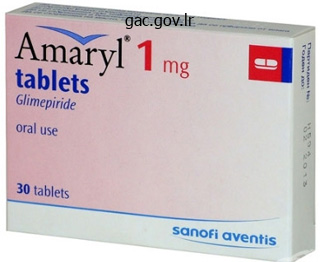
Purchase generic glimepiride from india
Once the primary enzyme in the sequence is activated, it generates a serine protease that cleaves and activates the next part in the pathway. The cascade additionally amplifies the response such that activation of a few early complement proteins triggers a bigger subsequent response. Furthermore, cleavage of the complement proenzyme reveals an active enzyme and a small membrane binding site. This causes the activated complement proteins to adhere to the membrane of the invading microorganism, rather than diffusing into the bloodstream. The classical, different and lectin pathways all converge on the cleavage of C3 into C3a and C3b. C9 C6 C7 C6 C5b C7 C6 C5b C7 C8 Cytosol C8 n While C3b and C5b function main gamers of the complement cascade, C3a and C5a act as diffusible, proinflammatory anaphylatoxins that recruit white blood cells (such as neutrophils and monocytes). The potential harmful properties of complement proteins require sophisticated mechanisms to prevent destruction of regular tissues. First of all, complement proteins are secreted in inactive varieties and are only activated within the presence of an antigen. Thirdly, particular inhibitors exist within the blood to deactivate rapidly complement proteins to stop widespread destruction of regular tissue. These strategies act collectively to management the potency and action of the complement system. While B-cells can secrete antibodies that act at great distances away from the unique B-cell, T-cells are dependent on the interaction with the target cells inside a brief range. They work together with the target cells in certainly one of two fashions: either by killing the goal cell or using the target cell in a signaling cascade to recruit other cells and improve the immune response. Furthermore, these peptide sequences must be introduced to the T-cell by special molecules on the surface of the target cell. The T-cell receptor recognizes the peptide sequence certain to the special molecule advanced and subsequently initiates downstream responses. These mechanisms allow T cells to reply successfully towards intracellular microorganisms, such as viruses, and towards phagocytic cells which have engulfed foreign extracellular peptides. Other subtypes of helper T-cells had been named after their manufacturing of one major cytokine, these embody, Th5, Th6, Th7, Th9, Th17 and Th22 cells. Unlike antibody molecules, T-cell receptors only exist in the membrane-bound form. The variable segment extends into the extracellular area, whereas the fixed phase inserts into the cell membrane to allow signal transduction throughout the T-cell upon activation. These cells are primarily found in epithelial tissue, such as skin and intestine, and in nasal mucosa. This explains the challenges confronted in matching transplant donors and recipients to decrease rejection of the transplanted organ. However, they both have a peptide-binding groove that holds a small peptide of degraded overseas proteins. These genes are joined before transcription in a random method to give an almost limitless range of T-cell receptors. The immune system of the transplant recipient assaults the transplanted tissue, which it sees as "foreign. The 1 and 2 domains make up the variable amino acid sequences that bind the degraded peptide fragment. The chain consists of an 1 and an 2 subunit, whereas the chain is composed of a 1 and a 2 subunit. Pathogens that reside within a cell are immune to Intracellular pathogens take over the host cellular machinery to produce peptides and proteins. This protein punches a gap through the cell membrane of the infected cell resulting in cell death. These secondary molecules act as coreceptors to stabilize and strengthen the interaction.
Generic 1 mg glimepiride fast delivery
It is necessary that the knowledgeable consent be signed, dated, and witnessed to show that consent was obtained before therapy started. The Ethics of Clinical Trials When the subject of ethics in medical trials is raised, essentially the most polarizing concern is that of randomization, particularly if the comparison treatment is a placebo or nonactive drug. Random task to intervention teams offers all topics the same likelihood of receiving every attainable remedy, and it serves a quantity of necessary functions. It constitutes a method of assigning patients to therapies in a method that is free of personal bias, and it varieties the premise for the statistical tests that might be used to check the underlying hypotheses. Most importantly, randomization distributes the variables, both measured and unobserved (and presumably unknown) among the groups in an opportunity, and therefore in an neutral method. It is one other means of ensuring lack of bias and allowing unambiguous statistical evaluation and interpretation of group knowledge. It identifies three basic ethical principles for all human topic research-respect for persons, beneficence, and justice. The Surgical Clinical Trial There is little question that randomized managed clinical trials are the simplest methods to evaluate new pharmacologic interventions, but controversy still attends their position in nonpharmacologic interventions, such as surgery. In a traditional surgical trial, the affected person is randomized to Procedure A or Procedure B; randomizing sufferers to one of the process arms requires the assumption that the surgeon has equal expertise in the interventions beneath analysis and that all participating surgeons have comparable experience. Devereaux and colleagues in 2005 offered a cogent argument and multiple scientific illustrations of the ways during which the expertise-based design will enhance the validity, applicability, feasibility, and ethical integrity of randomized managed trials in surgical procedure. If, as Devereaux and colleagues reveal, surgeons with expertise in, for example, an endoscopic strategy treat 70% of the sufferers in both groups A and B and surgeons with expertise in, for instance, an open approach treat 30% of these in each groups A and B, the trial results might be biased in favor of the endoscopic approach. Devereaux refers to this sort of bias as differential experience bias and suggests that its potential is excessive in surgical trials for three causes. First, measures are hardly ever instituted to ensure that the number of participating surgeons with expertise in every procedure is equal. This is particularly true if new units or procedures are being in comparison with an current normal of care. Clinical Trial Phases Clinical trials are typically described when it comes to their phase, a descriptor that designates the overall function of the trial. Phase I trials are small trials of 20 to eighty patients designed to research the toxic and pharmacologic results of a brand new therapy that has been studied in an animal model but which now needs to be tested in humans. Usually performed as a multicentered trial, the therapy is in comparison with a regular of care remedy to determine which is simpler. Considerable controversy has surrounded some surgical trials, the place sham procedures have been carried out on sufferers randomized into the control group. Measurement of Outcomes There are several other ways to categorize end result measures. The broadest scheme describes outcome measures as being either generic or disease-specific. As their name implies, the generic measures are generalizable throughout well being situations, practice settings, and types of treatments. Disease-specific outcomes capture more detailed details about function immediately associated to the situation of interest and make it more simple to attribute change to treatment response. Some measures are linked temporally to the disease or intervention process and thus enjoy a excessive degree of specificity with respect to well being outcomes; restoration from a surgical procedure, for example, or modifications that occur early within the natural history of the disease. Other measures occur later within the course of and are extra subject to the affect of external factors; life satisfaction, for example, will certainly be affected by health standing but may even be influenced by unrelated social, economic, and psychological components. This being the case, the difference or change in disease-related high quality of life following intervention is the extra significant parameter to observe. Selecting an Outcome Measure When deciding on an end result measure for use in a examine, investigators ought to concentrate on several major traits of the instrument. Second, there should be proof of its responsiveness or the power to detect clinically and socially important variations when change has indeed occurred. Third, there should be evidence of its reliability or the power to present consistent values between and inside subjects. Further, the end result measure should have these characteristics defined inside the population of interest.
Syndromes
- Pernicious anemia
- Protein
- Reducing the amount of blood thinners or stopping aspirin
- They include: arginine, cysteine, glutamine, tyrosine, glycine, ornithine, proline, and serine.
- Sweaty areas such as the groin or armpit
- CT scan or MRI of the head
- Stroke
Buy glimepiride with visa
Consequently, technical and radiophysical traits of the system and their consistency limit the smallest collimator that safely and reproducibly can be utilized. This also illustrates the beam dimension effect and the penumbra (the area near the edge of the sector where the dose falls rapidly) associated with each of the four collimator helmet sizes. The distance at which the beams start to overlap depends on beam measurement for a given irradiation method, which means that the dose outside the goal periphery is dependent upon the beam dimension. This theoretically means that a comparatively high dose can be delivered exterior the goal to a big volume of normal tissue. Thus, as the beam dimension increases, at some point one can not claim that the remedy is selective, and one should find another irradiation approach. The dose absorbed in regular tissue adjoining to the target periphery is probably the most important issue limiting the largest volume that can be treated radiosurgically. Earlier, the remedy with gamma (photon) irradiation was described; nevertheless, protons, neutrons, and electrons are additionally used to deal with certain tumors. Fractionation is necessary in phrases of lowering normal tissue problems; and, in certain conditions (such as optic nerve tumors), fractionated stereotactic radiotherapy (rather than radiosurgery) is preferred. Therefore, by breaking up the radiation into relatively small dose fractions (usually 1. The right-hand aspect of the determine exhibits the isodose curves for the four, eight, 14, and 18 mm collimator helmets. Absence of sub-lethal injury restore is also the rationale why decrease doses are wanted with radiosurgery to management the disease. As an instance, it has been found that roughly eight to 12 Gy is the utmost tolerated dose of the optic chiasm when using a single fraction of radiation, such as in radiosurgery. However, if using fractionated remedy, the traditional tissue tolerance of the chiasm is roughly 54 Gy. This same concept holds true for tumor-control doses: greater whole doses are wanted in fractionated therapy to get the identical tumor control as a lower dose in a single therapy because of sub-lethal-damage repair by the tumor cells between fractions. Therefore, contemplating the patient with a meningioma encasing the optic nerve, if the tumor were handled with stereotactic radiosurgery, a dose of roughly 14 Gy can be wanted; nonetheless, the utmost dose that the optic nerve can tolerate is approximately eight to 12 Gy with out risking severe imaginative and prescient compromise. With fractionated treatment, a dose of fifty four Gy to the tumor could be enough to management the tumor, and the optic nerve can tolerate 54 Gy if given in 1. However, the accuracy of radiation supply by way of a hard and fast headframe in a single session compared to multiple classes with no fixed head-frame additionally ought to be thought of. Therefore, each patient should be evaluated to determine whether or not radiosurgery or fractionated radiotherapy is probably the most applicable treatment. This is because sub-lethal-damage restore can happen throughout an extended radiation publicity, if the dose price is low enough. There has been some concern relating to the growing older sources in Gamma Knife models, and whether or not the lower dose price affects tumor management rates. Kondziolka and colleagues, reviewing the University of Pittsburgh therapy outcomes, found no clinical difference in tumor control rates during the first 9 years of use of their first gamma knife unit, previous to reloading. It was felt that this will have been due to the higher-dose fee in the course of the first year of operation of their gamma knife unit. However, there was no research to date to show a detrimental effect on tumor control due to decreasing dose fee from getting older Gamma Knife sources. In addition to dose, dose fee and fractionation, the quantity of the radiated subject is essential. With vestibular schwannomas, treating with a plan by which the dose tightly conforms across the tumor is important, as the goal can be to spare as much normal tissue as potential. However, for brain metastases, for instance, the precise borders of the goal could also be tough to outline and treating the goal with some margin of surrounding normal tissue could also be simpler in eradicating the tumor than a good dose across the target. Also, certain tumors may be located in additional critical areas, and it could be extra necessary to have a rapid dose fall-off to spare normal tissues (such because the brainstem) rather than a excessive dose to the tumor. In addition, the clinical results of the therapy (including late facet effects) might not happen for months or even years following the irradiation. The organic response to radiation is also dependent on the sort of cells which are irradiated. Some cell types are more radiosensitive than others; and, therefore, a decrease dose may be wanted to control one sort of tumor versus one other. Also, with the example of benign cranium base tumors corresponding to vestibular schwannomas, meningiomas, or paragangliomas, few cells are actively dividing at the time of treatment. Therefore, the primary effect will not be in destroying these more radiosensitive dividing cells, however rather a longer-term lower in the vascular supply to the tumor.
Cheap glimepiride online
Since is lacks a leus, which is that this portion known as the pars flacfibrous layer, tensa. The with its related muscle tissue, mesotympanic, and hypotympanic rior portion tube, and the vascular system. The eustachian of the tympanicvascular of the middle eustachian tube, and is thatmembrane is referred hypotympanic portion the portion system. The Middle Ear portion is that portion of the center hypotympanicof the middle portion of the various hypotympanic portion is that ear incorporates middle this portion ear that lies inferior to the aperture of of eustaear house between the tympanic membrane and bony trabeculae and to bony overlaying the eustaThe that lies inferior the the aperture of thethe jugchianbulb. This bony floor the oval and spherical home windows, the jugular bone, thethe hypotympanic windows, the with a convex superior rimmuscle bean formed stapes bulb the stapedius and a exposing the stapes bone, in stapedius muscle posteriorly, and the canal for the tensor tympani posteriorly, and the canal for the tensor tympani concave inferior a small channel (the inferior region. The oval windowinisplace by kidney footplate of the stapes oval window tympanic canaliculus) transmits held is kidney beanannular ligament. In numerous window, ear footplate ofgurations by bonehorizontalround winfootplate superiorly that obscure the in place by place of brane confi the stapes the is held is proscribed of the stapes bone is held inportion by the annular ligament. In facial Posteriorly, in the mesotympanum there the two window, the footplate of theimportance. The area facial a deep known as usually coated chronicspace facial canal is called the facial recess. Posteriorly in the mesotympanum(ie, of the in which theeminence)wall is preserved (ie, intact during which the ear canal wall is preserved there are (pyramidal ear canal accommodates the tendon intact two bony muscle earlier than its insertion into the the canal wallrecesses of scientific significance. These anteriorly anterior portion of eustachian tube and two recesses are necessary the middle ear house is identified as the7). The bony roof ofAthe epitympanum is formedtegmen canal roof of bony projection fromthe by the bony center ear known as the facial tymtation. This bony landmark is steady pos(pyramidal bony landmarkand the tendon of the prominence of the lateral is steady posteriorly because the tegmen its insertion into the neck teriorly muscle before properly because the epitympanic stapediusas the tegmen mastoidea. The most anterior portion of wall of the epitympanum is formed by the bony of the the the facial (fallopian) canal. The bony portion of epitympanum is shaped by the top prominence ear the lateral and superior semicirprominence of the lateral and superior semicirthe middle of house is called the protympanum and neck of the malleus and its articulation with culariscanal ampullaeprocess by the portion of the cular canal ampullae as nicely as the epitympanic and physique and brief as welland the orifice bordered superiorly as a epitympanic the portion of the of the (fallopian) canal. Thefor the portion of the facialanteriorly by the most ofhead eustachian tubefacial (fallopian) canal. These two ossicular the center ear referred to as the tegmen two ossicular for the the epitympanum. These the epitymmassesspace communicates ligaments anteriorly plenty are held in place by ligaments anteriorly bony landmark is steady posteriorly because the panic are held in place by posteriorly through and posteriorly to called the an axisad antrum to and posteriorly to present an axis the rotation tegmen mastoidea. The epitymthe central mastoid the of prominence of the panic space communicates posteriorly through panic house communicates posteriorly by way of a slim opening known as the aditus ad antrum to a slim opening referred to as the aditus advert antrum to the central mastoid tract of the mastoid cavity. The head a neck of mucous leus and its barrier, which may body and brief membrane barrier, which may completely(see membrane articulation the protympanum or pneumatization from with the completely or process8). The separate epitympanic house can also be incompletelya separate of thetwo compartments. Sound strain prolonged transmitted from the tympanic membrane called from the protympanum. Anteriorly, the epitympatransmittedof the the tympanic body of theacross the pinnacle from the tympanic membrane incus transmitted from malleus and membrane throughout num is separated atby the ossicular the cochleariform course of the middle earunit suspendedossicular chain within the function as a space the center ear house by the by ligaments comchain comfrom an the malleus,tip of the longof variable 9). The displacement process of the initiated by medial tip of the epitympanic house epitympanum. Sound strain tympanic transmittedislarger than that of the stapes footplate membrane isfrom the tympanictheratio by various larly elevated. Maintaining this stapes footplate membrane bigger than that of membrane across the a center 25 to space sound stress essential by a ratio of 25 strategies sound stress density in reconstructive by ratio of earto 1, the constitutes an density in 1, the by the ossicular chain comprised of the ear surgery. Maintaining this in the oval body of theThe stapes bone as constitutes anstirrup with reconstructive methods a unit suspended by ligadow. The the cruraproprincipleneck, and footplate the tip ofstapes therea head, in epitympanum.
Glimepiride 1mg amex
And, the traditional audiogram remains useful in summarizing the outcomes of fundamental hearing evaluation. Clinical audiology, nevertheless, now additionally contains different behavioral and electrophysiological take a look at procedures. A variety of speech and non-speech behavioral measures and a variety of other cortical auditory evoked responses can be found for medical evaluation of central auditory nervous system dysfunction and related auditory processing issues. The otorhinolaryngologist is in a pivotal position to facilitate identification of kids and adults in danger for listening to loss. Otorhinolaryngologists work carefully with audiologists in diagnostic hearing evaluation, and contribute to well timed and applicable medical or surgical intervention. In this article, we summarize present strategies and techniques for listening to evaluation of adults. We emphasize the applying of a take a look at battery strategy that maximizes diagnostic accuracy and effectivity, whereas minimizing take a look at time and costs. At the tip of the chapter, we outline in a glossary widespread terms and abbreviations essential in listening to evaluation and rehabilitation of listening to loss. Findings for the right ear represent a typical sensorineural audiometric pattern, whereas left ear findings typify a conductive hearing loss. The intensity of any sound is outlined by a ratio of its sound strain (or sound intensity) in comparability with a reference sound stress (or sound intensity). The reference sound pressure is the quantity of stress towards the eardrum attributable to air molecules when a sound is presented that vibrates the eardrum and may simply be detected by a standard human ear. Briefly, the relationship for sound intensity is described as dB = 10 log (sound intensity/reference intensity), or for sound stress as dB = 20 log (sound pressure/reference pressure). This is the standard for the intensity stage that corresponds to the common regular hearing threshold degree, the minimal detectable intensity for each take a look at frequency for a big pattern of normal hearers. In grownup listening to assessment, air-conduction hearing thresholds for tonal or speech signals are measured individually for every ear with earphones. They supply distinct benefits over the standard supra-aural earphones, including elevated comfort, lowered chance of ear canal collapse, higher inter-aural attenuation, disposability (aural hygiene), and higher acceptance by younger kids. The normal region for children is extra restricted as a result of even mild hearing loss can intervene with speech and language acquisition. The essential frequencies for understanding speech are inside the area of 500 through four,000 Hz. However, higher frequencies additionally contribute importantly to discrimination between certain speech sounds. The mechanism for the crossover is presumably bone-conduction stimulation caused by vibration of the earphone cushion in opposition to the skull at high stimulus depth ranges. With boneconduction stimulation, inter-aural attenuation may be very restricted, at most 10 dB. Clinically, one must assume cautiously that inter-aural attenuation for bone-conducted signals is 0 dB. That is, any sound presented to the mastoid bone of one ear by a bone-conduction vibrator could also be transmitted by way of the cranium to both or both internal ears. Masking is the audiometric approach used to get rid of participation of the non-test ear every time air- and bone-conduction stimulation exceeds inter-aural attenuation. The optimal masking signal is slim band noise for pure-tone alerts and speech noise for speech signals. With adequate masking, any sign crossing over to the non-test ear is masked by the noise. The otolaryngologist ought to always attempt to confirm that appropriate masking was utilized in interpreting hearing check results. With the sloping configuration, listening to is better for low frequencies and poorer for higher frequencies. High-frequency deficit listening to loss is the most common pattern related to a sensorineural listening to impairment. A rising configuration reveals comparatively poor listening to for decrease frequency stimuli and higher listening to for the high frequencies. One exception to the standard association of conductive listening to loss with rising configuration is Meni�re disease, which is discussed in Chapter 28, "Meni�re Disease, Vestibular Neuronitis, Benign Paroxysmal Positional Vertigo, Superior Semicircular Canal Dehiscence, and Vestibular Migraine.
Glimepiride 4mg otc
Value of skull radiography, head computed tomographic scanning, and admission for remark in circumstances of minor head harm. A additional contribution to the sensory system of the seventh nerve and its neuralgic situations. Gadolinium-enhanced magnetic resonance imaging of the facial nerve in herpes zoster oticus and Bell palsy: Clinical implications. Herpes zoster auris related to facial nerve palsy and auditory nerve symptoms: a forty four. Endoscope assisted surgery of the trigeminal, facial, cochlear or vestibular nerve. Trigeminal neuralgia: problems as to cause and consequent conclusions regarding remedy. Trigeminal neuralgia treated by microvascular decompression: a long-term follow-up examine. Posterior fossa reexploration for persistent or recurrent trigeminal neuralgia or hemifacial spasm: surgical findings and therapeutic implications. Total facial nerve decompression in recurrent facial paralysis and the Melkersson� Rosenthal syndrome: a preliminary report. A comparative research of the fallopian canal on the meatal foramen and labyrinthine section in young youngsters and adults. Labyrinthine segment and geniculate ganglion of the facial nerve in fetal and adult temporal bones. Surgical exposure of the internal auditory canal and its contents via the middle cranial fossa. Hypoglossal-facial nerve interpositional bounce graft for facial reanimation with out tongue atrophy. Facial reanimation with jump interpositional graft hypoglossal facial anastomosis and hypoglossal facial anastomosis: evolution in administration of facial paralysis. Temporalis muscle for facial reanimation: a 13-year experience with 224 procedures. Temporalis tendon transfer as part of a comprehensive strategy to facial reanimation. Long-term follow-up of nerve conduction velocity in cross-face nerve grafting for the treatment of facial paralysis. Free neurovascular muscle transposition for the remedy of facial paralysis utilizing the hypoglossal nerve as a recipient motor supply. It became apparent that the most direct approach to sure inaccessible intracranial tumors was by way of the skull base somewhat than by way of the calvarium. Therefore, in the first half of the twentieth century, surgeons averted the complexity of the cranium base and most well-liked the simpler resolution of working via the thin calvaria, even though injurious levels of brain retraction were often required. In the Nineteen Sixties, William House and others utilized trendy working microscopes and highspeed drills to resurrect a number of skull-base approaches that had beforehand been deserted as impractical. The term skull base surgical procedure is considerably of a misnomer, as only a few surgical procedures are literally carried out to resect lesions intrinsic to the cranium base. In actuality, the vast majority of neurotologic cranium base procedures are performed for lesions located beneath the cerebral cortex or adjoining to the brainstem, and the removing of bone allows exposure while minimizing cerebral and cerebellar retraction. This article will focus on the posterior fossa which is bordered by the clivus anteriorly, the posterior surface of the petrous portion of temporal bones laterally and the occipital bone posteriorly. It is bordered by the posterior floor of the temporal bone anteriorly, the anterior surface of the cerebellum posteriorly, the inferior olive medially, the inferior border of the pons and cerebellum superiorly, and the cerebellar tonsil inferiorly. The petrous apex is pyramidal in shape, with anterior, posterior, and inferior surfaces. The anterior floor varieties a portion of the center cranial fossa and the posterior floor marks the anterolateral extent of the posterior fossa. The lateral extent of the petrous apex is demarcated by the inside ear and intratemporal portion of the carotid artery.
Predigested Spleen Extract (Spleen Extract). Glimepiride.
- Dosing considerations for Spleen Extract.
- Infections, enhancing immune function, skin conditions, kidney disease, and rheumatoid arthritis.
- What is Spleen Extract?
- How does Spleen Extract work?
- Are there safety concerns?
Source: http://www.rxlist.com/script/main/art.asp?articlekey=96976
Buy glimepiride 4mg mastercard
The hiatus of the facial canal additionally accommodates the vascular provide to the geniculate ganglion area. The tympanic, or horizontal, phase of the nerve is approximately 11 mm long, running between the lateral semicircular canal superiorly and the stapes inferiorly, forming the superior margin of the fossa ovale. Between the tympanic and mastoid segments, the nerve gently curves inferiorly for about 2 to 3 mm. The mastoid, or vertical, phase is the longest intratemporal portion of the nerve, measuring roughly 13 mm. As the nerve exits the stylomastoid foramen on the anterior margin of the digastric groove, an adherent fibrous sheath of dense vascularized connective tissue surrounds it. The importance of the endoneurial sheath, within the context of nerve harm and repair, is that it offers a continuous tube by way of which a regenerating axon can grow. This layer accommodates the vasa nervorum, providing the blood provide in addition to the lymphatic vessels. Facial-Nerve Injury It is important to evaluation the types of nerve harm to understand better electrodiagnostic testing of the facial nerve, prognosis for restoration, and the event of synkinesis, in addition to the rationale for facial-nerve decompression. Table 34-2 summarizes the Sunderland and Seddon classifications of nerve injuries. A letter grading system was chosen to keep away from confusion with the House-Brackmann classification. This evoked electromyogenic response is recorded with surface electrodes positioned over the perioral (nasolabial) muscle tissue since a large consultant population of facial-nerve fibers can be sampled by recording the evoked response from this group of muscle tissue. A supramaximal bipolar stimulation (galvanic) is provided to saturate the nerve and produce a whole and synchronous depolarization. The galvanic stimulation is typically delivered as rectangular pulses, with a pulse duration of 200 �s and an interpulse interval of 1 second. The amplitude of the evoked response is plotted as a perform of time after stimulation. Both the conventional and affected sides are examined, and the amplitude of the responses is compared. The share of degenerated fibers is calculated arithmetically, as follows: better prognostic outcome. With resolution of the neural injury, in a nerve that has undergone axonotmesis, the axon will regenerate via the intact neural tubule, potentially allowing complete return of motor function to the muscle fiber innervated by that nerve fiber. The extra severely disrupted neural tubule injury of neurotmesis has the potential to regenerate in an unsuccessful manner and may thereby result in misdirection of fibers, clinically causing synkinesis and incomplete return of motor operate. The severity of the injury can be inferred from the speed of degeneration after damage. More speedy wallerian degeneration is related to neurotmesis, whereas nerves that degenerate extra slowly usually tend to exhibit axonotmesis. With a known complete transection of the facial nerve (eg, traumatic injury), 100 percent wallerian degeneration occurs over three to 5 days as the distal axon slowly degenerates. Therefore, early testing, within three days of paralysis, is in all probability not consultant of the degree of harm, and as outlined above, the time course of degeneration may mirror the diploma of harm. An necessary technical detail to which attention ought to be paid is the necessity to stimulate the nerve on the stylomastoid foramen 10 to 20 instances earlier than making an amplitude measurement. The initial stimulation will improve the synchronization throughout the nerve and, therefore, improve the reliability of the test. With more extreme injuries, axoplasmic disruption (axonotmesis) or neural tubule disruption (neurotmesis) will lead to wallerian degeneration distal to the site of damage. Needle electrodes are positioned into the orbicularis oculi and orbicularis oris muscles, and the patient is requested to make voluntary contractions. If voluntary contractions happen during the first two weeks after the onset of paralysis, early deblocking of the neural conduction block has taken place, and an excellent restoration of facial function will most probably observe. First, a system for monitoring facial-nerve perform in the course of the operation should be employed. The largest diamond bur that the operative website can safely accommodate ought to be used when the surgeon is near the fallopian canal.
1mg glimepiride with visa
Crossover Sound stimulus introduced to the take a look at ear travels across the head by air conduction or by way of the cranium by bone conduction to stimulate the other non-test ear. A decibel scale referenced to accepted standards for normal listening to in which zero dB is average regular hearing for each audiometric check frequency (audiometric zero). A decibel scale used in auditory brainstem response measurement referenced to common behavioral threshold for the press stimulus of a small group of normal hearing subjects. A take a look at of vestibular function by which nystagmus is recorded with electrodes positioned close to the eyes during stimulation of the vestibular system. Myogenic exercise recorded from the facial muscles, often within the nasolabial fold, in response to electrical stimulation of the facial nerve as it exits the stylomastoid foramen. Inter-aural attenuation Insulation to the crossover of sound from one ear to the other provided by the pinnacle. Inter-aural attenuation varies relying on whether or not the signal is presented by air conduction or bone conduction. Masking (masker) Carefully selected background noise introduced to the non-test ear in an audiometric process to forestall a response from the non-test ear due to crossover of the stimulus when interaural attenuation is exceeded. The level of masking noise necessary to overcome the conductive part and adequately mask the non-test ear exceeds inter-aural attenuation levels. The masking noise could then cross over to the test ear, and masks the sign (eg, pure tone or speech). An audiometric process which compares a threshold response with masking noise offered in-phase versus out-of-phase with a pure-tone or speech signal. Release from masking is a traditional phenomenon reflecting auditory brainstem integrity. Sounds measured within the exterior ear canal associated with power produced by the outer hair cells within the cochlea. Word lists developed first within the late Nineteen Forties containing all the phonetic elements of general American English speech that occurs with the approximate frequency of their prevalence in conversational speech. A measure of speech recognition or understanding reported in percent appropriate scores as a perform of the depth level of the speech sign. The arithmetic common of hearing threshold ranges for 500, 1,000, and 2,000 Hz, or the speech frequency region of the audiogram. A variation of the open fit listening to help design with the receiver located inside the exterior canal quite than the body of the listening to help. Rollover A lower in speech recognition efficiency in p.c appropriate at excessive sign depth ranges versus decrease ranges. An audiometric procedure developed by James Jerger (1970) for assessing bone-conduction listening to in sufferers with serious conductive hearing loss. Airconduction thresholds are decided with out masking after which with masking introduced by bone conduction to the brow. The measurement of the masked shift in hearing thresholds corresponds to the diploma of conductive hearing loss part. The lowest depth degree at which a person can detect the presence of a speech signal. A measure of central auditory function involving identification separately of a closed set of 10 syntactically incomplete sentences introduced simultaneously with a competing message. A measure of central auditory operate developed by Katz that makes use of spondee words offered within the dichotic mode. A scientific process developed by Jerger for assessing the power to detect a 1 dB increase in depth. The signal-to-noise ratio is the distinction between the intensity level of a sound or electrical event and background acoustic or electrophysiological power. The lowest depth stage at which a person can accurately determine a speech sign (eg, two syllable spondee words). This possibility may be utilized to cut back the presence of feedback while the wearer is utilizing the telephone.

Purchase glimepiride line
Using a dissecting instrument Endoscopic Surgery for Sellar and Parasellar Lesions: A Tailored Approach. This maneuver has to be accomplished carefully and solely, if circumferential inspection of the dissection airplane was attainable, to not tear and injure adherent surrounding neurovascular constructions. A new modified speculum guided single nostril technique for endoscopic transnasal transsphenoidal surgery: an evaluation of nasal problems. Mononostril versus binostril endoscopic transsphenoidal strategy for pituitary adenomas: a scientific evaluate and meta-analysis. Binostril versus mononostril approaches in endoscopic transsphenoidal pituitary surgery: medical evaluation and cadaver examine. Endoscopic versus microscopic transsphenoidal pituitary adenoma surgery: a meta-analysis. Comparison of sinonasal high quality of life and health standing in sufferers present process microscopic and endoscopic transsphenoidal surgery for pituitary lesions: a prospective cohort study. Endoscopic transsphenoidal strategy: adaptability of the process to totally different sellar lesions. A novel reconstructive method after endoscopic expanded endonasal approaches: vascular pedicle nasoseptal flap. Expanded endoscopic endonasal method for anterior cranial base and suprasellar lesions: indications and limitations. Extended endoscopic endonasal transsphenoidal method for the elimination of suprasellar tumors: Part 2. The increasing function of the endonasal endoscopic method in pituitary and cranium base surgery: a 2014 perspective. Variations on the standard transsphenoidal strategy to the sellar region, with emphasis on the prolonged approaches and parasellar approaches: surgical experience in a hundred and five cases. Olfactory groove and tuberculum sellae meningioma resection by endoscopic endonasal approach versus transcranial method: A systematic review and meta-analysis of comparative studies. Raza S, Effendi S, DeMonte F, Tuberculum sellae meningiomas: evolving surgical strategies. A simple scoring system to predict the resectability of skull base meningiomas by way of an endoscopic endonasal strategy. Outcomes after transcranial and endoscopic endonasal strategy for tuberculum meningiomas: a retrospective comparison. Endoscopic versus open resection of tuberculum sellae meningiomas: a choice evaluation. Of these, pituitary adenomas are the most common in adults, accounting for over 50% of all lesions in this region. However, the frontotemporal transcavernous method can be used for tumors with extensive invasion. This approach defines the most intricate and superior cranial base method,3 requiring elite surgical expertise, and offering a maximal surgical corridor to adequately access the lesions. The anatomy of the sellar area and its contents is necessary to perceive the idea of cranium base strategy in surgical administration of the lesions in these areas. Given the complicated anatomy of the sellar region and the adjoining cavernous sinuses, bounded closely by the bony structures and the optic nerve pose a problem in obtaining access while working on lesions in the area. Anatomy Located within the sphenoid bone, the sella turcica lies behind the chiasmatic groove and the tuberculum sellae, forming part of the middle cranial fossa. The sella turcica homes the pituitary gland that rests on the hypophyseal fossa (the seat of the sella). Posteriorly, the bony sella is steady with the clivus, which terminates laterally to form the posterior clinoid process. Drilling should be done across the lateral floor of the higher wing of the sphenoid bone as close to the frontal course of as possible. For publicity of the ground or the temporal base, groove is drilled to forestall interference with retracted temporal muscle.
Discount glimepiride 3mg with amex
The pneumotachometer may be connected to a head out displacement-type physique plethysmograph. The disadvantage is that this apparatus is cumbersome and that considerable patient cooperation is required. The anterior methodology of measurement locations a tube on the nasal vestibule of 1 facet, which is occluded whereas the patient breathes through the other nasal cavity. The posterior methodology of measurement involves placement of the tube in the oropharynx, handed through closed lips between the tongue and the palate. In this methodology, the measurement outcome can differ to a fantastic extent as a end result of the measured stress difference might simply be affected by the position of the soft palate. In energetic rhinomanometry, the affected person actively breathes through one nasal cavity whereas the narinochoanal (naris to choana) strain distinction is assessed within the contralateral nasal cavity. New gear has to be calibrated by the producer, and in addition by the operator earlier than measurements are taken on a given day. A normal preformed resistor must be used by the operator earlier than and after studies. Rhinomanometric measurements are obtained with the patient in a sitting position after an adaptation period of 20 minutes. As the measurements are taken, the info points for every breath which are displayed on the monitor display screen type a sigmoid pressure-flow curve in actual time. When a collection of breaths show common repetition of the curve, information acquisition is activated to sample two consecutive breaths. In the usual strain and airflow graph obtained from trendy rhinomanometry gadgets, airflow is recorded on the "y"-axis and strain on the "x"-axis. The "mirror" picture utilizing 4 quadrants of the graph is accepted as the usual representation in energetic anterior rhinomanometry. The greater the nasal resistance (the ratio of transnasal pressure to airflow), the closer the curve will be to the strain axis. Nasal congestion could be quantified when it comes to complete or unilateral nasal airway resistance. For fourphase rhinomanometry, resistance is decided for part 1 (ascending inhibitory phase) and section four (descending expiratory phase) by use of the best attainable move at a pressure of one hundred fifty Pa. Factors that affect nasal resistance embody postural modifications, exercise, and temperature of the air. However, it has the disadvantages that it requires training of the operator, and the potential for variation in results because of movement of the taste bud and due to bulky equipment. In addition, manipulation of the nasal mucosa with a catheter may be a confounding issue. The medical use of rhinomanometry has been limited, but it is a superb device for research. Rhinomanometry can be used for measuring the change in resistance before and after the usage of a decongestant in illness states such as allergic or nonallergic rhinitis. If the decongestant causes less than a 35% lower in resistance, a structural cause could additionally be thought of as the reason for nasal blockage. The anterior technique of measurement entails placement of a tube on the nasal vestibule of one facet, which is occluded while the affected person breathes via the opposite nasal cavity. In energetic rhinomanometry, the patient actively breathes by way of one nasal cavity whereas the narinochoanal pressure difference is assessed within the contralateral nasal cavity. The strain and move indicators are transferred to a pc to be analyzed, displayed, saved, and printed. In this graph, the curve on the best of the circulate axis represents the change in inspiration, and the curve on the left is the change in expiration. The right nasal cavity is represented on the higher part of the strain axis, and the left nasal cavity on the decrease part of the pressure axis. This method provides supplementary data; the ascending and descending parts of the curves during inspiration and expiration are additionally displayed (Adapted from reference 24. The system is held horizontally, and the masks ought to type an air-tight seal around the nostril.

