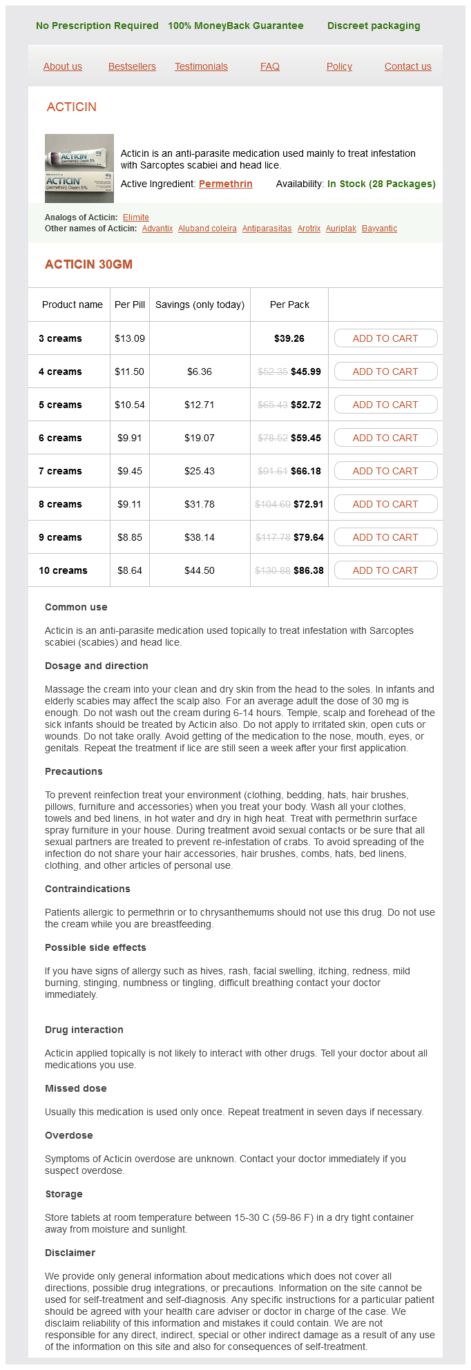
Acticin dosages: 30 gm
Acticin packs: 3 creams, 4 creams, 5 creams, 6 creams, 7 creams, 8 creams, 9 creams, 10 creams

Discount acticin 30 gm fast delivery
Pharmacotherapy: A Pathophysiologic Approach, 10e > Chapter 18: the Arrhythmias Cynthia A. Close monitoring is required of all of those medicine to assess for adverse results as nicely as potential drug interactions. This drug is efficient in terminating and preventing a extensive variety of symptomatic supraventricular and ventricular arrhythmias. The heart has two basic properties, particularly, an electrical property and a mechanical property. The synchronous interaction between these two properties is complicated, exact, and comparatively enduring. The research of the electrical properties of the guts has grown at a steady rate, interrupted by periodic salvos of scientific breakthroughs. Certainly, the brand new era of molecular biology and mapping of the human genome promises even greater insights into mechanisms (and potential therapies) of arrhythmias. The scientific use of drug therapy started with the usage of digitalis and then quinidine. A theme of drug discovery throughout this decade was initially to find orally absorbed lidocaine congeners (such as mexiletine and tocainide); later, the emphasis was on drugs with extremely potent effects on conduction (ie, flecainide-like agents). This chapter evaluations the rules involved in both regular and abnormal cardiac conduction and addresses the pathophysiology and treatment of the extra commonly encountered arrhythmias. Certainly, many volumes of full textual content might be (and have been) dedicated to primary and clinical electrophysiology. Consequently, this chapter briefly addresses those principles necessary for clinicians. Most tissues inside the conduction system also possess varying levels of inherent automatic properties. The conducting tissues bridging the atria and ventricles are referred to because the junctional areas. From the bundle of His, the cardiac conduction system bifurcates into a number of (usually three) bundle branches: one right bundle and two left bundles. These bundle branches further arborize right into a conduction community referred to because the Purkinje system. The conduction system as a complete innervates the mechanical myocardium and serves to initiate excitationcontraction coupling and the contractile process. As the electrical wave front strikes down the conduction system, the impulse finally encounters tissue refractory to stimulation (recently excited) and subsequently dies out. Prior to mobile excitation, an electrical gradient exists between the within and the surface of the cardiac cell membrane. In atrial and ventricular conducting tissues, the intracellular area is approximately -80 to -90 mV with respect to the extracellular environment. For instance, this specific pump (in addition to different systems) makes an attempt to preserve the intracellular sodium concentration at 5 to 15 mEq/L and the extracellular sodium concentration at a hundred thirty five to 142 mEq/L, in addition to the intracellular potassium concentration at 135 to a hundred and forty mEq/L and the extracellular potassium focus at three to 5 mEq/L. The action potential curve results from the transmembrane motion of particular ions and is divided into completely different phases. Phase zero or initial, rapid depolarization of atrial and ventricular tissues is attributable to an abrupt improve in the permeability of the membrane to sodium influx. This fast depolarization greater than equilibrates (overshoots) the electrical potential, leading to a brief preliminary repolarization or section 1. Calcium begins to move into the intracellular house at about �60 mV (during section 0), inflicting a slower depolarization. Calcium influx continues all through part 2 of the motion potential (plateau phase) and is balanced to some extent by potassium efflux. Calcium entrance (only via L channels in myocardial tissue) distinguishes cardiac conducting cells from nerve tissue and supplies the crucial ionic link to excitation-contraction coupling and the mechanical properties of the guts as a pump. The membrane remains permeable to potassium efflux throughout section three, leading to cellular repolarization. Phase four of the action potential is the gradual depolarization of the cell and is related to a continuing sodium leak into the intracellular area balanced by a reducing (over time) efflux of potassium. The slope of section 4 depolarization determines, in giant part, the automated properties of the cell.
Buy acticin 30 gm low price
Emotional well-being is a measure of coping capability and reflects the expertise of feelings starting from enjoyment to distress, and social well-being reflects the standard of relationships with family and associates, in addition to wider social interactions. Health utilities assess the value assigned to specific well being states by specific populations using standardized methods and are normally represented as a quantity between 0 and 1, with zero indicating demise and 1 indicating perfect health. Lower well being utility scores and better physique mass index were strongly associated with higher levels of lymphedema (stages 2 and 3). The authors found that most cancers survivors reported lower adjusted lymphedema well being utilities than those with major lymphedema and concluded that decreasing lymphedema in cancer survivors is essential. Hull22 famous in a cohort of patients with breast cancer-related lymphedema that the lymphedema led to issues in a wide range of every day actions, including: problem sleeping owing to positioning of the swollen limb; issue carrying objects, such as heavy pots or groceries; challenges with many forms of exercise and even walking; and problematic fitting and comfort of clothes. In this population of patients, swelling of the pinnacle and neck area can have main useful implications, such as dysphonia or an inability to swallow. In a research of 103 head and neck most cancers survivors, Deng and colleagues23 reported that exterior and inside lymphedema also affected nutritional consumption, resulting in weight loss in plenty of sufferers. In addition, inner lymphedema was recognized in 68% of the sufferers utilizing endoscopy and was also linked to self-reported voice-related signs. The results of the investigation of qualitative assessments revealed constant themes such as a big lack of understanding of and information about lymphedema by well being professionals; fear, shock, annoyance, and body picture issues as frequent emotional problems; and a big influence of remedy on healthcare costs and free time. The quantitative assessments described in the evaluate indicated that lymphedema sufferers endure from larger useful impairment, poorer psychological adjustment, and greater anxiety and 196 Principles and Practice of Lymphedema Surgery despair than the general inhabitants. Nine of the 19 research recognized within the literature targeted solely on breast cancer-related lymphedema, and 7 of the remaining studies included some patients with breast cancer and others with main lymphedema or lymphedema following remedy for other malignancies. The general EuroQol-5D score can then be used to calculate quality-adjusted life years for costeffectiveness analyses. Retrospective 7 Lower extremity/ Pedicled omento(2005)97 ilioinguinal plasty Campisi et al. Prospective 50 Upper extremity Lymphatic venous (2006)98 microsurgery Dardarian et al. Retrospective 29 Lower extremity/ Saphenous vein (2006)99 ilioinguinal sparing Takeishi et al. Randomized sixty four Lower extremity Omentoplasty (2008)101 controlled trial Boccardo et al. Prospective 19 Upper extremity Lymphatic venous (2009)102 anastomosis Boccardo et al. Randomized 55 Upper extremity Preventative (2009)103 managed trial education/early management protocol Torres Lacomba Randomized 120 Upper extremity Manual lymph drainet al. Retrospective 18* Lower extremity/ Lymphatic venous (2013) ninety three ilioinguinal anastomosis Morotti et al. Prospective 8 Lower extremity/ Lymphatic venous (2013)105 ilioinguinal anastomosis Subjective/magnetic 10% vs. This 22-item device is scored utilizing a seven-point Likert scale and has been validated among folks with various sorts of cancer. Participants indicate how nicely they agree or disagree with the 22 objects with scores ranging from 1 to 7. When whole scores are used, the topic could be categorized into two groups: low high quality of life (T scores 49) and high quality of life (T rating <49). The findings point out that it is a promising device to screen gynecologic most cancers sufferers for early indicators and signs of lymphedema. Although this instrument is still within the early phases of improvement, its psychometric properties are favorable for figuring out early lymphedema in head and neck most cancers survivors. Operative treatment of peripheral lymphedema: A systematic meta-analysis of the efficacy and safety of lymphovenous microsurgery and tissue transplant. Changes within the body image and relationship scale following a one-year power coaching trial for breast cancer survivors with or in danger for lymphedema. Psychosocial impact of lymphedema: A systematic review of literature from 2004 to 2011. The bodily penalties of gynecologic cancer surgical procedure and their impact on sexual, emotional, and high quality of life issues. Reflections on findings of the most cancers outcomes measurement working group: Moving to the subsequent section.

Order discount acticin
The superficial circumflex iliac artery and the vein are ligated on the degree of their origin. Dissection is restricted to the medial border of the femoral artery to stop any additional injury to the lymphatic vessels draining the decrease limb. Once the vascular anastomoses are performed the blood perfusion within the distal edge of the lymphatic groin flap is once more evaluated. The distal fringe of the flap is tunneled to the higher extremity alongside the vessels, reaching the proximal brachium and stuck with a single transfixation suture. Postoperative Care Patients obtain steering from the physiotherapist to actively mobilize the shoulder. The compression therapy is all the time continued for a minimum of six months after surgical procedure. However, most patients still need to use compression after that, at least in physically strenuous conditions. Depending on the extent and length of preoperative lymphedema, compression may be needed for up to two to three years, or completely. It is our follow to start manual lymphatic drainage as quickly as possible and continue remedy in the early postoperative interval, to theoretically help the spontaneous regrowth of the lymphatic vasculature within the axilla. From experimental research, we know that the lymphatic vascular growth and maturation process might take two to six months after the surgical procedure. Outcomes the primary objective of including a lymph node flap to breast reconstruction is to enhance lymphatic vessels flow perform and to release lymphedema patients from utilizing stigmatizing and uncomfortable compression clothes. In particular, sufferers with recurrent erysipelas infections or neuropathic pain of the arm appear to benefit from lymph node transfer. However, larger randomized research are wanted to clarify the therapeutic effects of lymph node switch. In our own beforehand revealed paper, one-third of our lymph node switch patients confirmed improvement of the lymphatic circulate function in postoperative lymphoscintigraphy. It has been proven that newly fashioned lymphatic vessels are being stabilized and maturated into true collecting lymphatic vessels spontaneously over the course of six months. To present extra information about the lymphatic function after lymph node transfer, additional imaging methods are growing, corresponding to magnetic resonance imaging lymphography. In fact, there are previous studies that suggest that instant breast reconstruction reduces the danger of postmastectomy lymphedema14 and that delayed breast reconstruction might cut back lymphedema signs of the affected arm. However, lymph node transfer presents possibilities that conventional breast reconstruction and different reconstructive choices are missing. In a super scenario, the lymphatic, immunological, and sentinel capabilities would all be restored. Summary Currently, lymph node transfer continues to be thought of as experimental surgical procedure. We do not know what the effects are on the lymphedema limb volume in the long run. Incidence of unilateral arm lymphoedema after breast most cancers: A systematic evaluation and metaanalysis. Anatomy of the superficial lymphatics of the belly wall and the upper thigh and its implications in lymphatic microsurgery. Therapeutic differentiation and maturation of lymphatic vessels after lymph node dissection and transplantation. From lymph to fat: Liposuction as a remedy for complete reduction of lymphedema. Reduced incidence of breast cancerrelated lymphedema following mastectomy and breast reconstruction versus mastectomy alone. Positive impression of delayed breast reconstruction on cancer therapy associated arm lymphedema. Preoperative ultrasound is a priceless device to consider the number of out there lymph nodes and node location previous to flap harvest.
Acticin 30 gm with visa
The thoracoacromial pedicle must be recognized early, on the undersurface of the muscle, and should be protected. Proximally, minimal muscle must be left over the pedicle, and, actually, the pedicle can be completely dissected from the muscle with appropriately delicate technique, to decrease bulk in the upper chest and lower neck. The medial and lateral pectoral nerves are additionally divided to maximize the flap arc of rotation. The skin between the donor-site incisions and the top and neck defect is elevated. Caution must be used when elevating the neck pores and skin away from the exterior jugular vein and the subclavian blood vessels to keep away from inadvertent vascular damage, particularly within the radiated neck. The clavicular head of the pectoralis muscle can be divided along the trail of flap rotation to maximize flap reach and minimize proximal bulkiness. When the myocutaneous flap is used, several tacking sutures are employed to decrease tension on the skin paddle. Myo-osseous or Osteomyocutaneous Variants Both the fifth rib and the outer desk of the sternum have been used together with the pectoralis major muscle and overlying pores and skin to reconstruct composite mandibular defects. When using the sternal variant, the skin island is designed along and over the sternum. The pectoralis main muscle attachments to the anterior part of the sternum are preserved. A longitudinal incision through the outer table and the cancellous portion of the sternum is made with a reciprocating noticed and osteotome. The fifth rib could additionally be included with the pectoralis main flap by equally preserving all of the pectoralis main muscle attachments. A subperiosteal elevation of the rib is performed after making medial and lateral osteotomies. If a small pleural tear happens, the lungs are insufflated, a small catheter is inserted through the tear and is placed under suction, and a major repair is attempted as the catheter is withdrawn. Skin grafts over the costal cartilages and ribs could take poorly and healing could additionally be extended. He had previously undergone an belly aortic aneurysm restore, aortobifemoral bypass grafting, and a left carotid endarterectomy. Because of these situations, we wished to avoid a microvascular free flap procedure. Case Example A 64-year-old male offered with a left retromolar trigone squamous cell carcinoma, stage T4N0M0. These vessels must be ligated somewhat than cauterized to avoid damage to the blood supply to the pores and skin paddle. Note the long reach of the flap owing to design of the pores and skin paddle over the fourth intercostal house. Bulkiness in the proximal neck is minimized by together with solely a modest amount of muscle across the proximal pedicle. Pearls and Pitfalls � For the longest arc of rotation, the pores and skin paddle of the pectoralis main myocutaneous pedicled flap ought to be centered over the fourth intercostal area, which is where multiple musculocutaneous perforating blood vessels enter the skin. Such length could be advantageous in preventing restriction of neck mobility due to contraction of the muscular portion of the flap postoperatively, a frequent complication related to this flap. Otherwise, consideration ought to be given to performing a pectoralis muscle flap covered with a skin graft as an alternative. Further experiences with the pectoralis main myocutaneous flap for instant repair of defects from excisions of head and neck cancers. A one-stage correction of mandibular defects utilizing a split sternum pectoralis major osteomusculocutaneous switch. The position of sternum in osteomyocutaneous reconstruction of major mandibular defects. Conversion of pedicled to free flap for salvage of the compromised pectoralis major myocutaneous flap in head and neck reconstruction. Surgical strategies and results of lateral thoracic cutaneous, myocutaneous, and conjoint flaps for head and neck reconstruction. Three-dimensional anatomical vascular distribution within the pectoralis main myocutaneous flap. New methodology of making ready a pectoralis major myocutaneous flap with a skin paddle that includes the third intercostal perforating branch of the internal thoracic artery. Plast Reconstr Surg 2009; 123:1220�1228 29 Supraclavicular Artery Island Flap Michael W.
Generic acticin 30 gm on line
Because the muscle Pearls and Pitfalls � the latissimus dorsi muscle free flap is most usually used for scalp reconstruction. They are most commonly discovered 8 to 10 cm under the posterior axillary fold and a pair of to 3 cm posterior to the lateral border of the muscle, at the identical horizontal level as the tip of the scapula. The transversely oriented paddle donor website is often easier to close and ends in less scar widening. One-stage switch of the latissimus dorsi muscle for reanimation of a paralyzed face: a new alternative. Skoracki Introduction the primary anatomical examine of the scapular fasciocutaneous flap was published by dos Santos1 in 1980 primarily based on bilateral dissections of 35 cadavers. Gilbert and Teot2 published the first scientific collection in 1982, utilizing the flap for ankle resurfacing in 4 patients. Swartz et al3 introduced the inclusion of a vascularized segment of the lateral scapular bone in 1986, and this osseocutaneous free flap was subsequently modified and improved by Coleman and Sultan. The osseocutaneous scapular free flap is an efficient alternative for osseous reconstruction of mandibular or maxillary defects where the fibular free flap is contraindicated or unavailable. Anatomy Landmarks the fasciocutaneous flap from this donor website can be oriented horizontally (scapular flap), or vertically (parascapular flap), or each ways if two skin islands are dissected based mostly on the 2 terminal cutaneous branches of the circumflex scapular artery. The axis of a scapular flap pores and skin island extends horizontally from the triangular area (bound by the long head of the triceps muscle laterally, the teres main inferiorly, and the teres minor and subscapularis superiorly), which is located on the lateral border of the scapula two-fifths of the means in which from the scapular spine to the scapular tip, to the posterior midline. The vertical dimension of the flap spans the distance between the scapular spine and the scapular angle. The parascapular flap skin island is centered vertically or obliquely alongside a line between the triangular space and the posterior superior iliac spine. Skin flap dimensions of 5 to 7 cm in width and 10 to 15 cm in size can normally allow for easy primary closure. A vascularized bone segment from the lateral scapular border, which receives blood from the circumflex scapular artery both immediately or through its musculoperiosteal branches to the teres minor and main, may be harvested from this area, alone or in combination with a fasciocutaneous flap compo- Advantages 1. Long and large-caliber vascular pedicle, especially if the subscapular vessels are included with the pedicle. A great amount of bone may be harvested from the scapula, together with the medial border, lateral border, and the scapular tip. Various chimeric flap configurations (including pores and skin, bone, muscle flaps) allow freedom in orientation of the completely different flap parts to tailor for composite defects. A segment of bone (usually ~ 3 � eleven cm in size) from the scapular angle to within 1 cm of the articular capsule could be harvested. The scapular tip (3 � 20 cm with intrinsic curvature) based mostly on the angular branch is another choice when the thoracodorsal pedicle is included. The circumflex scapular artery passes posteriorly into the triangular area and is three to 4 cm lengthy. In the triangular house, the circumflex scapular artery provides off subscapular, infrascapular, and direct muscular branches to the teres major and teres minor muscular tissues. The infrascapular branch passes between the scapula and the infraspinatus muscle, forming a collateral anastomotic network with the suprascapular artery, a department of the thyrocervical trunk. Beyond these branches, the artery is identified as the descending branch of the circumflex scapular artery, and as it emerges from the trian- gular area, it divides into its terminal branches; the cutaneous scapular artery (traveling horizontally) and the cutaneous parascapular artery (descending vertically). The scapular tip is provided by the angular branch that typically originates from the thoracodorsal artery (see below). Fasciomusculoperiosteal branches arising from the scapular cutaneous artery as it travels via the infraspinatus muscle provide the medial scapular border. The angular artery arises from the latissimus dorsi branch in 51% of circumstances, from the serratus anterior branch in 25%, because the third department of a trifurcation of the terminal thoracodorsal vessel in 20%, and as a branch of the thoracodorsal vessels arising proximal to its bifurcation into latissimus and serratus branches in 4%. Limited shoulder abduction of as a lot as 6 months can be anticipated following harvest of an osseous element, however thereafter the vary of motion should normalize. Similarly, a history of prior axillary surgical procedure, such as axillary lymph node dissection, and prior axillary radiation therapy, or higher limb lymphedema ought to immediate use of the contralateral scapula. Otherwise, the ipsilateral scapula is usually used for convenience of patient positioning and flap configuration.
European Beaver (Castoreum). Acticin.
- How does Castoreum work?
- Menstrual abnormalities, anxiety, sleeping disorders, and other conditions.
- What is Castoreum?
- Are there safety concerns?
- Dosing considerations for Castoreum.
Source: http://www.rxlist.com/script/main/art.asp?articlekey=96336
Buy generic acticin 30 gm
An end-to-side anastomosis, often via an interposition graft, is one way of reducing the morbidity of this process. In end-to-side grafting, the epineurium is regionally removed and a few (25 to 30%) of the hypoglossal nerve axons are transected in order that they may grow into the distal facial nerve. Speech and swallowing morbidity in addition to synkinesis and mass motion with tongue motion have been shown to be significantly lowered using an end-to-side approach. A process that has more lately turn out to be in style is the use of the masseteric branch of the trigeminal nerve for dynamic facial reanimation. The nerve usually takes an oblique course within the deep substance of the muscle, touring from posterior superior to anterior inferior. Following the nerve distally permits adequate 152 I Topics in Head and Neck Reconstruction of facial motion impartial of neck and shoulder operate seems to be very tough. The phrenic nerve has also been used for facial reanimation but may cause marked contraction with coughing, laughing, and deep inspiration, and use of the phrenic nerve is contraindicated in sufferers with pulmonary disease. In our practice, the spinal accessory and phrenic nerves are thought-about last resorts. Cross-facial nerve grafting has the advantage of offering pure emotional activation with out retraining. In this procedure, one or more nerves on the normal facet are sacrificed and linked to a nerve graft, which is tunneled subcutaneously to the affected facet of the face. Exposure of the contralateral regular nerve is usually performed via a facelift incision and elevation of a pores and skin flap; nerve branches are recognized and mapped as they exit the anterior fringe of the parotid gland and journey towards the muscle tissue of facial expression. The sural nerve is the most commonly used donor nerve for cross-facial nerve grafting owing to its size, availability, minimal donor-site morbidity, axonal density, and ease of harvest. Intraoperative mapping of the contralateral regular facet is carried out utilizing a nerve stimulator to establish redundant branches of the facial nerve that innervate the identical groups of muscular tissues. Grafting of zygomatic and buccal nerve branches have been described most incessantly, again due to their relatively extra valuable capabilities and contribution to symmetry in repose. The course of the interposition nerve grafts is often throughout the upper lip or lower lip soft tissue. Cross-facial nerve grafting has been described as both a one-stage or a two-stage process. Singlestage procedures cut back the number of surgical procedures required and patients could benefit from faster reinnervation. In a two-stage process, nerve graft(s) are sutured to the donor nerve department on the traditional facet and tunneled to the affected facet and left there. Axonal growth is usually estimated to be ~ 1 mm per day, which supplies a rough guide for the period of time needed earlier than the second stage is carried out. For practical functions, usually 6 to 12 months elapse before the second stage is carried out. In the second stage, the distal finish of the nerve graft is uncovered, the nerve is trimmed sharply to take away any neuroma, after which the nerve is sutured to the distal portion of the severed facial nerve on the paralyzed side. The nerve exits the cranium base, passes between the condyle and coronoid process of the mandible, and lies on the deep floor of the masseter muscle. The masseteric nerve has a robust motor impulse, which provides strong muscular activation and a quick reinnervation time, normally inside 3 months. Unlike the hypoglossal nerve, the masseteric nerve is situated near the route of the facial nerve, which often implies that interpositional nerve grafting is unnecessary. Patients can be taught to activate the facial muscles by specializing in tongue or jaw contraction after hypoglossal or masseteric nerve transfer, respectively. With both procedure, synkinesis can occur, as can unwanted activation throughout mastication and speech. Cerebral cortical adaptation can happen after rehabilitation with masseteric nerve switch, and emotional nerve activation has been reported. Additionally, control 10 advantage of two-stage procedures is that the severed facial nerve may be grafted to the masseteric or hypoglossal nerves as babysitter nerves whereas ready for cross-facial axonal development. The primary disadvantage of cross-facial nerve grafting is that outcomes are inconsistent, not only between surgeons, but also for single surgeons using the same approach. Undesirable weakening of the traditional contralateral facet is a potential danger that can be averted by finding and using nondominant branches as donor nerves.
Order line acticin
These vessels are normally preserved throughout a selective neck dissection however may be ligated or injured throughout a modified radical or radical neck dissection. The transverse cervical artery arises medially within the neck from the thyrocervical trunk, or occasionally from the subclavian artery immediately. The transverse cervical vein drains into the external jugular vein or the subclavian vein. The omohyoid muscle, which is recognized lateral to the posterior border of the sternocleidomastoid muscle and simply superior to the clavicle, is a surgical landmark for the transverse cervical artery and vein, which are deep to the muscle inside the loose supraclavicular fatty tissue. The cephalic vein can be a wonderful supply of venous drainage in head and neck microvascular surgery. Advantages of the cephalic vein are that it requires only a single venous anastomosis, offers an extended pedicle, lies outdoors the zone of radiation or prior surgical procedure, and is related to a generous diameter for microvascular anastomosis. The use of the inner mammary vessels for head and neck surgical procedure has not commonly been reported but is well-known in breast reconstruction with autologous free tissue transfer. Inferior to the third rib, the caliber of the vein diminishes considerably and is often lower than 1. Use of the pectoral department of the thoracoacromial trunk for end-to-end anastomoses obviously prevents use of the pedicle pectoralis main muscle or myocutaneous flap as a secondary flap for reconstruction. However, in circumstances the place the pectoralis major muscle flap has already been transferred and sufficient therapeutic has taken place, division and use of the pectoral branches, which stay nicely preserved within the perivascular fat pad, as recipient vessels has been reported. Use of the distal pedicle of one flap for anastomosis to a second flap has been described. Some authors have speculated that thromboembolism from the primary anastomosis impacts the second anastomosis, others have instructed that a steal phenomenon would possibly play a task in lowering perfusion to the second flap, and still others really feel that flap loss is due to the problem of positioning the second flap without kinking of the pedicle. Transfer of a second flap onto a branch of another free flap pedicle has also been reported without complications. The microvascular surgeon should be cognizant of surgical trauma, proximal ligation, and the potential of vessel occlusion related to radiation fibrosis or vascular disease. Vessels could have to be trimmed again proximally to an area of adequate caliber and patency. The exterior jugular vein has been associated with larger rates of venous thrombosis in some research, however not in others. If the exterior jugular vein is to be used, it ought to be checked for patency with heparinized saline irrigation, and it must be dissected fully from the floor of the sternocleidomastoid muscle to avoid potential pivot factors the place the vessel could kink during head movement. In addition, all sources of potential compression, such as comfortable tracheostomy ties, ought to be prevented during the postoperative therapeutic interval. Injury to the marginal mandibular branch of the facial nerve, great auricular and different cervicospinal sensory nerves, hypoglossal nerve, ansa cervicalis, spinal accent nerve, and vagus nerve are all potential problems of neck recipient vessel preparation. The spinal accent nerve originates posterior to the jugular vein, enters the medial floor of the sternocleidomastoid muscle, and exits its posterior border just superior to the great auricular nerve after giving off motor branches to the muscle. Sacrifice of the spinal accessory nerve is debilitating, resulting in shoulder drop, pain within the glenohumeral joint, and weakness, with restricted motion in the shoulder due to loss of trapezius muscle function. The risk of a chyle leak from both the thoracic duct within the left neck or the accessory thoracic duct in the right neck also exists, particularly with dissection of the transverse cervical artery and vein inside the supraclavicular region of the neck. If a chyle leak is recognized, an try could additionally be made to restore it with microvascular suture. When a chyle leak is acknowledged after surgery as an accumulation of milky fluid beneath the skin flap, an try could also be made to deal with it conservatively with closed suction drainage and dietary modifications. A massive inadvertent venotomy might end in not solely extreme hemorrhage, but also a life-threatening air embolism. Ligation of the internal jugular vein can outcome in severe facial edema, although that is often transient when the ligation is only unilateral. Bradycardia usually ceases when manipulation of the carotid bulb stops, however it can also be remedied by periadventitial injection of 2% lidocaine. Ann Plast Surg 2004;52(2):148�155, discussion 156�157 Shima H, von Luedinghausen M, Ohno K, Michi K. End-to-side venous anastomosis with the internal jugular vein stump: a preliminary report. Superficial temporal vessels as a reserve recipient web site for microvascular head and neck reconstruction in vessel-depleted neck. Further clinical use of the interposition arteriovenous loop graft in free tissue transfers.
Order acticin in united states online
This adipofascial tissue is proximal to the skin flap and is nicely positioned underneath the mandible during flap insetting. The donor website requires pores and skin grafting, with potential risks of tendon publicity, tendon adhesions, and unfavorable aesthetic outcomes. Anesthesia within the proximal thenar area from sacrifice of the lateral antebrachial cutaneous nerve is common, and occasionally radial sensory nerve injury can happen, resulting in numbness within the radial side of the fingers. However, in most head and neck reconstructions, the recipient vessels are inside quick reach. In reality, an extended pedicle will normally require careful looping and is less desirable. This is particularly true when the flap needs to cover the uncovered mandible intraorally to substitute gingival loss, although instant flap thinning could be safely carried out to cut back the majority. The advantage of the anterolateral thigh flap is the minimal donor-site morbidity, and flap elevation can be simultaneously carried out with tumor resection by the first surgical staff, whereas the forearm flap is normally elevated after tumor resection. In addition, numerous amounts of the vastus lateralis muscle may be harvested to cowl the mandible and to eliminate the submandibular and higher neck dead area. The Pectoralis Major Pedicled Flap the pectoralis major muscle flap could be harvested with ease and turned over the clavicle to reach the higher neck and flooring of mouth. All the muscle tissue across the vascular pedicle near the clavicle are divided to reduce the majority and improve the arc of rotation. The transversely oriented clavicular portion of the pectoralis muscle can also be divided to enhance pedicle size. If needed, a phase of the clavicle could be resected to permit the vascular pedicle to move through and then can be changed with mini titanium plates. The disadvantages of using the pectoralis major flap embody unfavorable aesthetic ends in the neck and attainable neck contracture from fibrosis of the muscle after radiotherapy. In our follow, the pectoralis main flap is usually reserved for high-risk sufferers, corresponding to those of superior age and people with extreme medical comorbidity. A thick flap for floor of mouth reconstruction may obliterate the labial sulcus, causing drooling and obstruction of tongue mobility. Instead, the flap edge ought to be sewn to the labial tissue on the base of the labial sulcus; Reconstruction of Glossectomy Defects the most typical defects in the oral cavity requiring reconstruction are glossectomy defects. These defects, even if they involve less than onethird of the tongue, are best reconstructed with a flap to decrease the risk of an infection and fistula formation, which might potentially delay essential adjuvant remedy. The most common defects in our follow are hemiglossectomy defects, which account for 65% of all glossectomy defects that require reconstruction. Because the tongue is a highly functional organ responsible for speech and deglutition, each effort must be made to maximize its useful preservation while providing reliable coverage. Reconstruction of Partial (Hemi-) Glossectomy Defects Goals of Reconstruction the tongue is a extremely useful organ with complex mobility. Therefore, the objective of reconstruction is to protect the mobility of the remaining tongue along with offering fundamental soft tissue coverage. Excess bulk in the oral cavity might impede the mobility of the remaining tongue, resulting in poor operate. The lateral arm flap can be a sensible choice in thin sufferers, with the potential for sensory reinnervation and first closure of the donor website. The pectoralis main muscle or myocutaneous flap may present primary protection; nevertheless, its bulk and potential to result in tongue tethering and neck contracture are disadvantages for hemiglossectomy reconstruction, as mentioned above. The practical outcomes following pectoralis main pedicle flap reconstruction are often compromised. Therefore, this flap is normally reserved for salvage after a failed free flap reconstruction or for use in high-risk cases deemed unsuitable free of charge flap reconstruction. The lingual nerve is a better recipient nerve for sensory reinnervation of the neotongue compared to different nerves, such as the inferior alveolar nerve and the larger auricular nerve. After evaluation of the defect, a Dobhoff feeding tube is inserted into the stomach, if it has not been positioned already by the ablative surgeons. Surgical Techniques Evaluation of the Defect the size of the surgical defect is measured from the tip of the remaining tongue to the posterior limit, which can be in the base of the tongue or instantly above the epiglottis. The width of the defect is tough to measure precisely due to the lack of three-dimensional construction.
Real Experiences: Customer Reviews on Acticin
Jaroll, 55 years: After it bifurcates from the posterior tibial artery, the peroneal artery takes a lateral position and runs in very close apposition to the fibula, periodically giving rise to nutrient blood vessels that penetrate the cortex of the bone, in addition to periosteal blood vessels and muscular, musculocutaneous, and septocutaneous perforating blood vessels that offer the encircling leg muscles and pores and skin. Tumors of the superior ramus rarely invade the temporomandibular joint, and if sufficient of the upper ramus can be spared, the optimal reconstruction is to fixate the bony free flap to the mandibular remnant.
Benito, 59 years: A second study revealed comparable findings but worse neurological outcomes were noted when epinephrine administration time exceeded 10 minutes. Circumferential pharyngoesophageal reconstruction with a supraclavicular artery island flap.
Vasco, 29 years: Patients normally remain in hospital for 3�5 days and are then discharged, with the recommendation to use a compression stocking and to elevate the extremities for the next two weeks. The flap is then mirrored cranially to expose the attachments of the posterior leaf to the transverse colon, which are taken all the means down to isolate it on the attachments of the anterior leaf.
Tippler, 23 years: Vancomycin Vancomycin requires multicompartment fashions to completely describe its serum-concentrationversus-time curves. Clinical expertise has shown that hematoma formation from persistent recipient site oozing could be the principle cause of vascular pedicle compression.
Porgan, 48 years: General Approach to Treatment Most patients ought to be positioned on each life-style modifications and drug therapy concurrently after a prognosis of hypertension. Once an early infection is suspected, incision and drainage in the operating room with adequate exposure must be carried out, adopted by thorough d�bridement and irrigation.
Kirk, 64 years: Heart Sounds the standard "lub-dub" sound of the normal coronary heart consists of the first coronary heart sound (S 1), which precedes ventricular contraction and is due to closure of the mitral and tricuspid valves, and the second coronary heart sound (S2), which follows ventricular contraction and is as a end result of of closure of the aortic and pulmonic valves. Only 20 different amino acids, in varied preparations, kind the essential units of all the proteins within the human body.
Curtis, 21 years: The artery lies near the iliac bone within a fibro-osseous tunnel along the inside (deep) surface of the iliac bone, several centimeters inferior to the bony rim. Neck wound an infection is the results of extended wound publicity and oral contamination during surgical procedure.
10 of 10 - Review by T. Kliff
Votes: 124 votes
Total customer reviews: 124
