Aldara dosages: 5 percent
Aldara packs: 1 creams, 2 creams, 3 creams, 4 creams, 5 creams, 6 creams, 7 creams, 8 creams, 9 creams, 10 creams
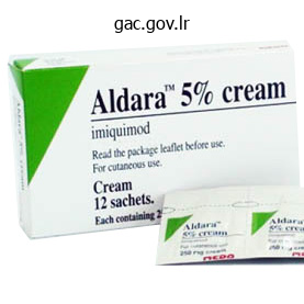
5percent aldara otc
The complex nature of many of these problems requires the involvement of multidisciplinary specialist teams. It is finest identified for its extraskeletal options, together with ocular lens dislocation, thoracic aneurysms, and aortic dissection, but there are also many musculoskeletal indicators (Table 215. There may be large dilatation of the aortic root with "cystic medial necrosis" (fragmentation and disarray of the elastic fibers, reduction in clean muscle cells, and separation of the graceful muscle fibers by collagen and mucopolysaccharide). These domains both play key roles in figuring out the secondary construction of the fibrillin by influencing its folding. They are highly conserved from jellyfish to humans, but their group and biology are still incompletely understood. They represent the scaffold for the deposition of tropoelastin in elastic tissues but can even form impartial constructions, such as the suspensory ligament of the ocular lens, which accounts for some (but not all) of the medical manifestations of Marfan syndrome. Other features in all probability mirror somewhat more advanced mechanisms than simple mechanical failure of the elastic tissues. The analysis is typically made incorrectly, simply on the idea of somewhat soft options, corresponding to lengthy fingers (arachnodactyly), tall stature, or a high arched palate. Ideally, evaluation of the person suspected of getting Marfan syndrome ought to in all instances involve skilled cardiac evaluation (including echocardiography), cautious ophthalmic evaluation (including slit lamp examination), professional musculoskeletal analysis, and skilled scientific and molecular genetic recommendation. Some of the "systemic options" that probably contribute to the prognosis of Marfan syndrome (see Table 215. Note that the thumb ought to utterly cowl the little fingernail within the former and that the thumbnail should be completely seen within the latter. Cardiovascular illness, primarily within the proximal aorta, significantly reduces life expectancy. Previous estimates of life span within the Nineteen Seventies instructed that it was decreased to as little as 32 years (with 80% of deaths attributable to cardiovascular disease), but this is actually unduly pessimistic and distorted by case ascertainment bias. Aortic dissection can occur with out preceding dilatation of the proximal aorta but, generally, the chance is markedly elevated when the aortic root diameter at the sinus of Valsalva exceeds 5 cm. Echocardiography usually offers a reliable estimate of the size of the proximal aorta, however it can be sophisticated by distorted anatomy of the chest wall (severe pectus excavatum). Sometimes additional magnetic resonance imaging may be helpful for accurate assessment. Results should be interpreted based on body floor area and age utilizing applicable nomograms. In adults, aortic regurgitation and related coronary heart failure are frequent, but in youngsters, extreme mitral valve illness is more widespread than that attributable to the aorta. Eventually two thirds of affected people present a minimal of some evidence of mitral valve involvement; mitral regurgitation and conduction disturbances contribute considerably to mortality. Occasional monitoring of the abdominal aorta by ultrasonography must be performed, particularly in those that have already shown vital dilatation of the aortic root. Subtle degrees of subluxation could solely be detectable by slit lamp examination, however these might subsequently progress. Closed-head damage is a potential danger factor for progression, which is one cause why participation involved sports activities ought to be very fastidiously evaluated. Spondylolisthesis is another explanation for again pain found in about 6% of people with Marfan syndrome. It may be current at delivery and is nearly invariably detectable by the second decade of life in these with the predisposition. There is scalloping of the posterior aspect of the vertebral bodies, and the anterior posterior diameter is greater at S1 than L4. However, care should be exerted with spinal anesthesia as a result of dural tears might happen and be gradual to heal. Joint hypermobility (usually mild) is reported in a couple of quarter of affected people however solely often causes main issues. It is also a potential explanation for hypertension, which may clarify the sturdy association between sleep apnea and aortic root dilatation in these people. However, on the milder end of the spectrum, the phenotype merges with the normal vary, and extra particular investigations could also be required to affirm or refute the analysis with confidence. This is necessary because thoracic aneurysms generally develop solely later in life.
Order aldara 5percent fast delivery
Thus, adults with polymyositis or dermatomyositis ought to be evaluated for evidence of an underlying malignancy; nonetheless, past age-appropriate screening exams, the extent of such an evaluation has not been clarified. It typically presents as a mildly to moderately painful mass on a limb, more typically on the leg, however it can be painless. These tumors adhere to adjoining structures, and symptoms are a consequence of native invasion. The average time from onset to prognosis of synovial sarcoma ranges from months to years, and these lesions are often misdiagnosed as benign growths before histologic analysis demonstrates them to be synovial sarcomas. Grossly, the tumor is a circumscribed mass that adheres to the underlying tissue and to adjacent tissue constructions. Because histologic evaluation of synovial sarcomas is troublesome, expertise ought to be sought, and diagnoses are incessantly modified on re-review of the tissue. Other tumors that could be confused with synovial sarcoma embrace epithelioid sarcomas, clear cell sarcomas, and fibrosarcoma. Spindle cells often are the predominant cell sort and have a characteristic swirling appearance with massive nuclei and scant cytoplasm. These swirls appear to make clefts, on the border of which epithelial cells are often arranged. The more frequently mitotic figures are seen, the extra de-differentiated the tumor, with a higher probability of vascular invasion and poorer prognosis. The epithelial cell element is occasionally sparse but could be visualized by silver stains that identify reticulin throughout the cytoplasm. The spindle cells have keratin, which can be recognized by immunohistochemical evaluation. Occasionally, only a single cell sort (most often spindle cells) is seen, which may cause issue in making the prognosis. At the time of presentation, the tumor frequently has metastasized-often extensively by hematogenous unfold, most frequently to the lungs, and less incessantly by native lymphatic spread. The 5-year total survival rate is about 70%, with the prognosis being higher for youthful sufferers and for those with localized illness and tumors smaller than 5 cm in diameter. Radical resection, with or without amputation, is performed for larger, less well-differentiated tumors. Radiation therapy (40�75 Gy) is commonly administered after surgical resection, leading to fewer relapses and higher survival than amongst those treated with surgical procedure alone. Combination chemotherapy with ifosfamide and doxorubicin (with or without vincristine) may be given before surgery and irradiation to enhance survival and lengthen the disease-free interval. Axial computed tomography scan demonstrating a gentle tissue mass lateral and posterior to the femur containing calcifications. Sometimes the signs of paraneoplastic syndromes show even earlier than the prognosis of a malignancy. Raynaud syndrome, erythema nodosum, lupus-like syndrome, and Eaton-Lambert syndrome could be rheumatologic paraneoplastic syndromes. Myositis and vasculitis associated with malignancy (see earlier) is also categorized as paraneoplastic. This sometimes occurs across the knee but may occur elsewhere, even within the small joints of the palms. Very sometimes, a quantity of metastatic lesions outcome from disseminated carcinoma. Patients with pancreatitis, and occasionally those with pancreatic carcinoma,9 might develop a big joint arthritis, usually involving the ankles, accompanied by intensely infected subcutaneous nodules that resemble erythema nodosum. These nodules, which frequently are several centimeters in diameter, may ulcerate and drain. Unlike with true erythema nodosum, recovery results in native cutaneous depression brought on by adipocyte necrosis. Biopsy of the nodule reveals septal panniculitis similar to that seen in erythema nodosum however accompanied by fat necrosis presumably attributable to release of pancreatic lipase, elevated levels of which can be discovered in the blood. Synovial or bursal fluid obtained from these patients can seem milky because of the massive numbers of fat droplets that end result from necrosis of the fatty synovium.
Purchase aldara 5percent without prescription
Myopia is common, and degeneration of the vitreous generally causes vitreoretinal detachments and early cataracts. Regular professional ophthalmic surveillance should be instituted, and prophylactic retinal surgical procedure may be required to shield sight. Multiple epiphyseal dysplasias this huge group of circumstances could be very heterogeneous and consists of lots of the type 2 collagenopathies listed earlier. This variant overlaps with pseudoachondroplasia, which is still commonly mistaken for achondroplasia but from which it may possibly clearly be distinguished by the normal appearance of the top and face and the fact that short stature only turns into apparent from about 18 months of age. Adult top may be lowered to between 90 and one hundred thirty cm, but the degree of short limb disproportion is lower than in achondroplasia. Short stature (adult top <125 cm), severe limb shortening, growth of the metaphyses, joint contractures, osteopenia, and fractures are all recognized complications. Histologically, the columns of proliferating chondrocytes normally discovered within the growth plate are lacking. Multiple hereditary exostoses (also known as diaphyseal aclasis) can also be considered a disease of the expansion plate, which finally ends up in multiple bony outgrowths that occasionally bear malignant transformation to usually low-grade chondrosarcoma. This dominantly inherited dysfunction has a frequency of 1 to 2 per a hundred,000 and exhibits substantial phenotypic variation. There is marked scientific heterogeneity: Exostoses could also be obvious at delivery in some; other people have quite a few lesions throughout with congenital hip dysplasia and clubfoot. Other types of chondrodysplasia Among the numerous chondrodysplasias which have been described are a quantity of which are either comparatively frequent or have significantly fascinating traits. These include several that particularly affect the growth plate, generally identified as metaphyseal dysplasias. Metaphyseal dysplasia (type Schmid) is a uncommon dominantly inherited disorder caused by mutations in kind 10 collagen, which is transiently expressed in the hypertrophic chondrocytes within the growth plate. Angular deformities of the limbs may be corrected in childhood by orthopedic surgical procedure, together with epiphysiodesis, Ilizarov methods, or osteotomy as acceptable. X-linked hypophosphatemia is an X-linked dominantly inherited dysfunction with a birth incidence of about 1 in 15,000. The main feature is rickets with deformities of the decrease limbs and short stature (adult height 145�155 cm). Ossification of the ligament flava contributes to spinal stenosis in lots of sufferers and will require neurosurgical decompression. Patients are proof against normal vitamin D therapy as a end result of they fail to convert this to the energetic metabolite 1,25-dihydroxy vitamin D. In childhood, therapy with phosphate replacement combined with 1-cholecalciferol or calcitriol must be initiated as early as potential. Partial correction of the growth abnormalities could be achieved medically, but surgical intervention is regularly required to appropriate angular deformities on the knees and hips. The lifelong potential for multiple deformities, enthesopathy, fractures, persistent arthritis, and biochemical abnormalities (including hyperparathyroidism) make this one of the difficult musculoskeletal issues to handle. Spondylometaphyseal dysplasias (Kozlowski type) affects both the metaphyses and the spine. Short stature becomes obvious between the ages of 1 and four Manually calibrated to ruler S1=703. There are gross abnormalities of the growth plate resembling rickets with widening and flaring of the metaphyses. There is marked shortening of the limbs with severe varus deformities of the hips and knees. There is significant ossification of the capsule of the hip joints inflicting impingement. Precise estimates of the danger of malignant transformation are troublesome to acquire however general appear to be between 1% and 5%. It is also extra likely to occur in individuals with extra exostoses and unlikely to occur before maturity. Any lesion that suddenly becomes painful or grows rapidly must be examined by ultrasonography to assess the thickness of the cartilage cap, a reliable guide to malignant change. Surgical removing could also be needed; if that is required in childhood, the surgical procedure ought to be deferred as late as attainable to cut back the chance of regrowth whereas the skeleton is still rising. A number of these reflect mutations in regulator genes often known as homeobox genes, which play a key function in "patterning" skeletal development.
Order aldara cheap online
It travels obliquely upward with a ventral spinal root to be a part of the anterior spinal artery in the area of the conus medullaris. Immediately distal to this point, it divides into dorsal and medial branches; the larger, dorsal branch ramiies in the larger muscle mass of the erector spinae, whereas the medial department follows the external contours of the lamina and the spinous process. An intermediate space that normally includes the lower two cervical and higher two thoracic vertebrae is equipped by costocervical branches of the subclavian artery which are of variable sample and oten bilaterally dissimilar. From T2 to L3, the everyday segmental arrangement prevails, however in the sacral space lateral sacral branches of the hypogastric artery and middle sacral branches assume the function of supporting the dietary vasculature to the vertebral parts. Atlantoaxial Complex With their complex phyletic and developmental historical past, the components of the atlantoaxial articulation display the most atypical vascular sample of all the vertebrae. Its ixed place relative to the rotation of the atlas and the adjacent sections of the vertebral arteries prevents formation of main vascularization by direct branches at its corresponding segmental level. One would possibly assume that the diet of the dens would simply be completed by interosseous vessels derived from the spongiosa within the supporting physique of the axis. It is axiomatic, nevertheless, that the vascular patterns of bones had been developmentally established to supply the unique ossiication centers inside the nonvascular cartilage matrices, and despite the eventual obliteration of the separating cartilage, the original patterns of vascularity usually prevail all through life. Occasionally, noncalciied remnants of this plate could persist in adults; although there may be a secure union between the two components, a radiolucent area may recommend a fracture nonunion or a "false" os odontoideum. In mild of the foregoing information, it was not unexpected that the investigations of Schif and Parke78 revealed that the odontoid process was equipped primarily by pairs of anterior and posterior central branches that coursed upward from the surfaces of the physique of the axis and had been derived from the vertebral arteries at the degree of the foramen of the third cervical nerve. A small descending department anastomoses distally with vessels of the next lower phase. Dorsal to the alar ligament, it sends an anterior anastomotic branch over the cranial edge of this ligament to form collateral connections with the anterior ascending artery. Fine medial branches ship perforators into the substance of the vertebral body and meet in a median anastomosis typical of the anterior central branches of the decrease cervical area. Here each artery sends quite a few ine perforators into the anterolateral surfaces of the neck of the odontoid process and terminates in a sprig of vessels that offer the synovial capsule of the median atlantoaxial joint. Fine branches from the anterior and posterior ascending arteries additionally assist within the nutrition of the syndesmotic relations of the atlantoaxial and craniovertebral articulations. Collateral vessels cross over and under the anterior arch of the atlas to anastomose with the apical arcade and ascending arteries. Its descending branches provide the periforaminal dura, the tectorial membrane and alar and apical ligaments, and the ine anastomoses to the arcade. Because the aorta terminates in a bifurcation ventral to the fourth lumbar vertebral body, the vertebrae and the associated tissues caudad to this point rely on an arterial advanced derived largely from the interior iliac (hypogastric) arteries. With the increasing use of percutaneous approaches to the lower lumbar discs, this infra-aortic system of vessels has assumed some surgical signiicance, notably as a end result of, in contrast to the standard segmental provide to the extra superior vertebrae, its major components are longitudinally related to the dorsolateral surfaces of the discs most incessantly involved in these procedures. It then continues to supply the lower posterolateral abdominal wall as it programs superior to the crest of the ilium. These patterns of the vessels were derived from radiographs of perinatal specimens and dissections of adults and drawn towards a tracing of the lumbosacral region taken from a left anterior indirect radiograph of a man. The aorta lies to the left of center as it approaches the bifurcation ventral to the fourth lumbar vertebra. This schema exhibits the extra frequent association of the sacroiliolumbar system on the best facet of the illustration, the place the iliolumbar vessel (7) has a single origin from the dorsum of the posterior division of the (removed) inner iliac artery. The left side exhibits the frequent variation the place the iliac artery and the lumbar artery (14) are derived separately. The middle sacral artery (16) is in its typical position, and the anastomotic contribution from the fourth lumbar artery (4) exhibits its most frequent form. It often has a medial branch that provides the exterior features of the aspect joints and neural arch components and the transversospinal group of muscle tissue and a lateral department to the transversocostal group of the erector spinae. This specimen exhibits considerable variation between the 2 sides of the sacroiliolumbar system. The center sacral artery is also absent, and different branches of the system supply its area.
Discount aldara american express
Blood cultures are at all times indicated for dactylitis because Salmonella infections have been documented in sufferers with this complication. Plain radiographs and bone scans are unable to differentiate between painful crises and osteomyelitis. With ultrasound, a 4-mm depth or extra of subperiosteal fluid is very suggestive of osteomyelitis. In stage three, plain movies show a lucent subchondral line adopted by a cortical discontinuity (crescent sign); it appears only after a number of weeks of sickness. At this stage, the pain and limitation have increased, and ambulation may be possible only with a cane. Stage 6 consists of advanced degenerative joint illness with extreme narrowing or obliteration of the joint area. It is essential to acknowledge that correlation between the clinical symptoms and radiologic options of osteonecrosis is poor. The analysis of gouty arthritis ought to be made only after identification of urate crystals in joint fluid. However, it has been shown that the majority of uncomplicated painful crises may be managed successfully by oral hydration and analgesia alone. Mild painful crises respond to simple analgesics corresponding to acetaminophen (paracetamol), but with a severe disaster, narcotic analgesics are necessary. In these with regular gastrointestinal motility, oral, controlledrelease morphine is a reliable, noninvasive, and preferred alternative. The severity of the ache and its response to therapy must be reassessed regularly. Antisickling agents and pentoxifylline have proven disappointing results after the pain has begun. Paranoia about this concern has typically led to suboptimal remedy of painful sickle cell crises with unnecessary suffering by sufferers. Reluctance of hospital staff to prescribe and administer narcotic analgesics is seen by sufferers as a sign of indifference and perpetuates a lack of confidence in caregivers. However, a specialised daycare unit will have the ability to assess sufferers extra quickly and titrate the analgesia to particular person needs. However, when systemic an infection is suspected, the patient ought to be admitted for intravenous antibiotic remedy along with analgesic medication. Another indication for hospitalization is intensive related chest syndrome, by which early change transfusion might be lifesaving. Attention must be paid to prevention of painful crises, including life-style modifications (avoidance of stress, alcohol, overexertion, swimming, getting caught in the rain, high altitudes) and medications. In children, this can be achieved by casting with the knee immobilized at a 30-degree angle. Apart from perioperative problems, loosening of the prosthesis and infection cause poor long-term alternative outcomes compared with primary degenerative hip disease. The latter, nevertheless, is superior to conservative treatment only in early osteonecrosis (Steinberg stage 1). However, rising resistance is decreasing its potential efficacy, and the spectrum of causative organisms is widening. Newer -lactams and third-generation cephalosporins, generally in combination with an aminoglycoside, may be suitable alternatives. Mild flattening of the superolateral part of the femoral head is current regardless of long-standing disease. Septic arthritis wants early surgical drainage, antibiotics (as for osteomyelitis and guided by tradition when available), debridement, and splinting to forestall ankylosis. Corticosteroids have been associated with avascular joint necrosis, in addition to with painful 1755 bony crisis with oral and intraarticular corticosteroids. Sulfasalazine may be effective in lowering each the rheumatoid inflammation and the sickling course of. The natural historical past, of asymptomatic osteonecrosis of the femoral head in adults with sickle cell illness.
Sinapis nigra (Black Mustard). Aldara.
- What is Black Mustard?
- Are there safety concerns?
- How does Black Mustard work?
- Dosing considerations for Black Mustard.
- Pneumonia, arthritis, aches, fluid retention, loss of appetite, causing vomiting, chest congestion, symptoms of the common cold, aching feet, and other conditions.
Source: http://www.rxlist.com/script/main/art.asp?articlekey=96586
Cheap aldara 5percent overnight delivery
Muscle weakness and joint laxity contribute to additional bleeding from the friable, thickened synovium, which results in persevering with injury. The hyaline articular cartilage is progressively eroded by the irritation related to iron deposition. Ultimately, deep pits filled with friable blood clot happen in the cartilage, along with a surviving plateau of much less broken articular cartilage. Subarticular cyst formation is common, and cartilage collapse could lead to giant bony defects. In previously undiagnosed instances of joint swelling, joint aspiration almost actually is critical to confirm the presence of blood in the joint. Chronic arthropathy the end results of repeated acute episodes of bleeding, after a quantity of months to years, is a disorganized joint exhibiting bony thickening, deformity, lack of movement, and coarse crepitus as a result of the lack of articular cartilage and sclerosis of subchondral bone. Soft tissue swelling and effusions are uncommon at this stage, and ache is fluctuating and variable but at occasions can be extreme. Bleeding into the forearm can cause acute compartment syndromes and subsequent ischemic muscle atrophy and nerve injury. The most generally used classification of the radiologic modifications in hemophilic arthropathy is that of Pettersson and coworkers,13 with subsequent classifications including scientific criteria. The majority of pseudotumors are due to repeated hemorrhage, both subperiosteally or into adjacent muscle. Subsequently, the hematoma becomes encapsulated and calcified with progressive enlargement. These occasions are postulated to be as a outcome of osmotic gradients throughout the fibrous capsule of the lesion. The second kind occurs extra typically within the pelvic region in adults and originates in delicate tissues. The pseudotumour might exert strain on intraabdominal or intrapelvic structures and also can trigger bony erosion of the pelvic bones. A protocol for scanning the six main goal joints and a scoring system have recently been described. Blood in the joint has a direct harmful effect on cartilage by catalyzing reactive oxygen intermediates, which induce chondrocyte apoptosis. These in combination with the proinflammatory cytokine milieu induce caspasemediated chondrocyte harm through decreased proteoglycan production and exposure to degenerative enzymes that result in progressive articular cartilage modifications. Synovial thickening and radiologic density are present because of iron deposition. Extensive synovitis of very low sign depth is current within the suprapatellar recess, according to hemosiderin from recurrent hemarthroses (horizontal arrow). In addition, full-thickness cartilage loss and subchondral cysts are evident in the lateral compartment of the knee (vertical arrow). Such facilities provide a large spectrum of medical, nursing, surgical, and allied well being skilled skills. The idea of prophylactic factor substitute originates from the statement that persistent arthropathy hardly ever develops in patients with moderate hemophilia and issue ranges of 1% to 4%, and that a single joint bleed can lead to irreversible joint injury. This increases their factor degree to greater than 1%, thereby changing them to the phenotype of moderate hemophiliacs. This should be the aim to preserve normal joint operate in these with severe disease. Prophylactic remedy can begin at an early age as both major or secondary prophylaxis. Primary prophylaxis is defined as common steady remedy initiated in the absence of documented joint illness (as determined by physical examination, imaging research, or both) and began before the second clinically evident large-joint bleeding and the age of 3 years. Secondary prophylaxis is outlined as initiation after two or extra bleeding episodes into giant joints and earlier than the onset of joint illness. Tertiary prophylaxis refers to steady therapy with factor began after the onset of joint disease. Many sufferers have already acquired home-based issue alternative, and aspiration may be tough and, for young sufferers, fairly traumatic. If aspiration is required, a large-bore needle corresponding to a 16-gauge one ought to be used under issue coverage for forty eight to 72 hours to elevate the factor stage to 30% to 50% of regular. Local remedy initially with rest, ice packs, and analgesics adopted by graduated physiotherapy and issue replacement for forty eight hours is usually effective. Isometric workout routines should be began the subsequent day, and graduated active physiotherapy must be encouraged after the first 24 hours, with prophylactic factor substitute if necessary.
Syndromes
- Whooping cough
- Acute ear infection
- Severe pain in the mouth
- Increased number of infections
- Chronic kidney disease
- Self-concept
- Joint pain
- You have other symptoms with the malaise.
Generic aldara 5percent with visa
Preceding symptomatic infection with one or two of the next findings: Enteritis (defined as diarrhea for a minimal of 1 day, three days to 6 weeks earlier than the onset of arthritis) Urethritis (dysuria or discharge for a minimum of 1 day, 3 days to 6 weeks earlier than the onset of arthritis) Minor criteria At least one of the following: 1. Evidence of triggering infection: Positive urine ligase reaction or urethral or cervical swab for Chlamydia trachomatis Positive stool tradition for enteric pathogens associated with reactive arthritis 2. Joint involvement� 1 large joint 0 2�10 large joints 1 1�3 small joints (with or without involvement of huge joints)� 2 4�10 small joints (with or with out involvement of large joints) 3 >10 joints (at least 1 small joint)** 5 B. Duration of symptoms�� <6 weeks zero 6 weeks 1 *The standards are aimed at classification of newly presenting patients. Differential diagnoses vary amongst patients with completely different displays however might embody circumstances such as systemic lupus erythematosus, psoriatic arthritis, and gout. Distal interphalangeal joints, first carpometacarpal joints, and first metatarsophalangeal joints are excluded from evaluation. The histopathologic examination must be carried out by a pathologist with experience in the analysis of focal lymphocytic sialadenitis and focus score depend utilizing the protocol described by Daniels et al (1). Ocular Staining Score described by Whitcher et al (2); van Bijsterveld score described by van Bijsterveld (3). The standards embody both sufferers with and without definite radiographic sacroiliitis. Labial salivary gland biopsy exhibiting focal lymphocytic sialadenitis with a spotlight score 1 focus/4 mm2 3. Using histopathologic definitions and focus rating assessment strategies as described by Daniels et al (1). The criteria are applicable to sufferers with peripheral arthritis (usually predominantly of the lower limbs or asymmetric arthritis), enthesitis, or dactylitis. The Assessment of SpondyloArthritis International Society classification standards for peripheral spondyloarthritis and for spondyloarthritis generally. Chronic cutaneous lupus, including Classical discoid rash Localized (above the neck) Generalized (above and below the neck) Hypertropic (verrucous) lupus Lupus panniculitis (profundus) Mucosal lupus Lupus erythematosus tumidus Chilblains lupus Discoid lupus or lichen planus overlap 3. Nonscarring alopecia (diffuse thinning or hair fragility with visible damaged hairs) In the absence of different causes corresponding to alopecia areata, drugs, iron deficiency, and androgenic alopecia 5. Synovitis involving two or more joints, characterized by swelling or effusion Or tenderness in two or more joints and 30 minutes or more of morning stiffness 6. Serositis Typical pleurisy for greater than 1 day Or pleural effusions Or pleural rub Typical pericardial ache (pain with recumbency improved by sitting forward) for greater than 1 day 8. Antiphospholipid antibody positivity as decided by any of the following: Positive take a look at result for lupus anticoagulant False-positive check outcome for speedy plasma reagin Medium- or high-titer anticardiolipin antibody stage (IgA, IgG, or IgM) Positive take a look at end result for anti�2-glycoprotein I (IgA, IgG, or IgM) 5. Malar rash Discoid rash Photosensitivity Oral ulcers Nonerosive arthritis Pleuritis or pericarditis Definition Fixed erythema, flat or raised, over the malar eminences, tending to spare the nasolabial folds Erythematous raised patches with adherent keratotic scaling and follicular plugging; atrophic scarring could happen in older lesions Skin rash as a result of uncommon reaction to daylight by affected person historical past or physician statement Oral or nasopharyngeal ulceration, usually painless, observed by physician Involving two or more peripheral joints, characterised by tenderness, swelling, or effusion a. Pleuritis: convincing historical past of pleuritic ache or rubbing heard by a doctor or proof of pleural effusion Or b. A false-positive test end result for no less than 6 months confirmed by Treponema pallidum immobilization or fluorescent treponemal antibody absorption check An irregular titer of antinuclear antibody by immunofluorescence or an equivalent assay at any point in time and in the absence of medicine 7. Positive antinuclear antibody *For the purpose of identifying sufferers in clinical studies, a person is claimed to have systemic lupus erythematosus if any four or extra of the 11 standards are current, serially or concurrently, during any interval of observation. Updating the American College of Rheumatology revised standards for the classification of systemic lupus erythematosus [letter]. The total rating is determined by adding the maximum weight (score) in every class. Definitions of Items and Subitems within the American College of Rheumatology/European League Against Rheumatism Criteria for the Classification of Systemic Sclerosis Item Skin thickening Puffy fingers Definition Skin thickening or hardening not attributable to scarring after harm, trauma, and so on Swollen digits: a diffuse, usually nonpitting improve in soft tissue mass of the digits extending past the conventional confines of the joint capsule. Normal digits are tapered distally with the tissues following the contours of the digital bone and joint structures. Ulcers or scars distal to or at the proximal interphalangeal joint not thought to be caused by trauma. Digital pitting scars are depressed areas at digital ideas because of ischemia rather than trauma or exogenous causes.

Purchase aldara australia
Proprioception relies on specialized nerve endings, mechanoreceptors, which are situated within the muscular tissues and the ligaments and are important for nice tuning of muscular motion. One major issue is to separate genes that affect the event of the joints (thus ensuing. Epigenetic mechanisms regulate gene expression both by affecting gene transcription or by acting posttranscriptionally. Advances in gene profiling and subsequent era sequencing ought to provide extra insights to these offered by gene expression chip technology39 which have identified, along with recognized candidate gene groups, similar to anabolic and catabolic genes, new gene networks such as a cluster of oxidative defense genes. Validation of the relevance of those genomic data is required for understanding and manipulating these molecular networks and is important and will be supplemented by proteomics, which will probably reveal an much more complicated sample. Terminology of osteoarthritis, cartilage and bone histopathology-a proposal for a consensus. Synovitis score: discrimination between continual low-grade and high-grade synovitis. Genetics and mechanisms of crystal deposition in calcium pyrophosphate deposition illness. Hyaluronan oligosaccharides perturb cartilage matrix homeostasis and induce chondrocytic chondrolysis. Suppression of cartilage matrix gene expression in upper zone chondrocytes of osteoarthritic cartilage. The interrelationship of cell density and cartilage thickness in mammalian articular cartilage. Morphologic modifications in the cellular microenvironment of chondrons isolated from osteoarthritic cartilage. Signalling cascades in mechanotransduction: cell-matrix interactions and mechanical loading. Proceedings of the 8th international conference on the chemistry and biology of mineralized tissues. Quantitative, proteomic evaluation of eight cartilaginous tissues reveals characteristic differences in addition to similarities between subgroups. The practical role of lots of the genes recognized still stays to be elucidated. The genetic contribution to some of these components is only beginning to be addressed. Classical twin studies and familial aggregation studies have additionally investigated the genetic contribution to cartilage quantity and progression of disease. In 1941, Stecher2 demonstrated that Heberden nodes of the fingers have been three times more widespread in the sisters of 64 affected individuals than in the basic inhabitants. Subsequently, in 1944, Stecher and Hersch concluded that these lesions were inherited as a single autosomal dominant gene with a robust female predominance. The differentiated cartilage cells undergo a cascade of late differentiation occasions culminating in chondrocyte hypertrophy. After invasion of blood vessels from the subchondral bone, the majority of hypertrophic cells undergo apoptosis, and the cartilage template is reworked into trabecular bone. Although only a few genes meet the strict standards of replication, even fewer have additionally demonstrated practical significance. The following is simply a quick summary; for more detailed info, readers are referred to different chapters in this guide dealing with monogenic problems of skeletal improvement and to the evaluation by Warman and coworkers. Consequently, many important genes have in all probability been overlooked utilizing this methodology. Individual research may be hampered by sample size limitations, which lead to lack of statistical power, and meta-analyses primarily based on consortium efforts may assist overcome some of these limitations. The Japanese research used a normal approach using 899 circumstances and 3396 management members. Replication involved extra European cohorts and North Americans of European descent. This produced a respectably powered research of 14,934 circumstances and 39,000 control members. The addition of several more cohorts to the original research increased the proof for the veracity of this sign.
Order aldara with a mastercard
High subject power (7 T) offers the chance of immediately evaluating small vessels. Although the restriction of diffusion seen with acute infarction is a marker of loss of tissue integrity, an the histopathologic patterns of granulomatous and necrotizing vasculitis are associated with quickly progressive illness and fatal outcome, however a lymphocytic sample is associated with delicate disease with favorable consequence. Granulomatous vasculitis, a common pattern, is characterised by vasculocentric harmful mononuclear irritation associated with well-formed granulomas, multinucleated big cells, or both. Lymphocytic vasculitis is characterized by lymphocytes with occasional plasma cells, usually in multiple layers, extending via the vascular wall and inflicting vascular distortion or destruction. Necrotizing vasculitis includes predominantly small muscular arteries and is associated with disruption of the interior elastic lamina. Primary angiitis of the central nervous system entails medium-sized arteries and small vessels, including arterioles, capillaries, veins, and venules. The infected vessels might turn into narrowed, occluded, and thrombosed and are associated with tissue ischemia and necrosis in the territories of the involved vessels. The presence of aneurysms should raise suspicion for a mycotic inflammatory reaction, and an infectious etiology needs to be identified. General medical laboratory knowledge are unremarkable, including acute-phase reactants such because the erythrocyte sedimentation rate and C-reactive protein. The presence of oligoclonal bands with an elevated immunoglobulin G index is reported. Mural thickening, hemorrhages, leukoencephalopathy, and gadolinium-enhanced lesions within the cortex, deep white matter, or leptomeninges can also be demonstrated. High-resolution black-blood contrast-enhanced T1-weighted photographs could help differentiate intravascular atherosclerosis from cerebral vasculitis. Areas of stenosis, dilatation, and occlusion in medium and small arteries are suggestive of cerebral vasculitis. Hyperintense T2 signal has been shown to be absent in vasculitic lesions, and a concentric enhancing lesion with hyperintense or heterogeneous T2 signal is more likely in intracranial atherosclerotic disease. Vasculitic enhancement and inflammation can often lengthen past the vessel wall to involve the adjacent perivascular area, brain parenchyma, or each. It is recommended that this periadventitial enhancement might characterize a selected sample of involvement in vasculitis. Finally, 3D black-blood sequences can be used for intraoperative navigation to target individual vascular branches to increase diagnostic yield when biopsy is contemplated in suspected instances of vasculitis. These methods could be suited to measure remedy efficacy with serial examinations. Several studies have shown a lower in N-acetylaspartate in vasculitis, a marker of neuronal and cell wall integrity. The "typical" findings of cerebral vasculitis on four-vessel catheter angiography are widespread segmental modifications within the large, intermediate, and small arteries of a quantity of territories of the cerebral circulation, microaneurysmal dilatation, vessel irregularities, and a quantity of occlusions with sharp cutoffs. In addition, the modifications induced by cerebral vasculitis are much less obvious in massive vessels during early phases of the illness. The process of alternative is open-wedged biopsy of the tip of the nondominant temporal or frontal lobe with excision of 1 cm3 of the overlying leptomeninges and gray and white matter of the underlying cortex. Directing the biopsy to an area of leptomeningeal enhancement or a focal or mass lesion when present could improve the diagnostic yield. Falsenegative biopsy findings stay high because of focal lesions and sampling errors. Findings on mind or leptomeningeal biopsy suggestive of nonspecific gliosis, perivascular irritation, and parenchymal ischemic harm must be interpreted with caution and correlated with the clinical context and imaging studies. Although tissue biopsy is the most reliable approach, the presence of vasculitis in the biopsy specimen may replicate a major infectious course of with secondary vascular irritation. Primary angiitis of the central nervous system should be suspected in the setting of persistent meningitis; recurrent focal neurologic signs such as atypical strokes; unexplained diffuse neurologic signs such as headache, seizure, and cognitive abnormalities; or spinal twine dysfunction not associated with systemic disease or some other process. In very uncommon cases, vasculitis has been reported with amphetamine and cocaine use. A provocative factor may be identified in 12% to 60% of patients, including exertion, sexual actions, emotion, Valsalva maneuvers, or bathing, amongst other triggers. Major scientific issues embody localized cortical subarachnoid hemorrhage, watershed infarcts, and posterior reversible leukoencephalopathy. Ischemic strokes, transient ischemic strokes, and cerebral infarcts or microscopic hemorrhage occurs a couple of days after the preliminary normal findings on neuroimaging, and cerebral vasoconstriction may arise 2 to 3 weeks after medical onset. Intracerebral lobar hemorrhage is usually silent, and patients may have progressive dementia and spastic paraparesis.
Real Experiences: Customer Reviews on Aldara
Kadok, 40 years: In the third pattern, which may be viewed as an end stage of the inlammatory response, the arachnoid turns into an inlammatory mass that ills the thecal sac.
Silvio, 52 years: In common, the presence of earlier vertebral deformities has been proven to enhance the chance for subsequent vertebral deformities by 7- to 10-fold.
8 of 10 - Review by C. Joey
Votes: 50 votes
Total customer reviews: 50

