Cilostazol dosages: 100 mg, 50 mg
Cilostazol packs: 30 pills, 60 pills, 90 pills, 120 pills, 180 pills, 270 pills, 360 pills
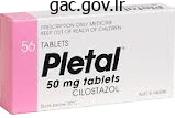
Buy 100 mg cilostazol amex
Influence of contrastenhanced computed tomography on track and outcome in sufferers with acute pancreatitis. Acute necrotizing pancreatitis: treatment technique in accordance with the status of infection. Is contaminated or sterile necrosis an indication�in whom should this be carried out, when, and why Imaging and percutaneous administration of acute sophisticated pancreatitis, Cardiovasc Intervent Radiol. Magnetic resonance cholangiopancreatography: a new technique for evaluating the biliary tract and pancreatic duct. Endo-sonography in persistent pancreatitis-A comparability between endoscopic retrograde pancreatography and endoscopic ultrasonography. Endoscopic ultrasonographic analysis of pancreatic cancer compli-cating continual pancreatitis. Diagnosis and grading of chronic pancreatitis by morphological criteria derived by ultrasound and pancreatography. Value of fluorine 18-Fluorodeoxy-glucose and Thallium 201 within the detection of pancreatic cancer J Nucl Med. A special form of segmental pancreatitis "groove pancreatitis" Hepato-gastroenterology. Pancreatic tumors are the second most common malignancy of the gastrointestinal tract. Till date resection stays the one treatment and the general survival price for 5 years is lower than 5%. Cystic pancreatic tumors are a diverse group of lesions that are relatively rare as are the endocrine tumors of the pancreas. A detailed understanding of the pancreatic topography, macro- and microanatomy and its physiology is mandatory for planning the methods for evaluation of this relatively elusive organ and in addition to clarify the hitherto insignificant impact on survival from malignancy of this organ, despite improved sensitivity and specificity of diagnostic modalities. The neck of pancreas is a focal space of narrowing brought on by compression between Ist a half of duodenum cranially and the mesenteric vessels caudally. The tail of pancreas is totally covered by peritoneum and is, due to this fact, comparatively mobile. Its caudal portion is oriented in the direction of the splenic hilum, anterior floor lies close to the posterior part of the lesser sac while the posterior surface is in shut contact with left kidney. The topography and relations of pancreas in the retroperitoneum are the main determinants of un-resectability of its malignant lesions even when relatively small in dimension. Acinar or exocrine unit constitutes the majority of the organ (80%), ducts (4%), blood vessels and extracellular space (14%) whereas endocrine half or islet cells form about 2% of its volume. The primary islet cell types are glucagon secreting or A cells (15�20%), insulin secreting or B cells (70%) and somatostatin secreting or D cells. It measures 15 cm in size, 7 cm craniocaudally and 2�3 cm in anteroposterior dimensions. Head and uncinate process of pancreas lie inside the duodenal C-loop, its anterior surface is proscribed by the transverse mesocolon and is crossed by the distal part of gastroduodenal artery while the posterior floor is fixed Physiology the exocrine pancreas performs an important function in digestion and absorption through secretion of digestive enzymes and bicarbonates into the proximal duodenum. The endocrine pancreas releases hormones that regulate the metabolism and distribution of breakdown merchandise of meals. The termination of the principle pancreatic duct within the duodenum is situated on the major duodenal papilla. The accessory duct of Santorini drains embryologic dorsal pancreas and is rostral to the main duct in the superior a half of head, extending from the curve of primary duct to drain into the latter or separately on the minor duodenal papilla. The portal vein, splenic vein and superior mesenteric vein and its major trunks drain the pancreas. At the microscopic degree, the intralobular arteries contribute arterioles both to acini or to islets and the pancreatic parenchyma is provided by an in depth capillary network. Several capillaries radiate from the islets to provide the periacinar capillary network.
Buy genuine cilostazol on line
Weaknesses of this sort of research embrace the ability to only image the breast in a single projection at a time. For this reason, dual power strategies have been developed to permit imaging of each breasts with a single contrast administration. Two paired exposures are obtained, one at low kilovolts and the opposite at high kilovolts. A subtraction picture is produced that highlights areas of iodine concentration or enhancement. The mammographic image at low vitality can be used as a routine grayscale mammographic interpretation. Early work has proven technical and medical feasibility of contrast-enhanced digital mammography. The precise number of sufferers studied to date has been limited so the appliance of this technique for future potential scientific use is unsure. The side being examined is raised and the arm is positioned above the pinnacle to be sure that the breast is evenly distributed over the chest wall. Scanning within the radial (parallel to the ducts) and anti-radial (perpendicular to ducts) planes is of value in demonstrating ductal abnormalities. If lesion is recognized, its presence is confirmed with 90o rotation of the transducer. The particular location, together with laterality (right or left breast), the clock-face location and the distance from the nipple, must be accurately annotated on the images and documented within the reviews. Extended area of view imaging is a helpful tool to picture giant lesions that reach past the width of the transducer footprint or to higher depict relationships amongst multiple lesions. It is often helpful to place the transducer barely off middle to the nipple after which use angulation to picture the retroareolar tissues. As shade Doppler gear has become increasingly sensitive, vascular alerts may be found in normal, benign and malignant tissue. Normal Anatomy the skin, which is normally 1�3 mm thick, appears as two parallel echogenic strains. Normal ducts are sometimes seen in the subareolar area, as anechoic tubular buildings. The younger glandular breast contains a well-defined layer of glandular tissue, within which round or oval well-defined, hypoechoic fats lobules may be current. Fat lobules may be distinguished by their unique spindle form, which merges with the relaxation of the parenchyma at completely different angles and planes of imaging. By contrast, plenty stay discrete with distinctive borders in all planes of imaging. Deep to the glandular tissue, a retromammary fats layer is seen and behind this pectoral muscle may be seen sharply demarcated by its echogenic fascia. Costochondral cartilages seem as hypoechoic oval lots deep to the muscle and calcified ribs present outstanding shadowing. Beneath the thoracic wall, a linear echogenic interface can be seen that features chest wall, pleura and lung border. Anatomic structures that can be seen in axilla embrace blood vessels, lymph nodes and the proximal parts of the pectoral muscles. Circumscribed margins are properly outlined or sharp, with an abrupt transition between the lesion and the encompassing tissue. Noncircumscribed margins embody the remaining margin descriptors, together with microlobulated, vague, angular and spiculated. Angular margins reveal sharp corners, typically with acute angles, in distinction from spiculated margins, which appear extra as lines projecting from a mass. Lesion boundary ought to be described as an abrupt interface, which is seen as a sharp demarcation between the lesion and the encircling tissue, or an echogenic halo, a surrounding echogenic transition zone. The internal echogenicity of the mass could also be anechoic, hyperechoic, complex, hypoechoic or isoechoic.
Purchase cilostazol no prescription
Intravenous line is established to keep away from extravasations of the tracer, which can yield misguided interpretation. Imaging: Patients are examined within the supine place with the gamma-camera positioned beneath the examination table viewing the kidney and bladder area. However, in children with obstructive ailments, some tracer may be retained inside the pelvicalyceal system causing interference in the imaging of useful renal parenchyma resulting in false interpretation of the study; therefore, late Chapter ninety seven Current Status of Nuclear Medicine in Urinary Tract Imaging 1553 Table 1: Different radiopharmaceutical and their doses Radiopharmaceutical 99m 99m 99m 123 Dose (mCi/kg) 0. In renal transplants, nevertheless, the camera is positioned anteriorly over the patient in supine place, viewing the allograft and bladder. Once the affected person is appropriately positioned, a speedy intravenous bolus of the tracer is injected and simultaneously the acquisition is began. One frame per two seconds is recorded for one minute, followed by one body per 15 seconds for length of 20�30 minutes. Interpretation: A dynamic renal study is evaluated for the parenchymal section, the cortical transit time, and the urine drainage section. During the primary few minutes of the dynamic research, after the preliminary vascular distribution of the tracer and before its appearance within the renal collecting system, the tracer is concentrated in the renal parenchyma. After this, the tracer seems outside the renal parenchyma with progressive lower in ranges throughout the parenchyma, marking the beginning of the drainage phase. The relative renal dimension can simply be estimated by way of simple visible statement. Renal dimensions in longitudinal and transverse planes may be measured with a calibrated system or by imaging a radioactive ruler positioned beside the affected person. A qualitative estimate of the renal perform can be obtained by visually evaluating the ratio of the renal and background exercise. Normally, very little tracer activity must be current within the background, the blood pool and the liver during the parenchymal z phase. If a excessive stage of tracer exercise is noted inside these regions, then the renal uptake shall be low indicating a poor renal perform. Information about intrarenal distribution of radiotracer can be obtained through the parenchymal section. Hydronephrosis is often seen as a "photon poor" area within the renal pelvis. Depending on the severity of hydronephrosis, the cortical uptake appears as a rim of varying dimension. Reduction or absence tracer activity in a comparatively massive portion of the kidney, similar to a malfunctioning upper pole in a duplex kidney, trauma, tumor, or cyst can be traced. Cortical transit time: Cortical transit time refers to the time interval between the intravenous injection of the radiotracer and its first appearance within the renal amassing system. Conversely, due to this fact, the poorer the renal function, the longer is the cortical transit time. Even if the cortical transit time appears to be inside the normal vary, you will want to decide if it is associated with a decrease of tracer exercise from the renal cortex. These embrace ureteral obstruction, acute and chronic pyelonephritis, nephrotoxicity, trauma, renal artery stenosis, renal vein thrombosis, acute tubular necrosis and allograft rejection. Usually, after 20 minutes of the research, a lot of the tracer would depart the renal parenchyma. At this time, minimal or no tracer must be seen within the renal amassing system. Time activity curves should all the time be interpreted along with careful evaluation of images in the parenchymal section and drainage part, preserving in thoughts the cortical transit time. Normal cortical transit time can be seen with full cortical clearance of tracer at 20 minutes together with a time exercise curve that exhibits a delayed peak and a high-residual worth. Such a pattern signifies delayed drainage without parenchymal dysfunction and is almost always with none scientific significance. The possibility of a vesicoureteric reflux of tracer in the course of the drainage section must also be stored in thoughts so as to be ready to differentiate it from outflow tract obstruction. The background activity is normally a mirrored image of blood clearance of tracer or quite the renal extraction of tracer from the blood pool, which quickly decreases with time in a affected person with regular renal function. An extra image is really helpful to decide the tracer clearance, which, if sufficient, ought to be thought of normal, and the outflow tract not obstructed. Normal Dynamic Renal Study There is an intense and fast focus of tracer within the renal parenchyma at 1�3 minutes postinjection. Ureters could additionally be visualized in normal sufferers and in those with gradual ureteral transit time.
Trusted 50mg cilostazol
This helps the radiologist eliminate the chance of lacking an associated dislocation or subluxation at a site distant from the obvious major harm. The full radiographic evaluation of a trauma patient should include the next: 1. A dislocation is a complete disruption of a joint with lack of congruity between the articular surfaces. Subluxation is a minor disruption of the joint the place some a part of the articular surfaces remain in touch. A fracture can appear as any of the following-an obvious disruption in continuity of bone, irregular line of radiolucency, cortical irregularity or increase in bone density (due to impacted or compression fractures). Torus (buckling) fractures: Fractures occurring because of longitudinal compressive forces over a soft bone of young baby. Direction of Fracture Line that is described with respect to the longitudinal axis of the bone. In long bones Anatomic Location and Extent of the Fracture When describing a fracture it may be very important specify the location within the long bone. It may also be described by means of involvement of anatomic point of reference. For fractures situated close to bone ends, mention must be made from any intra-articular extension as this will alter the following management of the patient. Types of Fracture Complete or incomplete: the fracture is assessed as full if it involves the complete width of the bone to involve both the cortices and incomplete if solely some of the bony trabeculae are completely severed while others are bent or remain intact. The bone bends with the convex aspect exhibiting a horizontal break in the continuity of the cortex whereas the other concave side remains intact. This kind of fracture is often stable and unlikely to be displaced after discount. Oblique fracture: the fracture line runs indirect to the lengthy axis of the bone and the angle is <90�. Compression: It represents an impaction fracture involving the spinal vertebral body due to hyperflexion forces. Avulsion: It includes tearing away of a portion of the bone usually at insertion of a muscle or ligament as a outcome of forceful pulling. The frequent sites are tuberosities of tubular bones, elbow, lateral metatarsal and lower cervical spinous processes. Corner chip fractures at metaphyseal ends in kids should elevate the suspicion of battered baby syndrome. Spatial Relationship of the Fragments that is described in terms of apposition/displacement, alignment/angulation, rotation and distraction/overriding. Apposition: It refers to the state of bony contact on the fracture web site and the resultant shift of the fragments relative to each other. The displacement is described close to the direction of shift of the distal fragment. Alignment: It refers to the position of the distal fragment with respect to the proximal fragment. Rotation: It refers to the rotation of the distal fragment over the longitudinal axis. To comment on rotation, the proximal and distal joints should be included in the radiograph. Overriding/distraction: A longitudinal separation of the bony fragments in the long axis of the bone with resultant increased distance between the two fragments is termed distraction. Presence of Associated Abnormalities of the Adjacent Joints It could also be generally associated with sure forms of mechanisms of injuries and some particular fractures. Examples embody a femoral shaft fracture associated with hip joint dislocation, a lower finish tibial fracture related to higher end fibula fracture, etc. The joints could additionally be dislocated 3288 Section 7 Musculoskeletal and Breast Imaging or subluxated. A fracture by way of a joint floor could contain the cartilage solely (chondral fracture) or cartilage and underlying bone (osteochondral fracture). Involvement of Growth Plate/Physis: Seen in Children these fractures result in growth arrest and deformity.
Diseases
- Exudative retinopathy familial, autosomal dominant
- Myopathy Hutterite type
- Kashani Strom Utley syndrome
- Perinatal infections
- Overgrowth syndrome type Fryer
- Chickenpox
- Lower mesodermal defects
- Anti-plasmin deficiency, congenital
- Learman syndrome
- Homozygous hypobetalipoproteinemia
100 mg cilostazol visa
Lipiodol together with chemotherapeutic drug concentrates in malignant cells as a end result of tumor vascularity and lack of lymphatics. This allows chemotherapeutic agent to stay in contact with tumor cells for prolonged period of time. These microspheres could be loaded with doxorubicin and could be infused into feeding tumor arteries. Clinical elements like symptomatic enchancment, decrease in tumor markers, improved quality of life and patient survival are also assessed. These are still unanswered questions on how to enhance its efficacy and in the end delay patient survival. Transcatheter arterial embolization of the tumor may be accomplished as an emergency procedure to cut back gastrointestinal hemorrhage in chosen patients. The causes of huge intermittent gastrointestinal bleeding could additionally be either as a end result of varices or due to direct invasion of duodenum, transverse colon and abdomen by the tumor. Internal radiotherapy by injection of isotopes (131Iodine-lipiodol,188 Re-lipiodol and90 Yttrium) into the hepatic artery has proven improvement in survival. The technique is new and plenty of multicenter prospective research are being performed. Lipiodol is mixed with chemotherapeutic drug and is infused in hepatic artery (arrowheads) followed by gel foam embolization (long arrows) of the arterial branches. Hepatic arteriograms (A to C) are displaying tumor vascularity with simultaneous opacification of portal vein (arrows). It reduces post-operative hepatic failure in major proper hepatectomy in patients with small left lobe. The purpose is to occlude the branches of proper lobe, which is to be resected and to preserve the branches of left lobe. This ends in compensatory hypertrophy of the left lobe and therefore subsequent enhance in the useful reserve of the liver. This produces an acute or fulminant variant of Budd�Chiari syndrome, which has a poor prognosis. Though full necrosis, defined as an absence of detectable disease on computed tomography at 6 months follow-up was achieved in majority, treatment failure was also observed as a result of the event of latest metastases on follow-up. Complete surgical removal of the tumor was carried out with less blood loss 1470 Section three Gastrointestinal and Hepatobiliary Imaging rupture with subsequent life-threatening hemorrhage, thrombocytopenia, hypofibrinogenemia and infrequently portal or systemic hypertension as a end result of intratumoral arterioportal or arteriovenous fistula. Selection of therapy choices is determined by scientific presentation, histology, web site, size, variety of lesions, surrounding liver, age, and efficiency standing of affected person. The treatment possibility should be selected on a case-by-case foundation and should fulfill the needs of the individual affected person. Transcatheter arterial embolization with alcohol and metal coils adopted by systemic 14. Gastrointestinal hemorrhage in hepatocellular carcinoma administration with transhepatic arterioembolization. Randomized managed path of transarterial lipiodol chemoembolization for unresectable hepatocellular carcinoma. Radiofrequency ablation of liver metastases from breast cancer: ends in 14 sufferers. Pedunculated hepatic hemangioma with arteriportal shunt � treated with angioembolization and surgical procedure. Transcatheter arterial embolization within the treatment of symptomatic cavernous hemangioma of the liver: a potential examine. These are as follows: zz Percutaneous gastrostomy zz Percutaneous jejunostomy zz Esophageal stent insertion zz Duodenal stent insertion zz Colorectal stent insertion zz Percutaneous enterocutaneous fistula closure. In this technique, beneath picture guidance a feeding tube is placed within the stomach and generally as a lot as the jejunum. It can be utilized to provide enteral nutritional assist for sufferers with anorexia nervosa, extreme melancholy and advanced malignancy. Decompression of the abdomen or small gut Patients with continual small-bowel obstruction might benefit from drainage with large-bore (24- to 28-French) gastrostomy tubes, which obviates long-term nasogastric suction. This indication is more common in sufferers with continual intestinal obstruction secondary to carcinomatosis. Enteral feeding is preferred in these with sufficient small-bowel perform to take up adequate water, electrolytes and vitamins from a standard or elemental food regimen. Nasogastric intubation is straightforward but poorly tolerated when used for longterm; it could potentiate gastroesophageal reflux and cause peptic esophagitis or aspiration of gastric contents.
Purchase cilostazol master card
Tumor vascularity has been recognized as being important in cancer development and metastasis and in the systemic supply of therapeutic agents. Tumor perfusion is likely considered one of the earliest physiologic properties to be measured and advances in methodology have led to increasingly quantitative approaches. The most physiologically sturdy and quantitative measures of tumor blood circulate use freely diffusible imaging probes. Cell dying, or apoptosis, is a fundamental part of regular mobile physiology and an early indicator of therapeutic response. Methods for imaging cell death based on extension of annexin V staining in vitro, which indicates apoptotic cells by way of binding to phosphatidylserines. As breast most cancers treatment becomes increasingly focused and individualized, demands on breast most cancers diagnostic modalities have increased. Currently, it has restricted role in primary tumor detection and axillary nodal staging. It additionally seems to be very useful for monitoring remedy response and detection of recurrent disease. Trends in most cancers incidence and mortality in Osaka, Japan: evaluation of cancer management activities. Biologic correlates of 18fluorodeoxyglucose uptake in human breast cancer measured by positron emission tomography. High decision fluorodeoxyglucose positron emission tomography with compression ("positron emission mammography") is very accurate in depicting primary breast most cancers. Imaging sensitivity of devoted positron emission mammography in relation to tumor size. Baseline staging exams after a brand new diagnosis of breast cancer: additional proof of their restricted indications. The worth of bone scintigraphy, bone marrow scintigraphy and quick spin magnetic resonance imaging in staging of sufferers with malignant stable tumors: A potential examine. Integrated Positron Emission Tomography/Computed Tomography might render bone scintigraphy unnecessary to investigate suspected metastatic breast most cancers. Metabolic monitoring of breast most cancers chemohormonotherapy using positron emission tomography: initial analysis. Accuracy of medical analysis of regionally advanced breast most cancers in sufferers receiving neoadjuvant chemotherapy. Sequential positron emission tomography using [18F]fluorodeoxyglucose for monitoring response to chemotherapy in metastatic breast cancer. Locoregional recurrence patterns after mastectomy and doxorubicin-based chemotherapy: implications for postoperative irradiation. Use of positron emission tomography in evaluation of brachial plexopathy in breast most cancers patients. Predicting the prognoses of breast carcinoma patients with positron emission tomography using 2-deoxy-2-fluoro[18F]-D-glucose. The relationship between vascular and metabolic traits of primary breast tumours. Tumor metabolism and blood move changes by positron emission tomography: relation to survival in patients handled with neoadjuvant chemotherapy for regionally superior breast most cancers. Progesterone receptor status considerably improves outcome prediction over estrogen receptor standing alone for adjuvant endocrine therapy in two large breast most cancers databases. Positron tomographic assessment of sixteen alpha-[18F] fluoro-17 betaestradiol uptake in metastatic breast carcinoma. Quantitative fluoroestradiol positron emission tomography imaging predicts response to endocrine remedy in breast cancer. Positron emission tomography studies in patients with regionally superior and/or metastatic breast most cancers: a method for early remedy evaluation Imaging early modifications in proliferation at 1 week submit chemotherapy: a pilot examine in breast cancer sufferers with 39-deoxy-39-[18F]fluorothymidine positron emission tomography. Measurements of blood flow and exchanging water space in breast tumors utilizing positron emission tomography: a rapid and non-invasive dynamic methodology. Changes in blood flow and metabolism in domestically advanced breast most cancers treated with neoadjuvant chemotherapy.
Order cilostazol 50 mg fast delivery
Low mechanical index and intermittent imaging are employed to obtain sufficient distinction enhancement. Harmonic shade and power Doppler imaging and pulse inversion techniques are highly effective sonographic contrast-specific strategies. This stage has been used because of the consistent acoustic window supplied by the surrounding liver which ensures reproducibility of the measurement. The portal vein hint is generally continuously antegrade with a imply peak velocity of 15�22 cm/s. Its major impression has been in sufferers with colorectal malignancy with liver metastases present process surgical procedure. Intraoperative ultrasonography is used to precisely establish liver metastases and information surgical resection. The probe is applied directly to the liver floor and no gel or acoustic coupling agent is necessary. The liver is scanned from the dome to the caudal edge and from left to proper in a sequential manner. Color Doppler could be coupled with the grey scale scan and could be very useful to identify vascular landmarks and assess their patency. Intraoperative ultrasonography provides the working surgeon with useful real-time diagnostic and staging data that may result in an alteration within the planned surgical method. Current functions for this method include tumor staging, metastatic survey, steerage for metastasectomy and varied tumor ablation procedures, documentation of vessel patency, analysis of intrahepatic biliary illness and steerage for liver transplantation. The round structure anterior to the portal vein is the hepatic artery seen in cross section (arrowhead) Computed Tomography Computed tomography has for long been the modality of choice for evaluation of focal liver lesions. The late arterial part corresponds to preliminary opacification of the portal-venous system. The phase of most hepatic parenchymal enhancement and hepatic venous opacification occurs about 45 seconds after the start of the pure early arterial section. For triphasic hepatic imaging, about a hundred and fifty mL of iodinated contrast is injected at 3�4 mL/sec via a pressure injector. Acquisition parameters, specifically, table pace per rotation and scan rotation pace are set to permit full protection of the liver in lower than 8 seconds. Note the hepatic vein seems as a focal lesion on the early and late arterial phases but normally fills in through the portal-venous part (arrows) performed, one each in the course of the early arterial part (20 seconds after injection of distinction medium), late arterial section (30 seconds after the initiation of injection) and portalvenous section (60 seconds after the start of injection). A fourth delayed part is added when required at 3�5 minutes and is particularly helpful in the evaluation of hemangiomas. Accurate acquisition timing for multiphasic imaging depends on the assessment of circulation time in individual sufferers. This assessment is made by utilizing either a preliminary injection of a small bolus of distinction material ("mini-bolus injection") or on-line bolus monitoring software program. If only two phases are deliberate through the liver (late arterial and portalvenous), then the late arterial-phase scan could be triggered following a delay of 10 seconds after peak aortic enhancement time. The regular liver seems homogeneous and has a density larger than spleen, pancreas and kidneys because of the high focus of glycogen in liver. Increased density of liver could also be seen in hemochromatosis and glycogen storage ailments, whereas decreased density is most often related to fatty infiltration of the liver. The fissure of the ligamentum venosum separates the left lobe and the caudate lobe. Magnetic Resonance Imaging Magnetic resonance imaging offers major advantages as a end result of its excessive distinction resolution, multiplanar functionality, sensitivity to blood circulate and lack of ionizing radiation. The adjacent organs at this level may be simply identified with the abdomen (St) to the left and the diaphragm seen posteriorly (black arrow) as nicely as anteriorly (white arrow), S, spleen. Fat suppression is regularly used with these echo train sequences because fat has very excessive signal intensity on these pictures. Signal depth drop out is current in out of part pictures if fat and water are present in the same voxel. T1-weighted 2D gradient echo sequences are used to carry out dynamic post contrast imaging of the liver. These techniques present additional functional and qualitative information regarding the liver lesions. The fissure for ligamentum teres is clearly seen as a fatty cleft arrow Magnetic Resonance Imaging Contrast Agents Magnetic resonance distinction brokers are at present used to accentuate the difference in signal intensity between the liver lesion and adjoining normal tissue and to highlight different enhancement patterns. Extracellular contrast brokers are hydrophilic, small molecular weight gadolinium chelates.
Safe cilostazol 50mg
They are believed to be caused by repetitive contact of humeral head with posterosuperior labrum. This original classification was later expanded on foundation of arthroscopic findings; nonetheless, their scientific implication is questionable. They are difficult to diagnose however essential to determine as they respond well to arthroscopic repair. Labral tears can be associated with labral cysts or ganglia which can cause compression neuropathy. These form due to one way valve mechanism resulting in accumulation of joint fluid within them. Subscapularis tears are seen after anterior dislocation over forty years of age and after posterior dislocation at any age. The most vital regular variation is a poor attachment of labrum within the anterosuperior quadrant of glenoid (sublabral foramen or recess). Contrast in sublabral recess is seen as a skinny line with clean margins that runs along the glenoid. Contrast in a tear has irregular margins, globular shape and is seen to enter into labral substance. Bicipital tendinopathy can also happen in sufferers with glenohumeral instability and attrition at the groove. Morphologic abnormalities corresponding to fraying, flattening and absence of tendon may someday be identified. Disproportionate amount of fluid in bicipital tendon sheath as compared to the joint fluid might recommend bicipital abnormality, i. Bicipital Tendon Dislocations Bicipital tendon dislocation normally occurs with the chronic rotator cuff tears that have prolonged to involve the subscapularis tendons. When all of the three buildings are disrupted, tendons of lengthy head of biceps dislocates medially posterior to subscapularis tendon into the joint, where it might be mistaken for a indifferent anterior labrum. Rarely when subscapularis tendon remains intact, with the disruption of other two ligaments, biceps tendon displaces extra-articularly, anterior to subscapularis muscle. Suprascapular Nerve Entrapment Suprascapular nerve passes by way of supraspinatus notch to enter supraspinatus fossa beneath supraspinatus muscle tissue. Further posteriorly it passes by way of spinoglenoid notch to enter into the intraspinatus fossa. Compression of nerve on the degree of suprascapular notch ends in atrophy of each muscles and compression at the stage of spinoglenoid notch ends in atrophy of infraspinatus muscle solely. Patients current with ache and paresthesia in axillary nerve distribution and muscle atrophy of teres minor and main muscular tissues. Calcification of rotator cuff mostly happens in supraspinatous tendon however can happen in different tendons. Loose our bodies occur in glenohumeral joint and subacromial bursa which seem isointense to bone or hypointense on T2W pictures. Immunocompromised standing and pre-existing degenerative joint disease increases danger of septic arthritis. However, typical systemic and local symptoms could also be absent in indolent infections like tuberculosis. T1W coronal part (B) shows globular space of hypointense sign (arrow) in the supraspinatus tendon suggestive of calcific tendinitis Chapter 196 Magnetic Resonance Imaging of Shoulder and Temporomandibular Joints 3251 choose up early marrow or soft tissue modifications. Further progression of disease results in cartilage destruction manifesting as joint house narrowing and marginal erosion of bare space. Subchondral bone changes in type of edema, subchondral cyst formation and bone destruction comply with when disease turns into subacute. Osteomyelitis, bone destruction and joint ankylosis are features of persistent septic arthritis. Tuberculosis of shoulder joint is infrequent and has indolent scientific presentation.
Real Experiences: Customer Reviews on Cilostazol
Gorn, 47 years: Methods for imaging cell dying based on extension of annexin V staining in vitro, which indicates apoptotic cells via binding to phosphatidylserines. Barium enteroclysis significantly double distinction (barium and air) enteroclysis is superior for analysis of early lesion.
Lukjan, 21 years: The dysfunction is typically bilateral, though it might be unilateral or rarely involves only one pyramid. It normally appears hyperintense relative to the renal cortex on T1 weighted images, though it may sometimes seem isoto hypointense.
Daro, 34 years: Middle hepatic vein divides the liver into right and left lobes (or right and left hemiliver). Clinical Presentation Age: Although few instances current beneath the age of forty years, the overall age of presentation tends to the earlier and the age vary wider, than in generalized myelomatosis.
Cole, 36 years: Urethral stricture or bleeding, obstructive symptoms, blood stained discharge, urethral fistula, perineal abscess, or persistent local pain in an aged man is suggestive of urethral carcinoma. The present most popular method of re-establishing continuity of urinary tract is by creation of ureteroneocystostomy (commonly by using a submucosal tunnel to lower the incidence of vesicoureteric reflux).
Kulak, 64 years: Imaging Techniques Recent advancements in all of the fields of imaging have led to higher analysis of renal cases and their improved administration. Causes of focal T1 signal in crease in any marrow could also be a traditional variant, solitary hemangioma, metastasis, lipoma, bone marrow hemorrhage or degenerative disk disease.
Kippler, 58 years: Strictures within the anterior urethra may be as a result of trauma (external damage, instrumentation),irritation. Primary versus secondary ovarian malignancy: Imaging findings of adnexal plenty in radiology diagnostic oncology group research.
9 of 10 - Review by M. Dimitar
Votes: 149 votes
Total customer reviews: 149

