
Tadapox dosages: 80 mg
Tadapox packs: 30 pills, 60 pills, 90 pills, 120 pills, 180 pills, 270 pills
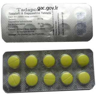
Buy tadapox 80mg fast delivery
It considers the modifications in extracellular matrix composition throughout regular being pregnant and term supply and people seen in preterm untimely rupture of membranes. Takashina S, Ise H, Zhao P et al 2004 Human amniotic epithelial cells possess hepatocytelike traits and functions. Tong X, Wang L, Gao T et al 2009 Potential perform of amniotic fluid in fetal growth � novel insights by comparing the composition of human amniotic fluid with umbilical wire and maternal serum at mid and late gestation. Neu J: Gastroenterol ogy and Nutrition: Neonatology Questions and Controversies, 2nd ed. These latter cavities abut on the embryonic bilaminar disc where the epithelial epiblast and visceral hypoblast are approximated. A fourth cavity, the allantois, will type as a hypoblastic diverticulum in stage 7. Only the epiblast will give rise to the embryo; all other layers produced so far are extra embryonic. The amnion and chorion (and surrounding mesoblast) are part of the extraembryonic somatopleure, whereas the yolk sac, allan tois and surrounding extraembryonic mesoblast represent extraembry onic splanchnopleure. At the junctional zone surrounding the margins of the embryonic space, the place the walls of the amnion and yolk sac converge, the somatopleuric and splanchnopleuric layers of extraem bryonic mesoblast are continuous. The phrases epiblast and hypoblast are used to make the excellence between the earliest bilaminar disc layers and the later embryonic layers. Epiblast and hypoblast include mixed populations of cells with little restriction, which establish the placental structures and extraem bryonic tissues earlier than the production of embryonic cell traces at gastrula tion. The older terminology depicting three germ layers that give rise to the pores and skin, gut lining and intervening tissues is thus incorrect for the bilaminar and trilaminar embryonic disc. The utility and retention of this aged terminology for the early phases of embryology proceed to cause confusion and inhibit the event of extra pertinent descrip tive language to describe these early events. The earliest three cell line ages, not layers, that give rise to the extraembryonic membranes and the embryo are trophoblast, hypoblast and epiblast; every expresses dif ferent lineagespecific genes for its institution, upkeep and differentiation (Artus and Hadjantonakis 2012). At early stage 6, the epiblast is producing extraembryonic mesen chyme from its caudal margin. With the looks of the primitive streak, a course of is begun whereby cells of the epiblast either cross deep to the epiblast layer to form the populations of cells inside the embryo, or remain on the dorsal aspect of the embryo to become the embryonic ectoderm. The primitive streak is the site of organizer cells analogous to these found in embryos that do endure gastrula tion. The appearance of the primitive streak therefore marks the start ning of a period when gross alterations in morphology and complicated rearrangements of cell populations happen. During this time, the epiblast will give rise to a posh multilaminar structure with a defined cranio caudal axis. By the top of gastrulation, cell populations from different, typically broadly separated, areas of the embryonic disc will turn into spatially related and the embryonic form may have been produced. Primitive streak and node Seen from the dorsal (epiblastic) facet, at stage 6, the embryonic disc appears elongated. The relative dimensions of the primitive streak and the fates of the cells that pass through it change with the developmental stage. Thus, the streak extends half method along the disc within the stage 6 embryo, reaching its greatest relative size in stage 7 and its maximum length in stage eight. Formation of the primitive streak is induced by the underlying vis ceral hypoblast, which remains beneath the streak even at later phases. Experiments clearly present the lip of the blastopore to be a dynamic wave front on which cells are carried into the inside to kind the roof of the archenteron, a situation analogous to ingression through the node of the prechordal plate and endoderm. The primitive streak similarly may be considered analogous to the coapted, or fused, lateral lips of the blastopore, and the cloacal membrane and its instant environs are thought of analogous to the ventral lip of the blastopore. At the primitive streak, epiblast cells undergo a interval of intense proliferation, the rate of division being much quicker than that of blasto meres throughout cleavage. Streak formation is associated with the local manufacturing of several cell layers, in depth disruption of the basal lamina, increase in adhesive plaques and hole junctions, synthesis of vimentin, and lack of cytokeratins by the rising cells.
Buy tadapox on line amex
The maxilla is carried downwards and forwards by expansion of the orbits and nasal septum and by sutural development, particularly at the fontanelles and zygomaticomaxillary and pterygomaxillary sutures. In the primary year, progress in width happens on the symphysis menti and midpalatal, internasal and frontal sutures; such development diminishes and even ceases when the symphysis menti and frontal suture shut through the first few years, although the mid-palatal suture persists until mature years. After sutural development, close to the tip of the second year, expansion of the facial skeleton occurs by surface accretion on the face, alveolar processes and palate, and resorption in the walls of the maxillary sinuses, the upper surface of the onerous palate and the labial facet of the alveolar process. Coordinated development and divergence of the pterygoid processes mirror deposition and resorption of bone on appropriate surfaces. Obliteration of the calvarial sutures progresses with age, beginning between 20 and 30 years internally, and somewhat afterward the exterior. Obliteration normally begins within the coronal or sagittal sutures after which extends into the lambdoid suture. The size of the mandible and maxillae diminish following the lack of teeth and consequent resorption of alveolar bone; this reduces the vertical depth of the face and increases the mandibular angles. Usually irregular in measurement and form, and most frequent within the lambdoid suture, they also occur at fontanelles, especially the posterior fontanelle. They might characterize a pre-interparietal element, a real interparietal, or a composite. One or extra pterion ossicles or epipteric bones could seem between the sphenoidal angle of the parietal and the greater wing of the sphenoid; they range significantly in measurement but are roughly symmetrical. Craniosynostosis Sutural progress makes an essential contribution to progress of the skull, particularly through the first few years of life. Premature closure of sutures (craniosynostosis) results in restriction of progress along the sutures, producing morphological modifications which will end in cranium deformity (Sharma 2013). Premature fusion may occur in one or more of the cranial sutures; when the sutures across the cranial base are concerned, extreme limitation of facial bone development will happen. Metabolic issues similar to rickets and familial hypophosphatasia can also end in synostosis. Raised intracranial pressure with or without hydrocephalus, visible deterioration and mental retardation may outcome. Scaphocephaly (sagittal craniosynostosis) is the most common and results in lengthening of the vault in an anteroposterior course; it can also occur along side other sutures. Coronal synostosis, both unilateral (plagiocephaly) or bilateral (brachycephaly/oxycephaly), is the subsequent most frequently seen and leads to lowered anteroposterior growth with marked supraorbital recession. Metopic craniosynostosis (trigonocephaly) and pansynostosis (turricephaly), where each the coronal and sphenofrontal sutures are concerned, are a lot less common. When vital orbital hypertelorism develops, a transcranial bipartitioning process is needed in order to convey the two orbits collectively. This is extremely encouraging, provided that research has shown that the right intercourse can often be predicted from the grownup dwelling face with solely 96% accuracy. Generally speaking, the male skull is extra strong and the feminine more gracile, although there are apparent genetic, and therefore ethnic, variations that have to be considered when trying to assign sex from a skull. The female forehead is generally higher, more vertical and extra rounded than that of the male, with retention of the frontal eminences. The male mandible is more strong and bigger than that of the female; it usually shows a larger height in the area of the symphysis menti, the chin is squarer, the condyles are bigger, the muscle attachments are more pronounced and the gonial angle is generally less than 125�. A male cranium has thicker and extra rounded orbital margins, pronounced supraorbital ridges, and infrequently a well-defined glabella that occupies the midline above the root of the nose. The temporal strains are extra pronounced in the male and the supramastoid crest generally extends posterior to the external acoustic meatus. Other websites of muscle attachment on the skull replicate the biomechanical requirement to keep the more robust male head erect. They include the mastoid course of for sternocleidomastoid, which is generally extra sturdy within the male and more gracile in the feminine, and the nuchal strains, especially the exterior occipital protuberance for the attachment of the ligamentum nuchae. The cranial base in the male is generally extra robust and the bone is thicker, which implies that this area of the cranium survives inhumation significantly nicely and is due to this fact of worth in intercourse identification from fragmentary stays. The relationship between chronological age and skeletal maturity is closest in the juvenile years, and due to this fact larger accuracy is achieved within the prediction of age from the juvenile than from the grownup cranium. Examples of further options that permit reliable age determination all through the subadult years are: development of the nasal spine (by yr 3), completion of the hypoglossal canal (by 12 months 4), formation of the foramen of Huschke (by yr 5), ossification of the dorsum sellae (by year 5), and fusion of the completely different elements of the occipital bone (by year 7). The fontanelles are usually all closed by the center of the second year; the posterolateral is the primary to close within the first 2 months after delivery, and the anterior fontanelle is the final to close across the middle of the second year. The mastoid course of appears within the second yr and the metopic suture between the 2 frontal bones is often closed by the tip of the first yr.
Purchase cheap tadapox on-line
The focal tight junctions, which align to turn into increasingly linear, are localized to the boundary between the apical and basolateral surfaces. After a sperm binds to the zona pellucida, the acrosome response takes place (see detail at top). The outer acrosomal membrane (blue), an enzyme-rich organelle within the anterior of the sperm head, fuses at many points with the plasma membrane surrounding the sperm head. Then these fused membranes kind vesicles, that are eventually sloughed off from the pinnacle, exposing the acrosomal enzymes (red). The enzymes digest a path via the zona pellucida, enabling the sperm to advance. Eventually, the sperm meets and fuses with the secondary oocyte plasma membrane and this triggers cortical and zona reactions. First, enzyme-rich cortical granules within the oocyte cytoplasm release their contents (yellow) into the zona pellucida, starting at the level of fusion and progressing right and left. Next, within the zona response, the enzymes modify the zona pellucida, reworking it into an impenetrable barrier to sperm as a guard towards polyspermy (multiple fertilization). Post-translational controls are enough and appear to involve regulation by way of protein phosphorylation. The means of compaction is essential for the era of cell range within the early embryo. As every polarized cell divides, it retains important elements of its polar organization, so that its daughter cells inherit cytocortical domains, the character of which displays their origin and group within the authentic father or mother cell in the eight-celled embryo. Thus, if the axis of division is aligned roughly at right angles to the axis of cell polarity, the extra superficially placed daughter cell inherits all of the apical cytocortex and some of the basolateral cytocortex and is polar, whereas the extra centrally placed cell inherits only basolateral cytocortex and is apolar. In distinction, if the axis of division is aligned approximately along the axis of the cell polarity, two polar daughter cells are shaped. In this fashion, two-cell populations are fashioned in the 16-cell embryo that differ in phenotype (polar, apolar) and place (superficial, deep). The number of cells in every inhabitants in anyone embryo will be decided by the ratio of divisions alongside, and at right angles to , the axis of eight-cell polarity. The theoretical and observed limits of the polar to apolar ratio are sixteen: 0 and 8: 8. In cleavage, the era of cell diversity, to both trophectoderm or inside cell mass, happens within the 16-cell morula and precedes the formation of the blastocyst. During the 16-cell cycle, the outer polar cells proceed to differentiate an epithelial phenotype, and show additional elements of polarity and intercellular adhesion typical of epithelial cells, whereas the internal apolar cells remain symmetrically organized. During the subsequent cell division (16�32 cells), a proportion of polar cells again divide differentiatively, as within the earlier cycle, every yielding one polar and one apolar progeny, which enter the trophectoderm and internal cell mass lineages, respectively. Although differentiative division presently is less common than at the 8- to 16-cell transition, it has the essential function of regulating an acceptable number of cells in the two tissues of the blastocyst. Thus, if differentiative divisions had been comparatively infrequent on the 8- to 16-cell transition, they are going to be extra frequent at the 16- to 32-cell transition, and vice versa. After division to 32 cells, the outer polar cells full their differentiation into a functional epithelium, display structurally full zonular tight junctions and begin to form desmosomes. Once the blastocyst types, the diversification of the trophectoderm and inside cell mass lineages is full, and trophectoderm differentiative divisions not occur. Formation of a morula and blastocyst inside the zona pellucida and blastocyst hatching from the zona pellucida. B, the blastocyst cavity is developing and the internal cell mass may be seen on one aspect of the cavity. Trophoblast oozes out of a small slit; many embryos type a figure-of-eight form bisected by the zona pellucida, especially if it has been hardened during oocyte maturation and cleavage. The outer cells of the blastocyst � the trophoblast or trophectoderm � are flattened polyhedral cells, which possess ultrastructural options typical of a transporting epithelium. The trophoblast covering the internal cell mass is the polar trophoblast and that surrounding the blastocyst cavity is the mural trophoblast.
Generic tadapox 80 mg otc
It types a tight compartment, which signifies that infections involving the nail bed or an increase in pressure. The blood vessels are organized longitudinally and show quite a few glomus our bodies, which are encapsulated arteriovenous anastomoses concerned within the physiological control of peripheral blood circulate in relation to temperature. Nail mattress cells differentiate in the path of the nail plate and contribute to its thickness ventrally. Hyponychium the hyponychium is the world under the free nail between the onychodermal band proximally and the distal groove. It is an epidermal ridge that demarcates the junction between the finger pulp and the subungual constructions. Nail development Nail development is determined by the turnover price of the matrix cells, which varies with digit, age, environmental temperature and season, time of day, dietary status, trauma and various ailments. Generally, the rate of development is said to the size of the digit, being fastest (approximately 0. Fingernails grow 3�4 occasions quicker than toenails, more quickly in summer season than in winter, and quicker in the young than in the old. A fingernail grows out in about 6 months, whereas a toenail is changed, on average, in 18 months. Genetic keratin problems (see Haines and Lane (2012)) could result in nail dystrophies similar to pachyonychia congenita, where the nails turn out to be grossly thickened. A, Note the assorted horizontal plexuses fed by direct cutaneous, fasciocutaneous and musculocutaneous arteries. At the bottom of the dermis a broad, flat arterial plexus supplies a extra superficial papillary plexus, which in turn gives off capillary loops that enter the dermal papillae. These drain right into a superficial plexus below the subpapillary venous plexus, which drains through collecting vessels into a deeper plexus at the junction of the reticular dermis and subcutis, and this, in turn, drains into bigger subcutaneous channels. It is richly innervated and can be involved with autonomic features corresponding to thermoregulation. Information concerning the exterior surroundings is relayed by way of receptors that are aware of numerous stimuli, which may be mechanical (rapid or sustained touch, pressure, vibration, stretching, bending of hairs, etc. Pacinian corpuscles subserve deep stress and vibrational sensation, and are positioned deep in the dermis or in the hypodermis, particularly of the digits. They are rapidly adapting mechanoreceptors and are liable for sensing mild touch. These receptors are particularly suited to detecting form and texture during active exploratory touch and are numerous in the finger pads. The major input is transmitted by neurones whose cell our bodies lie within the spinal and cranial ganglia, and whose myelinated or unmyelinated axons are terminally distributed, mainly inside the dermis. Efferent autonomic fibres are unmyelinated and can be either noradrenergic or cholinergic. They innervate the arterioles, arrector pili muscles, and the myoepithelial cells of sweat and apocrine glands. In the scrotum, labia minora, perineal pores and skin and nipples they also provide the graceful muscle fasciculi of the dermis and adjacent connective tissue. Except in the nipples and genital space, the exercise of the autonomic efferent nerves is mainly involved with regulation of heat loss by vasodilation and vasoconstriction, sweat manufacturing and pilo-erection (although this last is a minor operate in humans). On reaching the dermis, the nerve fasciculi branch extensively to kind a deep reticular plexus that innervates a lot of the dermis, including most sweat glands, hair follicles and the larger arterioles. Many small fasciculi cross from this plexus to ramify in another superficial papillary plexus at the junction between the reticular and papillary layers of the dermis. Branches from this move superficially into the papillary layer, ramifying horizontally and vertically, and terminate both in relation to encapsulated receptors, or as terminals reaching the extent of the basal lamina. In some cases, they enter the dermis as free endings which might be responsive to gentle pressure and touch sensation or to nociceptive stimuli. As these fasciculi terminate, they lose their epineurial and perineurial sheaths, leaving the Schwann cell�axonal complexes or bare axons enveloped solely by basal lamina, in direct contact with the matrix. These bare distal axonal terminals could also be susceptible to pathogens getting into via a skin abrasion. The construction and classification of sensory endings are described intimately on page 59. The segmental arrangement of the spinal nerves is reflected within the sensory supply of the skin: a dermatome is the area provided by all of the cutaneous branches of a person spinal nerve by way of its dorsal and ventral rami (p.
Discount tadapox 80 mg amex
The major glycosaminoglycans of embryonic and fetal pores and skin are glycuronic acid and dermatan sulphate. The progressive morphological differentiation of the dermis involves its separation from the subcutis at about the third month; modifications in composition and measurement of collagen fibrils and their organization into bundles, amongst which cells turn out to be comparatively fewer; downgrowth of epidermal appendages; the organization of nervous and vascular plexuses; and the relatively late appearance of elastic networks. The papillary and reticular regions are present as early as 14 weeks but the overall group of the dermis continues to develop postnatally. Eccrine sweat glands Eccrine (merocrine) sweat glands are one type of sudoriferous gland. Sweat gland rudiments seem in the second and third months of gestation as cell buds related to the primary epidermal ridges of the finger and toe pads of terminal digits. They elongate into the dermis and by sixteen weeks the decrease portion begins to type the secretory coil, within which, by 22 weeks, secretory and myoepithelial cells are present. The strong cord of cells connecting the coil to the epidermis turns into the intradermal duct, and the lumina of both are shaped by dissolution of desmosomal contacts between the cells. The intraepidermal duct is foreshadowed by a column of concentrically arranged internal and outer cells within which a lumen is shaped and opens on the surface at 22 weeks. Vascular supply and lymphatic drainage the dermal vasculature is generally thought to be developed in situ by the transformation of angiogenic mesenchymal cells. Closed endothelium-lined channels containing nucleated purple cells are current by 6 weeks underneath the ectoderm, and by the eighth week are organized in a single airplane parallel to the dermis; these finally type the subpapillary plexus. Both plexuses lengthen by budding and give rise to the final patterns of arterioles, venules and capillaries, which are established shortly after birth. Lymphatic vessels are fashioned by mesenchymal cells, which turn into organized to enclose pools of proteinaceous fluid leaking from the creating capillaries. Epidermal ridges the primary epidermal ridges develop as regularly spaced, small downgrowths of epidermal cells separated by corresponding dermal ridges during the second and third months of gestation. In the fifth month secondary ridges develop, with the pattern becoming evident on the surface; this sample is finalized through additional remodelling postnatally. Innervation Sensory cutaneous nerves (axons and Schwann cells) are derived from the neural crest (via dorsal root and cranial sensory ganglia). Small axons are present superficially at a stage when the epidermis is bilaminar, and by 8 weeks of gestation a functioning cutaneous plexus is already present. By the fourth gestational month, the dermal plexuses are richly developed and Meissner and Pacinian corpuscles have appeared. Nails Fields of proliferative ectoderm appear on the tips of the terminal segments of the digits. At the bilaminar stage, a continuous lamina densa is present, separated from the basal plasma membrane by a lamina lucida traversed by loosely fibrillar material; comparable filaments prolong from the lamina densa into the mesenchymal matrix. These immunocytochemical and morphological observations are of importance for the prenatal analysis of genetically determined ailments similar to epidermolysis bullosa. The basal lamina provides a bodily supporting substrate and attachment for the creating epidermis, and is assumed to be selectively permeable to macromolecules and soluble factors regulating epidermal�dermal morphogenetic interactions. It is estimated that the surface area of a premature neonate weighing 1505 g is approximately 1266 cm2, whereas a neonate of 2980 g has a floor area of 2129 cm2. In the untimely toddler the even thinner epidermal layer permits absorption of a selection of substances such as chlorhexidine and permits a considerably higher transepidermal water loss than happens in full-term neonates. With the exceptions of the palms, soles and nail beds, the pores and skin of the neonate has virtually no papillary loops. The earliest sweating happens on the brow, adopted by the chest, higher arm and, later, extra caudal areas. Acceleration of maturation of the sweating response occurs in untimely infants after delivery. Wrinkle traces Wrinkle traces are brought on by contraction of underlying muscular tissues and are often perpendicular to their axis of shortening. Occupational lines are creases produced by repeated muscular contractions related to explicit trades or abilities. Surface sample traces, tension lines and skin creases Externally seen skin lines are associated to numerous patterns of epidermal creasing, ridge formation, scarring and pigmentation. A easy lattice sample of lines occurs on all main areas of the physique aside from the thick skin of volar and plantar surfaces.

Potassium Acid Phosphate (Phosphate Salts). Tadapox.
- Sensitive teeth, heartburn, cleaning out the bowels as a laxative preparation for intestinal tests such as colonoscopy when sodium phosphates are used, and other conditions.
- Improving aerobic exercise performance.
- Preventing some types of kidney stones.
- High blood calcium, when sodium and potassium phosphates are used.
- Are there any interactions with medications?
Source: http://www.rxlist.com/script/main/art.asp?articlekey=96719
Tadapox 80 mg on-line
Other glioblasts retain an attachment (or kind new expansions) to the pia mater, the innermost stratum of the meninges, as pial end-feet. Glioblasts additionally line the central canal and cavities of the mind as generalized or specialized ependymal cells, but lose their peripheral attachments. Glia operate as perineuronal satellites, offering cellular channels that connect extracerebral and intraventricular cerebrospinal fluid, the cerebral vascular bed, the intercellular crevices of the neuropil, and the cytoplasm of all neural cell varieties. Significant advances in our understanding of the mechanisms that regulate the behaviour of early neural populations are primarily based on work on amphibian, chicken, mouse and fish embryos, using a mixture of genetic, embryological, biochemical and molecular strategies. Developing neurones produce axons that traverse nice distances to reach their goal organs. It incorporates the apical, nucleated parts of the pseudostratified columnar neuroepithelial cells and rounded cells undergoing mitosis. The early ventricular zone also accommodates a inhabitants of radial glial cells, whose processes cross from the ventricular surface to the pial floor, thus forming the internal and external glia limitans (glial limiting membrane). As growth proceeds, the early, pseudostratified epithelium proliferates and an outer layer, the marginal zone, devoid of nuclei however containing the external cytoplasmic processes of cells, is delineated. Multiple pouches type under these experimental circumstances, suggesting that the process of pouch formation per se (including morphogenesis and tough dimension determination) is driven domestically by an intrinsic self-organizing mechanism, whereas the particular place and variety of pituitary placodes are beneath the management of complex positional instructions from region-specific regulators (Sasai et al 2012). In the cerebral hemispheres, a zone termed the cortical plate is formed outside the intermediate zone by radially migrating cells from the ventricular zone. The most just lately formed cells migrate to the outermost layers of the cortical plate, so that earlier-formed and migrating cells become subjacent to those migrating at a later time. The subplate zone is an extra transient stratum found deep to the early cortical plate within the forebrain. The early neural epithelium, including the deeply placed ventricular mitotic zone, consists of a homogeneous inhabitants of pluripotent cells whose various appearances reflect totally different phases in a proliferative cycle. The ventricular zone is taken into account to be populated by a single primary type of progenitor cell and to exhibit three phases. The cells now become rounded close to the interior limiting membrane and bear mitosis. The cells so formed could then either start one other proliferative cycle or migrate outwards. This differentiation may be initiated as they pass outwards through the postmitotic resting period. The proliferative cycle continues with the manufacturing of clones of neurones and glioblasts. This sequence of occasions has been known as interkinetic nuclear migration; it will definitely declines. At the last division, two postmitotic daughter cells are produced and so they differentiate on the ventricular floor into ependyma. The progeny of a few of these divisions transfer away from the ventricular zone to form an intermediate zone of neurones. The early spinal twine and much of the brainstem show solely these three main layers, i. Here, cells proceed to multiply to provide additional generations of neurones and glia, which subsequently migrate into the intermediate and marginal zones. A population of cells resulting from the uneven division of stem cells at the ventricular zone transfer to the subventricular zone, where they divide symmetrically. The cells are Tbr2-positive and appear to migrate tangentially rather than radially in the growing cortex (Miyata et al 2010, Sessa et al 2010, Fietz and Huttner 2011). The origins and lineages of cells within the nervous system have been determined experimentally by means of autoradiography, microinjection or retroviral labelling of progenitor cells, in cell culture. Most neurones are formed prenatally in mammals however some postnatal neurogenesis does happen. Large neurones, similar to principal projection neurones, are most likely to differentiate before small ones, similar to local circuit neurones. However, their subsequent migration seems to be impartial of the times of their initial formation.
Syndromes
- An x-ray of the heart using dye to see how and where blood is flowing (angiogram)
- Viral infections (such as Herpes simplex)
- Double vision
- Recent chest surgery or endoscopy
- Small changes in potassium levels can have a big effect on the activity of nerves and muscles, especially the heart.
- Shallow or gasping respirations
- Is there a pattern to the occurrences?
Cheap tadapox 80mg on-line
Commissural connections are with the contralateral premotor, motor and superior parietal (area 5) cortex. Ipsilateral corticocortical connections with space 5 within the superior parietal cortex, and inferior parietal area 7b, are widespread to both dorsal and ventral subdivisions of the premotor cortex, and both send a serious projection to the primary motor cortex. The dorsal premotor space also receives fibres from the posterior superior temporal cortex and tasks to the supplementary motor cortex. The supplementary motor cortex receives connections from widespread regions of the ipsilateral frontal lobe, together with from the first motor cortex, the dorsal premotor space, the dorsolateral and ventrolateral prefrontal, medial prefrontal and orbitofrontal cortex, and the frontal eye area. These connections are reciprocal however the major ipsilateral efferent pathway is to the motor cortex. Parietal lobe connections of the supplementary motor cortex are with the superior parietal space 5 and probably inferior parietal space 7b. Contralateral connections are with the supplementary motor area, and motor and premotor cortices of the contralateral hemisphere. Subcortical connections, other than with the thalamus, cross to the striatum, subthalamic nucleus and pontine nuclei, the brainstem reticular formation and the inferior olivary nucleus. The supplementary motor space makes a substantial contribution to the corticospinal tract, contributing as much as 40% of the fibres from the frontal lobe. The supplementary motor area accommodates a illustration of the body in which the leg is posterior and the face anterior, with the higher limb between them. Its role within the control of motion is primarily in complicated tasks that require temporal group of sequential actions and within the retrieval of motor memory. Stimulation of the supplementary motor area in conscious sufferers has been reported to elicit the sensation of an urge to transfer, or of anticipation that a movement is about to happen. The perimeter of these areas is delineated by an interrupted line to point out uncertainty as to their exact extent. Areas 22 and 37 are considered by some to be respectively auditory and visuo-auditory areas associated with speech and language. All of those association connections are prone to be, or are recognized to be, reciprocal. Neuronal exercise in the premotor cortex in relation to each preparation for movement and motion itself has been extensively studied experimentally. In behavioural duties, neurones in the dorsal premotor cortex present anticipatory activity and task-related discharge as properly as path selectivity, but little or no stimulus-related modifications. The dorsal premotor cortex is probably important in establishing a motor set or intention, contributing to motor preparation in relation to internally guided movement. In contrast, ventral premotor cortex is more associated to the execution of externally (especially visually) guided actions in relation to a particular external stimulus. It receives its major thalamic projection from the parvocellular mediodorsal nucleus, with further afferents from the medial pulvinar, the ventral anterior nucleus and the suprageniculate� limitans complicated, and connects with the paracentral nucleus of the intralaminar group. The thalamocortical pathways to the frontal eye subject type part of a pathway from the superior colliculus, the substantia nigra and the dentate nucleus of the cerebellum. The frontal eye subject has intensive ipsilateral corticocortical connections, receiving fibres from several visual areas within the occipital, parietal and temporal lobes, including the medial temporal area (V5) and area 7a. There is also a projection from the superior temporal gyrus, which is auditory rather than visible in function. From inside the frontal lobe, the frontal eye subject receives fibres from the ventrolateral and dorsolateral prefrontal cortices. It projects to the dorsal and ventral premotor cortices and to the medial motor area, probably to the supplementary eye area adjoining to the supplementary motor space proper. It initiatives prominently to the superior colliculus, to the pontine gaze centre throughout the pontine reticular formation, and to other oculomotor related nuclei in the brainstem. Area 24 within the cingulate gyrus adjacent to space 6 accommodates several motor areas, that are termed cingulate motor areas. An additional useful subdivision, the pre-supplementary motor area, lies anterior to the supplementary motor space on the medial floor of the cortex. In the present dialogue, these extra medial motor areas are included with the supplementary motor cortex. The supplementary motor area receives its major thalamic input from the anterior a half of the ventral lateral nucleus, which in turn is the main recipient of fibres from the inner phase of the globus pallidus. In non-human primates, two subdivisions of the lateral prefrontal cortex are acknowledged: a dorsal area equal to area 9, and maybe together with the superior part of space forty six; and a ventral space, consisting of the inferior a half of area forty six and area 45. Both the dorsolateral and ventrolateral prefrontal areas receive their main thalamic afferents from the mediodorsal nucleus, and there are further contributions from the medial pulvinar, from the ventral anterior nucleus and from the paracentral nucleus of the anterior intralaminar group.
Purchase cheap tadapox online
Degrees of freedom Joint motion can be described by rotation and translation about three orthogonal axes. There are three attainable rotations (axial, abduction� adduction, flexion�extension) and three possible translations (proxi modistal, mediolateral, anteroposterior). A few joints have minor but pure translatory actions, but most joint movement is by rotation. Since there are three axes for independ ent rotation, joints could have as a lot as three degrees of freedom. This apparently simple classification is difficult by the complexity of joint structure and has consequent effects on motion. For a uniaxial hinge joint with a single degree of freedom, a single unchanging axis of rotation can be predicted. Coupled (or conjunct) movements occur as an integral and inevitable accompaniment of the main motion. Adjunct actions can occur independently and may or may not accompany the principal movement. At the shoulder, flexion is referred to an indirect axis through the centre of the humeral head in the airplane of the scapular physique, the arm transferring anteromedially forwards and hence nearer to the ventral aspect of the trunk. At the hip, which has a transverse axis, flexion brings the morphologically dorsal (but topographically ventral) floor of the thigh to the ventral side of the trunk. Description of flexion on the ankle joint is sophisticated by the fact that the foot is set at a proper angle to the leg. Elevation of the foot diminishes this angle and is usually termed flexion; nevertheless, it includes the approximation of two dorsal surfaces so might equally be known as extension. Flexion has also been outlined as the fetal posture, implying that elevation of the foot is flexion, a view supported by withdrawal reflexes in which eleva tion is at all times related to flexion at the knee and hip. Definitions primarily based on morphological and physiological concerns are thus con tradictory; to avoid confusion, dorsiflexion and plantar flexion are used to describe ankle actions. Abduction and adduction Abduction and adduction happen around anteroposterior axes besides at the first carpometacarpal and shoulder joints. The phrases usually indicate lateral or medial angulation, except in digits, the place arbitrary planes are chosen (midlines of the center digit of the hand and second digit of the foot), as a end result of these are least cellular in this respect. Similarly, abduction of the humerus on the scapula happens in the scapu lar airplane round an oblique axis at right angles to it. Axial rotation Types of joint movement 102 Joint surfaces move by translation (gliding) and angulation (rotation), normally in combination, to produce gross movements at the joint. Alternatively, the axis may be at an angle to the topographical axis of a bone, as in movement of the radius on the ulna in pronation and supination, where the axis joins the centre of the radial head to the bottom of the ulnar styloid course of. In these examples, rotations may be independent adjunct motions, constituting a degree of freedom, or compulsory (coupled) rotations, which always accompany another main movement as a consequence of articular geometry. Obligatory coupled motion is frequently combined with a degree of voluntary adjunct motion, the latter dictating what proportion of the movement happens above the minimum compulsory part. Circumduction Circumduction combines successive flexion, abduction, extension and adduction, and occurs when the distal end of an extended bone circumscribes the bottom of a cone that has its apex at the joint in query. Examples are circular movements of the hand and foot in regards to the shoulder and hip joints, respectively. This capability is important in a selection of cellular features, including loco movement, phagocytosis and mitosis. In muscle cells the filaments of actin, myosin and other related proteins are so plentiful that they virtually fill the inside of the cell. Moreover, they align predominantly in one course, so that interac tions at the molecular degree are translated into linear contraction of the entire cell. The ability of those specialised cells to change form has thus turn out to be their most important property. Assemblies of contractile muscle cells, the muscle tissue, are machines for converting chemical vitality into mechanical work. Muscle forces move limbs and drive most of the functions of the human body, and muscle tissue constitutes 40�50% of body mass. Capillaries (C) and nerves (N) lie between the fibres, oriented primarily in parallel and so are additionally sectioned longitudinally.
Order tadapox without a prescription
By stage eleven, the septum transver sum extends dorsally on each side of the developing gut and turns into steady with the mesenchymal populations that proliferate from the partitions of the pericardioperitoneal canals. Cells of the septum trans versum give rise to the sinusoids of the liver, the central portions of the diaphragm and the epicardium. Lateral plate is the time period for the early unsegmented mesoblast popula tion lateral to the paraxial mesenchyme. Mesoblastic cells, which arise from the center of the primitive streak (primary mesenchyme), migrate cranially, laterally and caudally to reach their locations, the place they revert to epithelium and type a continuous layer that adheres to the ectoderm dorsally and the endoderm ventrally. The epithelium faces a new intraembryonic cavity, the intraembryonic coelom, which turns into confluent with the extraembryonic coelom and provides a route for the circulation of coelomic fluid through the embryo. Once formed, the intraembryonic coelomic wall turns into a proliferative epithelium that produces new populations of mesenchymal cells. The mesenchymal population subjacent to the ectoderm is termed somatopleuric mesen chyme and is produced by the somatopleuric coelomic epithelium. It is important to notice that these phrases are related solely caudal to the third pharyngeal arch. This unsplit lateral plate is believed to contribute to the cricoid and arytenoid automobile tilages, the tracheal rings and the related connective tissue. Neural crest cells arise from cells that lie initially on the outermost edges of the neural plate, between the pre sumptive dermis and the neural tube, and are dedicated to a neural crest lineage before the neural plate begins to fold. After neurulation, neural crest cells kind a transient axial inhabitants and then disperse, in some cases migrating over considerable distances, to a selection of dif ferent developmental fates. Unlike mesoblast, which is produced from Somatopleuric mesenchyme produces a combined population of connec tive tissues and has a big organizing impact on the degree of the developing limbs. The sample of limb improvement is controlled by information contained within the somatopleuric mesenchyme. Regions of the limb are specified by interplay between the floor ectoderm (apical ectodermal ridge) and underlying somatopleuric mesenchyme; collectively, these tissues type the progress zone of the limb. The somato pleuric mesenchyme in the limb bud also specifies the postaxial border of the growing limb. Somatopleuric mesenchyme offers rise to the connective tissue components of the appendicular skeleton, together with the pectoral and pelvic girdles and the bones and cartilage of the limbs, and their associated ligaments and tendons. It also offers rise to the dermis of the pores and skin of the ventral and lateral body walls and of the limbs. The particular person tissue parts have been separated but are aligned in register through the numbered zones. It performs a patterning position in endodermal devel opment, specifying the region and villus kind within the intestine, and the branch ing pattern within the respiratory tract. The mesenchyme cells are organized as layers, one steady with the dorsal side of the par axial mesenchyme and the somatopleure, the other with the ventral facet of the paraxial mesenchyme and the splanchnopleure. As development proceeds, the intermediate mesenchyme forms a loosely packed dorsolateral wire of cells, which lengthens on the caudal end and ultimately joins the cloaca. Angioblastic mesenchyme Mesenchymal cells, which give rise to the mobile elements of the blood, the endothelium and the mesenchymal layers of the tunica externa and adventitia of blood and lymphatic vessels, come up from extraembryonic and intraembryonic sources. Evidence means that endodermal tissues are needed for endothelial differentiation, par ticularly the early foregut. Angioblastic mesenchyme varieties early within the third week of improvement from extraembryonic mesenchyme within the splanchnopleure of the yolk sac, in the body stalk (containing the allantois), and within the somatopleure of the chorion. The peripheral cells flatten as a vascular endothelium, whereas the central cells rework into primitive red blood corpuscles. Intraembryonic blood vessels are first seen at the endoderm�mesenchyme interface inside the lateral splanchnic mesenchyme on the caudolateral margins of the cranial intestinal portal. Angioblastic competence has been demon strated inside the ventral (splanchnopleuric) mesenchymes with which the endoderm interacts. Somites, derived from paraxial mesenchyme, have been proven to be a supply of angioblasts that both differentiate with the somite derivatives, or migrate to the neural tube, ventrolateral physique wall, limb buds, mesonephros and the dorsal a half of the aorta. The earliest angiogenic mesenchymal cells form blood vessels by vasculogenesis, a course of by which new vessels. Later vessels develop by angiogenesis, sprouting and department ing from the endothelium of preexisting vessels; this process is the means by which most other vessels develop. The ultimate pattern of vessels is controlled by the encompassing, nonangiogenic mesenchyme; vessels become morphologically particular for the organ during which they develop, and in addition immunologically specific, expressing organspecific proteins. Major restructuring of early vessels occurs as the embryo grows; anastomoses type and then disappear, capillaries fuse and provides rise to arteries or veins, and the direction of blood move may reverse several occasions earlier than the final association of vessels is accomplished.
Real Experiences: Customer Reviews on Tadapox
Chris, 31 years: After head folding, the eye fields increase as two lateral optic evaginations, which turn into optic vesicles, one on both sides of the early brain.
Ramon, 27 years: The anterolateral system (spinal lemniscus), medial lemniscus and the trigeminothalamic tracts all ascend through the brainstem to attain the thalamus.
Aldo, 26 years: The fastigial nucleus influences visceromotor methods via pro jections of the vestibular nuclei and connections with the catechol aminergic nuclei of the brainstem and the hypothalamus (Zhu et al 2006).
8 of 10 - Review by K. Lukar
Votes: 81 votes
Total customer reviews: 81