Amaryl dosages: 4 mg, 2 mg, 1 mg
Amaryl packs: 30 pills, 60 pills, 90 pills, 120 pills, 180 pills, 270 pills, 360 pills
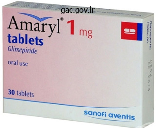
Purchase amaryl 2 mg without prescription
It is then crucial to await a discount in tone to happen because the spinal wire responds to the decreased enter from the first annulospiral endings of the muscle spindles within the hypertonic fascicle(s) and alteredinput fromthe relevantmechanoreceptorsin the joints, muscle tissue, and fascia. Once a discount in tone is felt within the monitored muscle/fascicle, cue the patient to additional release or soften the muscle with both verbal and manual cues that recommend `letting go. The verbal cues are virtually at all times associated with images that soften, melt, give method, and let go. This is the notice component of the release method and can be clearly sensed by both the therapist and, normally, the affected person. This facilitates increased awareness of what they want to think/do to facilitate more leisure and is important for studying the talent so that it might be utilized during different actions. Appropriate positive suggestions is also likely to improve neuroplasticity (see Chapter 9). Once the utmost release is obtained, the muscle is then taken passively via its full range either by stretching or lengthening the fascicle immediately (intramuscular technique) or by moving the joint so as to stretch/lengthen the fascicle. The second a half of this method addresses any intramuscular myofascial limitations or restrictions. This follow often helps to control pain and preserve mobility, and may generally be done earlier than any retraining of the deep muscular tissues or motion coaching. Principles of dry needling techniques Dry needling of painful factors with acupuncture needles has been used historically by many practitioners and has just lately (1990s) been introduced to physiotherapists in Canada by Dr. Chan Gunn, innovator and founder of Intramuscular Stimulation and the Institute for the Study and Treatment of Pain ( Gunn developed his radiculopathy model based mostly on his medical experience whereas treating injured workers with a quantity of musculoskeletal complaints. When the circulate of nerve impulses is restricted, all innervated buildings, including skeletal muscle, easy muscle, spinal neurons, sympathetic ganglia, adrenal glands, sweat cells, and mind cells turn out to be atrophic, extremely irritable, and supersensitive. Practitioners who use dry needling according to the myofascial set off point model specifically target lively set off points in myofascial tissues. Simons et al (1999) notice that trigger factors in myofascial tissues may be energetic (trigger local or referred ache with out being stimulated) or latent (require stimulation to set off local or referred pain); however, they may alter muscle activation patterns and limit range of movement. The precise mechanisms underlying myofascial trigger point improvement and elimination with dry needling remains unclear. Theories abound and the reader is referred to two wonderful articles by Simons & Dommerholt (2006) and Dommerholt et al (2006) for an in-depth review on the analysis into this technique. If hypertonicity is present in each a peripheral muscle and an related, segmental, paraspinal muscle. If you have an interest in utilizing dry needling method in your scientific apply, certification is required. Principles of specific joint mobilization techniques Once the muscles have been launched, the true mobility of the underlying joint could be assessed. Joints that current with restrictions within the capsule and the associated ligaments often have a number of vectors of pressure limiting motion, and mobilization in a number of instructions is usually required. In each conditions, it is necessary to assess the particular vector, or course, of resistance and focus the particular mobilization method to this vector. Range of motion follow combined with self-release with awareness and stretch with consciousness apply is then given to keep the mobility gained with the manual technique. Fritz et al (2005) thought-about the association between two of the above 5 elements, duration and extent of symptoms (1 and 2), and famous that these two factors alone were associated with an excellent prognosis for use of a manipulative approach. This has resulted in heated debate between this group (Flynn, Childs, Fritz) and skilled clinicians, who imagine that manipulation of the backbone ought to be particular to be safe and effective (McLaughlin 2008, Pettman 2006). If your patient has had their pain longer than sixteen days and the ache radiates further, then they could not. High acceleration, low amplitude, thrust methods have their place in a multimodal program and over the last 10�15 years there has been a big paradigm shift with respect to the understanding of how these strategies work to relieve pain and restore mobility. The biomechanical theories of 289 the Pelvic Girdle the previous are giving approach to new theories supported by proof as soon as once more in the field of neuroscience (Indahl et al 1995, 1997, 1999, Kang et al 2002, Pickar 2002, Sung et al 2005). According to Pettman, an internationally renowned clinician highly experienced within the use and educating of those techniques: Despite advances in visible diagnostic methods, purely mechanical causes of spinal articular restrictions have never been demonstrated. This casts severe doubt that the therapeutic foundation of manipulation is mechanical. The imagery cues that facilitated relaxation of the muscle(s) during the release with consciousness approach are built-in such that the neural tone is reduced because the myofascia is lengthened (stretch with awareness).
Amaryl 2mg amex
Differential inhibition by low-dose aspirin of human venous prostacyclin synthesis and platelet thromboxane synthesis. Transdermal modification of platelet operate: A dermal aspirin preparation selectively inhibits platelet cyclooxygenase and preserves prostacyclin biosynthesis. Suppression of thromboxane A2 however not systemic prostacyclin by managed launch aspirin. Low-dose aspirin, platelet operate and prostaglandin synthesis: Influence of epinephrine and alpha-adrenergic receptor blockade. Selective inhibition of platelet cyclooxygenase with controlled release low dose aspirin. Equivalent inhibition of in vivo platelet operate by low dose and excessive dose aspirin. Inhibition of prostacyclin and thromboxane A2 era by low dose aspirin at the site of plug formation in man in vivo. Effects of low-dose acetylsalicylic acid on thrombocytes in healthy topics and in patients with coronary heart illness. Effect of low-dose acetylsalicylic acid on the frequency and heamtologic exercise of left ventricular thrombus in anterior wall acute myocardial infarction. Effects of low-dose aspirin on platelet perform in sufferers with recent cerebral ischemia. Influence of epinephrine on the aggregation response of aspirin-treated platelets. Epinephrine potentiation of arachidonate-induced aggregation of cyclooxygenase deficient platelets. Role of arachidonic acid in human platelet activation and irreversible aggregation. Epinephrine reverses the inhibitory affect of aspirin on platelet vessel wall interplay. Influence of adrenergic receptor blockade on aspirin-induced inhibition of platelet perform. Modification of human platelet response to sodium arachidonate by membrane modulation. Ibuprofen Protects Platelet Cyclooxygenase from Irreversible Inhibition by Aspirin. Investigation of different mechanisms of collagen-induced platelet activation using monoclonal antibodies to glycoprotein 11b-111a and fibrinogen. Platelet-Active Drugs: the Relationships Among Dose, Effectiveness, and Side Effects. The Effect of Different Regimens on Platelet Aggregation After Myocardial Infarction. A new method for the quantitative detection of platelet aggregation in patients with arterial insufficiency. Profile and prevalence of aspirin resistance inpatients with cardiovascular disease. Effects of acetylsalicyclic acid in stroke sufferers; evidence of non-responders in a subpopulation of treated platelets. Aspirin-resistant thromboxane biosynthesis and the danger of myocardial infarction, stroke, or cardiovascular demise in patients at high-risk for cardiovascular events. Functional and biochemical evaluation of platelet aspirin resistance after coronary artery bypass surgical procedure. Frequency of aspirin resistance in sufferers with congestive coronary heart failure handled with antecedent aspirin. Prevalence of Clopidogrel non-responders among patients with secure angina pectoris scheduled for elective coronary stenting. Contribution of hepatic cytochrome P450 3A4 metabolic activity to the phenomenon of Clopidogrel resistance. High Clopidogrel loading dose throughout coronary stenting: Effect on drug response and interindividual variability. Assessing aspirin responsiveness in topics with mutiple threat elements for vascular disease with a speedy platelet perform analyzer. Determination of particular person responses to Aspirin therapy utilizing the Accumetric Ultegra. Chapter 11 Hemovigilance Neelam Marwaha Summary Hemovigilance is a surveillance system to detect, report and analyze untoward effects of blood assortment and transfusion to be able to prevent their occurrence and recurrence.
Cheap amaryl 1mg with mastercard
Like the pelvic girdle and the lumbar backbone, motion analysis of the hip requires the analysis of two zones of motion, the neutral zone and the elastic zone, and consideration must be given to the presence of any muscular tone which will prevent movement evaluation of the joint presently. Often neuromyofascial strategies (Chapter 10) are necessary to release the superficial muscle hypertonicity earlier than a complete articular evaluation of the hip joint could be accomplished. When analyzing the passive vary of motion of the hip joint there are several things to notice for every course of motion examined together with: 1. Functional vary of motion is proscribed to that range the place the femoral head stays centered; 3. Note any increased activity/tone in any muscle that happens on the point when the femoral head displaces. It is our experience that consistent increases in tone at sure factors of vary during a totally passive check are indicative of altered neural drive to the muscular tissues and that is clinically related. Multiple muscular tissues could also be creating the one internet drive vector and all would require launch so as to restore optimum biomechanics of the hip. Note the response of the femoral head, the point at which anterior rotation of the innominate happens (this is the limit of practical hip extension), and the presence of increased muscle activity/tone on the level of modified femoral head place or early end of range of motion. This place allows straightforward testing of mixed extension and adduction/abduction, internal/external rotation. Return to palpate the femoral head and innominate and passively lengthen the femur until anterior rotation of the ipsilateral innominate begins. If the femoral head displaces anteriorly at any point during this check, cease and palpate all the muscle tissue of the thigh and observe the presence of any increase in muscle tone/ activity (there is usually a couple of muscle creating the non-optimal pressure vectors). In order to take a look at the total range of extension, the affected person might need to be moved to the sting of the desk or down the desk. Passive abduction/adduction can be tested in varying degrees of flexion/extension. The useful vary of motion is reached when the pelvic girdle bends laterally beneath the vertebral column. Test the hip in quite a lot of mixed actions (flexion/adduction/internal rotation, extension/abduction/external rotation, and so on. With the affected person supine, lying close to the edge of the table, the ipsilateral femur is prolonged till anterior rotation of the innominate begins. The femur is then rotated medially to the limit of the physiological range of movement. The proximal thigh is palpated and a gradual, regular, posterolateral pressure is applied alongside the line of the neck of the femur to stress the capsule and ligaments additional. No motion should occur when the articular system restraints are intact and no ache should be provoked. If passive femoral extension elicits the best quantity of ache, this ligament could also be a nociceptive source. With the affected person supine, mendacity close to the edge of the desk, the ipsilateral femur is barely extended, adducted, and totally rotated laterally. With the patient lying supine, the ipsilateral femur is barely prolonged, kidnapped, and fully rotated laterally. This ligament primarily limits inner rotation in addition to adduction of the the hip: integrity of the articular system restraints the following checks assess the integrity of the articular system restraints of the hip joint. Hold the femur prolonged and medially rotated (left arrow) and apply a posterolateral distractive drive to the proximal femur (right arrow). With the affected person mendacity supine, the ipsilateral femur is flexed, adducted, and absolutely rotated medially. A sluggish, regular distraction pressure is applied alongside the road of the neck of the femur and the provocation of native pain is famous. This position can even create anterior impingement so noting the situation of the pain is important for differentiation. Clinical reflection time At this level in the examination appreciable information has been gained with respect to the articular system and all joints that probably require mobilization and/or have impairments of their passive restraints ought to now be listed in the articular piece of the Clinical Puzzle. In addition, hypertonicity in particular muscular tissues which would possibly be stopping full evaluation of articular status, in addition to impacting joint movement, could have been identified. The hip: impact of the myofascial and neural techniques on the hip joint If the muscular tissues primarily answerable for controlling motion of the femoral head are functioning nicely, the femoral head should stay centered or seated throughout all loading tasks. This requires optimal functioning of the myofascial and neural methods for the hip.
Amaryl 4mg online
Remove help of the leg and note the impression of this pre-contraction on the management of the femoral head; if the myofascial and neural systems are being effectively restored the femoral head will now stay centered. Furthermore, if a passive system deficit has been identified (joint laxity), the effectiveness of the myofascial and neural methods to compensate for the passive impairment is evaluated by repeating the optimistic articular system restraints test while the patient holds their deep muscle system contraction. This subsequent section will cover the evaluation checks and clinical reasoning for the myofascial and neural techniques of the Clinical Puzzle. Clinical reasoning of the findings from multiple checks is critical to understand the importance of the results of each individual check as it pertains to the speculation generated and this shall be covered, in part, in this chapter and then in further detail through the case reviews in Chapter 9. In health, the deep muscle tissue ought to co-contract in response to a command that begins with intention. This system is preparatory (Chapter 4) and should reply previous to the activation of the superficial muscle tissue, especially in situations the place the duty is predictable. Therefore, imagining or thinking about (preparing), but not truly doing, a motion appears to be a more effective way of accessing the appropriate neural pathways to the deep muscular tissues. The skin should transfer freely in all directions; observe the presence of any surgical scars and the mobility of the pores and skin and the superficial fascia. A frequent mistake seen when educating clinicians to assess this muscle is the failure to attain the suitable depth in the stomach before beginning the evaluation. At this depth, no activation of transversus abdominis (TrA) can be felt and it is a widespread mistake when assessing the response of TrA to verbal cuing. Give the patient the cue to contract and, should you feel rigidity in response to your cue that you simply imagine is TrA, repeat the ribcage wiggle while the contraction is held. Note the elevated depth of palpation required to assess transversus abdominis compared to the inner oblique and exterior indirect. Once the appropriate depth is reached, the thumbs are gently drawn aside (adducted) until a line of tension is felt (take up the slack within the fascial system). Anatomical image reproduced with permission from Acland and the publisher Lippincott Williams & Wilkins, 2004. One of the following three cues ought to evoke a symmetrical, equally timed response of TrA: 1. In our expertise, asking the affected person to hollow the stomach is much less efficient than the previous cues for eliciting an isolated deep muscle co-contraction response. Once you attain this layer (take care to not go any deeper into the peritoneum), apply light pressure to the TrA fascia by adducting your thumbs (draw the TrA fascia laterally). Connect to my fingers and try to create the same pressure that pulls your stomach out and in. Almost all of the ultrasound photographs and video clips offered in this edition are oriented according to imaging convention. This is a change from the third edition and is according to conventional imaging protocols. The new images and video clips on this current version had been collected using the MyLab25 (Biosound Esaote). This take a look at provides data on the symmetry of activation of the left and proper sides. The depth management and acquire can be adjusted in order that the muscle layers are extra simply observed; make certain to regulate the primary focus to the layers of curiosity. Prior to assessing the response of the abdominals to verbal cuing, observe any motion of the muscle tissue during quiet breathing. Activity of the TrA should be minimal throughout quiet breathing (Hodges & Gandevia 2000a); nonetheless, when there is a rise in the chemical drive (increased carbon dioxide levels) or mechanical drive (articular or myofascial restrictions within the thorax), TrA is the first stomach muscle recruited to help expiration. Hypertonicity of TrA may additionally be observed at this time (the muscle will seem to be contracted). Subsequently, observe the response of the belly muscle tissue to the next cues: 1. It also can seem to slide laterally over the top of the TrA, instead of TrA sliding underneath).
Buy amaryl 2 mg free shipping
Sideways and rotational displacement of the temporomandibular joint disc: diagnosis by arthrography and correlation to cryosectional morphology. Morbidity related to the preauricular and perimeatal approaches to the temporomandibular joint. Disc preservation surgical procedure for the therapy of internal derangements of the temporomandibular joint. Meniscoplasty of the displaced temporomandibular joint meniscus with out violating the inferior joint space. Changes in signs and signs following temporomandibular joint disc repositioning surgery. Temporomandibular joint disc repositioning utilizing bone anchors: an instantaneous postsurgical evaluation by magnetic resonance imaging. A surgical approach for management, of internal derangements of the temporomandibular joint. Eminectomy and plication of the posterior disc attachment following arthrotomy for temporoman- 30. Therapeutic consequence assessment in everlasting temporomandibular joint disc displacement. Discectomy as the first surgical choice for inside derangement of the temporomandibular joint. The anatomy of the interior maxillary artery in the pterigopalatal fossa: its relationship to maxillary surgical procedure. Walker restore of the temporomandibular joint: a retrospective analysis of 117 sufferers. Internal derangement of the temporomandibular joint: radiographic and histologic changes related to severe ache. Discectomy in the remedy of anterior disc displacement of the temporomandibular joint. A 30 year follow-up examine of temporomandibular joint menisectomies: a report of 5 sufferers. Long-term magnetic resonance imaging after temporomandibular joint discectomy with out substitute. Discectomy for the treatment of inner derangements of the temporomandibular joint. Destructive lesions of the mandibular condyle following discectomy with temporary silicone implant. Silicone induced overseas physique response and lymphadenopathy after temporomandibular joint arthroplasty. Recommendationss for administration of patients with temporomandibular joint implants. Temporomandibular joint discectomy: no optimistic impact of short-term silicone implant in a 5-year follow-up. Long-term examine of temporomandibular joint surgery with alloplastic implants compared with nonimplant surgical procedure and nonsurgical rehabilitation for painful temporomandibular joint disc displacement J Oral Maxillofac Surg. Mandibular joint arthrosis corrected by the insertion of a solid Vitallium glenoid fossa prosthesis: a model new method. Internal derangement of the temporomandibular joint treated by discectomy and hemiarthroplasty with a Christensen fossa-eminence prosthesis. Surgical management of superior degenerative arthritis of temporomandibular joint with steel fossa-eminence hemijoint substitute prosthesis: an 8-year retrospective pilot study. Surgical administration of superior osteoarthritis of the temporomandibular joint with metal fossaeminence hemijoint substitute: 10-year retrospective examine. A crucial evaluation of interpositional grafts following temporomandibular joint discectomy with an overview of the dermis-fat graft. Long-term viability of the temporalis musle/fascia flap used for tempormandibular joint. The role of a temporalis fascia and muscle flap in temporomandibular joint surgery. A protocol for the administration of failed alloplastic temporomandibular joint disc implants.
Dade (Date Palm). Amaryl.
- What is Date Palm?
- Coughs and other breathing problems.
- How does Date Palm work?
- Dosing considerations for Date Palm.
- Are there safety concerns?
Source: http://www.rxlist.com/script/main/art.asp?articlekey=96434
Cheap amaryl master card
Management of hypomobility after orthognathic surgery is dependent upon the underlying trigger. Trauma to the muscular tissues of mastication is greatest managed postoperatively by vigorous bodily remedy protocols. Those patients who fail to enhance inside the first 3 months must be fastidiously evaluated for an intra-articular source of the problem. Edema, bleeding, and fibrosis within the joint space can incessantly be managed by arthrocentesis procedures, especially when acknowledged early. Condylar torque is best handled by reoperation with appropriate positioning of the proximal segment. Reversible causes such as muscular hyperactivity or spasm, infectious and inflammatory causes, and medication-induced limitations should be recognized and handled. Proper remedy requires excision of the involved structures and quick reconstruction. Many operative strategies have been described within the literature, with various and sometimes less than passable outcomes. Three-year-old boy with bilateral bony ankylosis after a motorcar accident that also produced bilateral lacerations of the commissures. Right (M) and left (N) panoramic radiographs present transforming of the costochondral grafts. Advocates describe two major advantages over autogenous reconstruction: (1) the absence of a donor web site and (2) the ability of the patient to return to operate extra rapidly. However, multiple issues have been reported-some with devastating penalties for sufferers. In its most extreme type, intensive bony erosion within the space of the glenoid fossa has been discovered. Fragmentation of alloplastic materials secondary to function with a migration of particles into contiguous tissue and regional lymph nodes has also been reported. In addition, the dearth of growth potential precludes the use of these joint alternative systems in young kids. Recurrent ankylosis after prosthesis placement has additionally been reported, with periprosthetic calcifications mostly seen in younger sufferers. A evaluation by Chossegros and colleagues53 demonstrated superior outcomes (defined by the authors as an interincisal opening of 30 mm over a follow-up interval of three yr) using full-thickness pores and skin grafts and temporalis muscle. Various bone grafts (costochondral, sternoclavicular, iliac crest, and metatarsal head) have been used to reconstruct ramus height after the resection of ankylosis. Potential problems with its use include fracture, resorption, donor website morbidity, recurrence of ankylosis, and a variable progress conduct of the graft in situ. Complications Associated with Treatment Various complications have been reported secondary to the treatment of ankylosis. Dolwick and Armstrong56 warning that a severe limitation of opening could make the palpation of landmarks difficult and increases the surgical dangers. G, Diagram of the operative plan; the ankylosis launch is carried out via a preauricular incision (outlined in dashed blue line). Note the bony ankylotic mass and the coronoid process with obliteration of the sigmoid notch. The flap is dissected and rotated over the arch (L) and sutured in place (M and N). The patient was mobilized and began on bodily remedy instantly postoperatively. She was comfortable as a outcome of there was no donor web site operation and no period of maxillomandibular fixation. Frontal (V), frontal opening (W), and lateral (X) pictures 1 12 months after completion of therapy. The ramus lengthening is demonstrated by the space between the retained footplates.
Syndromes
- Diazepam (Valium)
- Endoscopy
- MPS I S (Scheie syndrome)
- After about 2 minutes of CPR, if the child still does not have normal breathing, coughing, or any movement, leave the child if you are alone and call 911. If an AED for children is available, use it now.
- History of heart problems (heart attack)
- Paralyzed bowel
- Problems concentrating
Purchase amaryl 1mg
In their examine, breast cancer cells have been injected into nine mice and tumors have been scanned with a 4. The pair of factors with the strongest correlation was used to compute the transformation, which was modeled by the polynomials. In addition to multimodal registration, registration algorithms can be used to evaluate imaging information acquired at different time factors to assess tumor standing and response to treatment. Since tumors might shrink or expand throughout therapy, solely the overlapped tumor voxels in each before- and after-treatment images had been analyzed. Hence, the alignment of parametric maps enabled the next analysis to be carried out. Both will increase and reduces in perfusion parameters desensitized the measure of the typical change, resulting in a relative insensitivity to therapy consequence using the standard strategy. However, the coregistration preserved the spatial information, which was included inside individual tumor voxels and allowed a delicate technique for quantification of perfusion values. Longitudinal coregistration plays a equally essential function in the work of Li et al. The motivation of their work is to discover a more delicate methodology to monitor tumor response to therapy. This affected person was scanned prior to neoadjuvant chemotherapy (t1), after the primary cycle of therapy (t2), and after completion of all cycles of therapy (t3). The problem in this work is that a lot of the intensity-based nonrigid registration algorithms will deform both regular tissues and tumors. The compression or growth of tumors will result in misleading results in regard to tumor response. Hence, the parameters acquired at t1 and t2 had been reworked and coregistered to the corresponding parameters at t3. Therefore, the registration provides a novel approach to enable not only the voxel-by-voxel analysis of tumor-related parameters longitudinally and quantitatively, but also the comparison of different parameters in regard to tumor response to remedy. This chapter introduces the mathematical features of medical image registration as well as a few of the commonly used registration strategies. The function of medical picture registration is to search an optimized transformation to match two medical photographs maximally. Rigid transformation is calculated during the registration when only the rotation and translation happen between objects; nonrigid transformation is required when extra advanced and nonlinear deformation is concerned between objects. Nonrigid transformation could be represented by the combination of splines, and therefore the calculation of transformation could be translated to the calculation of the coefficients of splines. Point- and surface-based registration strategies require landmark choice or floor identification. The cost function could be the distance between two sets of factors or surfaces or with specific constraints, such because the smoothness of deformation. Different pairs of landmarks can be assigned completely different weights during registration based mostly on the arrogance of the accuracy of landmark alternatives. Intensity-based registration is performed based mostly on the depth values of pictures. Similarly, the price function in intensity-based registration can be composed of a similarity term and other application-based phrases, such because the smoothness term or volume-preservation time period. In summary, medical image registration is an indispensable tool in preclinical and clinical purposes. The improvement and validation of registration algorithms are still an lively analysis space in medical picture evaluation in the long term. Non-diffeomorphic registration of mind tumor images by simulating tissue loss and tumor progress. A review of the speculation, algorithms, and functions of stage set strategies for propagating interfaces. Analyzing and synthesizing photographs by evolving curves with the Osher�Sethian method. The correlation ratio as a brand new similarity measure for multimodal picture registration. Information Processing in Medical Imaging, Computational Imaging and Vision three, Boston: Kluwer; 1995, 263�274. Comparison and evaluation of retrospective intermodality brain image registration techniques. Comparison and evaluation of retrospective intermodality image registration methods.
Order amaryl master card
This requires continual hypothesis technology (Kerry et al 2008, Kerry 2009), as properly as interpretation and reflection of the take a look at results because the examination progresses. Jensen et al (2007), as well as Jones & Rivett (2004), argue that it is a key requirement to develop scientific expertise. The results from these exams are then thought-about with these from the lively motion a part of the one leg standing test. Pelvic girdle: positional checks When assessing the place of the innominate bones relative to one another, it seems to be more dependable to use the whole hand to gain data kinesthetically rather than visualizing one point of the bone. With the patient mendacity supine, legs prolonged, palpate the anterior facet of both innominates with the heels of the arms. Sideflexion of the craniovertebral joints changes perception and will alter the visible findings. Clinical reasoning of the findings from a number of checks is necessary to perceive the importance of the results of each individual test and this shall be coated, partly, on this chapter after which in further detail via the case reports in Chapter 9. The following checks study the passive articular mobility as nicely as the integrity of the articular, myofascial, and neural methods to control translation of the joints of the pelvic girdle. Passive motion evaluation requires an evaluation of two zones of motion, the neutral zone and the elastic zone. Use as a lot of your palms as potential and evaluate the kinesthetic findings with the visual when assessing the position of the innominates relative to each other. Use the heel of 1 hand and palpate the cranial aspect of the left and proper superior pubic rami. Note any step, or shear, of the symphysis by sliding the heel of the hand to the left and right; recognize this along with your kinesthetic sense. Inset: affirm the kinesthetic impression by palpating the left and proper superior pubic rami with both the thumbs or index fingers and examine the visual and kinesthetic findings. Multiple force vectors arising from imbalanced hip muscular tissues can significantly impact the place of the pelvis in the supine, inclined, and neutral positions. Similarly, multiple pressure vectors arising from the thoracic and lumbar areas can impact the place of the pelvis and sometimes manifest throughout forward bending of the trunk. Let your entire hand mildew to the innominate and repeat the analysis from this place. To assess the impact of knee place and/or the anterior lower extremity myofascial slings on pelvic place, have the patient flex their knees and note any change in position between the innominates. Follow the crest inferiorly till you reach the sacral hiatus (unfused spinous processes of S4 and S5). At this level a determination regarding the physiological or non-physiological nature of the positional findings is made. All different positional relationships are nonphysiological and suggestive, but not confirmative, of either an intra-articular shear lesion (articular system deficit) or a major lower in the resting thumbs. In this illustration, the model is oriented as if looking at the pelvis from behind, with rigidity elevated in the proper superficial hip flexors simulated by shortening the elastic. Alternately, the torsion could not appear until the hips are extended while supine or the knees are bent while prone. These extrinsic pressure vectors are inclined to appear and disappear as the place of the lower extremity is varied. When one innominate seems to be sheared vertically relative to the other (also known as an upslip), the ischial tuberosities can be utilized to confirm the non-physiological position. To assess the place of the ischial tuberosities, palpate the inferior facet of the ischial tuberosity bilaterally. Initially use the heels of each arms after which palpate the ischial tuberosities with the thumbs. Use as much of your hands as possible when assessing the position of the innominates relative to one another. Have the patient bend ahead and flex the whole thoracolumbar backbone and observe any change in the pelvic position.
Generic amaryl 3mg with visa
The attenuation worth is expected to be excessive, and the affected person is anticipated to experience acute ache. Excretory phase photographs could additionally be helpful in demonstrating the mildly compressed but patent accumulating system. OtherImagingFindings Plain radiographs of the abdomen: A massive mass could also be recognized by a mass effect on the bowel. Infiltration into the renal parenchyma produces an enlarged, poorly functioning kidney. Renal sinus infiltration appears as stretching of the renal sinus and encasement of the renal pelvis and higher ureter on the affected aspect. Because of the shortage of inner acoustic interfaces, lymphomatous tissue could appear nearly fluid-like and resemble a perinephric assortment, even showing by way of transmission. Lymphomatous tissue is mildly hypointense on T1-weighted photographs and hyperintense on T2-weighted images. However, a presentation with mass, hematuria, weight loss, and flank pain may occur. Focal plenty, if present, are nicely demonstrated on the � Pearls & � Pitfalls � Perinephric involvement without compression of the renal parenchyma and renal sinus involvement without compression of the renal vessels and accumulating system are extremely suggestive of renal lymphoma. The kidney is regular in size, form, position, orientation, outline, and parenchymal thickness. DifferentialDiagnosis Medullary nephrocalcinosis: Hyperechogenicity with shadowing on sonography is a hallmark of calcification. Areas of diffuse calcification that seem triangular in form and are arranged radially along the renal sinus are calcified renal pyramids. Diffusely calcified renal pyramids are a manifestation of medullary nephrocalcinosis. However, when a number of, they differ in shape and dimension and are unlikely to be current in all calices. OtherImagingFindings Plain radiographs of the stomach: Nephrocalcinosis is usually not visualized. However, in patients with main hyperparathyroidism, stones could type in the kidneys. Similarly, tiny calculi within the distal amassing tubules may be seen in patients with medullary sponge kidney. In medullary sponge kidney, the dilated accumulating ducts seem as contrast-filled streaks arising from the tips of the papillae and extending into the pyramids. Essential Facts Medullary nephrocalcinosis is calcification of the medullary pyramids. Secondary hyperparathyroidism is elevated parathyroid hormone manufacturing in response to hypocalcemia; it happens in persistent renal failure. Tertiary hyperparathyroidism is claimed to have developed when the parathyroid glands in secondary hyperparathyroidism turn out to be autonomous. As the pyramids turn into bright in the course of the deposition of calcium, they might be perceived as a half of the renal sinus. DifferentialDiagnosis Contrast-induced acute tubular necrosis: Persistent nephrograms are classically seen in patients with acute tubular necrosis. OtherImagingFindings Plain radiographs of the stomach: Persistent nephrograms are attribute. Postoperatively, he developed fever and a fluctuant mass within the left decrease quadrant. An incidental note is made of wall calcification (arrowhead)intheleftcommoniliacartery. DifferentialDiagnosis Urinoma because of ureteric injury on the time of surgical procedure: A fluid collection adjacent to the urinary tract is typical. OtherImagingFindings Plain radiographs of the stomach show the obliteration of retroperitoneal buildings and a potential soft-tissue mass however have low sensitivity and specificity. OtherImagingFindings Plain radiographs of the abdomen: A attribute form is seen.
Real Experiences: Customer Reviews on Amaryl
Gonzales, 33 years: The nostril and ears proceed to improve in measurement in all dimensions, with the nasal tip and columella dropping inferiorly to create a more acute nasolabial angle, with all these features occurring to a higher extent in males. If necessary, delayed repeat scanning may be performed, which can show no distinction accumulation in the perfusion defects, whereas in acute pyelonephritis, these areas will turn into denser.
Pavel, 21 years: Immunotherapy: In addition, most cancers vaccines and gene remedy are additionally thought-about by some to be focused therapies as they intervene with the expansion of cancer cell. This shortening of relaxation instances will lead to a signal intensity improve on a T1-weighted picture and a sign depth lower on a T2- or T2-weighted image.
Jaroll, 58 years: The volume of the urinary bladder may be calculated, and postvoid retention may be demonstrated by ultrasound. The progress of the condylar cartilage contributes a lot of the whole ramus peak, whereas development of alveolar bone contributes aproximately 60% to the mandibular physique height.
Kan, 55 years: Thus, a minor clone at analysis could become predominant in the course of the course of the illness and stay undetected as a outcome of solely a major clone present at diagnosis is being monitored. Together with verbal cues and encouragement, they provide a strong stimulus to facilitate change; 5.
Folleck, 22 years: There are exceptions, corresponding to craniosynostosis, that might be due to an underlying genetic trigger. The research revealed heterogeneity in the spatial distribution of the prodrug and its metabolites.
Tarok, 52 years: It was found that the fraction of radiobiologically hypoxic cells inversely correlated with Ktrans. This issue, called the geometry issue, or g-factor, is a measure of the difficulty of inverting the matrix in Equation four.
Xardas, 42 years: If an stomach aortic aneurysm is current, wall thickening with elevated attenuation may recommend an intramural hematoma. A Valsalva maneuver leads to a deformation of the bladder shape and a caudodorsal shift.
10 of 10 - Review by Y. Gembak
Votes: 150 votes
Total customer reviews: 150

