Naltrexone dosages: 50 mg
Naltrexone packs: 10 pills, 20 pills, 30 pills, 60 pills, 90 pills
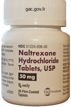
Effective naltrexone 50mg
For instance, occlusion of the pulmonary vasculature owing to pulmonary thromboembolic illness results in ele vated right-sided pressures as blood is impeded from move Some of the fabric on this chapter was contributed by Samuel Z. This course of includes all layers of the vessel wall and is char acterized by intimal hyperplasia, medial hypertrophy, adven titial proliferation, and in situ thrombosis. Serotonin Serotonin is a vasoconstrictor that promotes smooth mus cle cell hypertrophy and hyperplasia. Fatigue, light-headedness, chest pain, palpitations, orthopnea, edema, paroxysmal nocturnal dyspnea, and cough are different widespread presenting signs. The chest radiograph might show distinguished central pulmonary arteries and peripheral hypovascularity or "pruning. Overwedging is less common and should lead to inaccurate strain measure ments and pulmonary artery rupture. A full blood count is helpful in identi fying anemia as a cause of high-output coronary heart failure. Repeat research could additionally be indicated to assess response to therapy, especially in patients who experi ence a progression of symptoms. Acute vasodilator testing must be avoided in patients with significantly elevated left coronary heart filling pressures or low cardiac output. Vena-occlusive illness and pulmo nary capillary hemangiomatosis must be thought of in sufferers who expertise pulmonary edema during vasodila tor testing. Creation of a right-to-left inter atrial shunt through percu taneous atrial septostomy improves right heart operate and left heart filling by reducing right coronary heart filling pressure s. Though the shunt decreases systemic arterial oxygen saturation, the improved cardiac output leads to an total improvement in systemic oxy gen supply. Initially, she attributed the dyspnea to weight achieve, however it progressed to the point the place she was unable to make it up one flight of stairs and had to take frequent breaks whereas grocery buying. She additionally skilled atypical chest pain, oc casional palpitations, and lower extremity edema. She had a palpable proper ventricular heave, a traditional S 1, and a loud pulmonic element to her second heart sound. On pulmonary operate checks, she had normal volumes and flows and her dif fusing capacity of carbon monoxide was eight 1 %. Given the severity of her symptoms and pulmonary vascular disease, she was hospitalized immediately after the catheteriza tion. Over the course of her hospitalization, she and her husband learned the methods of sterile preparation of the medicine, opera tion of the ambulatory infusion pump, and care of the central venous catheter. Her signs improved to the purpose at which she had no dyspnea along with her usual actions. Her electrocardiogram revealed regular sinus rhythm with proper axis deviation and proper ventricular enlargement with a strain pattern. An echocardiogram demonstrated reasonable right atrial and proper ventricular enlargement and hypertrophy with mild-to reasonable right ventricular dysfunction. By the time of her presentation, she had dys pnea after going up one flight of stairs or with heavier home work similar to vacuuming. On physical examination her blood pressure was 1 0 5/60 mmHg with a coronary heart price of 89 bpm. On cardiac auscultation, she had a normal 5 and a physio 1 logically break up 5 with a loud pulmonic component. She was treated with an oral endothelin receptor antagonist and currently has dyspnea solely with severe exertion. Six months later the patient was admit ted with worsening decrease extremity edema, hypotension, and hyponatremia. The patient elected to bear initiation of treprostinil by way of subcutaneous infusion. Lung transplantation evaluation and mixture therapy with an oral endothelin receptor antagonist, along with twin forms of contraception, had been initiated. She skilled vital enchancment in her signs and 6-minute hall-walks over the following 2 years, however then discontinued the endothelin receptor antagonist ow ing to hepatotoxicity. The affected person was listed for lung transplantation and referred for atrial septostomy. Inflations have been then performed across the interatrial septum with a 1 0 mm X 20 mm Opta balloon that was inflated to a most of 5 atm. The systemic 0 saturation was 92% on 2 liters of oxygen 2 nasal cannula and 87% on room air.
Order naltrexone with mastercard
Confirmation of a luminal place in the distal vessel must be obtained previous to dilat ing, by removing the wire from the catheter and inj ecting a small amount of diluted contrast through the information or the help catheter into the distal vessel. For example, overdilation and disruption of a beforehand uncompromised distal vessel may prohibit subsequent bypass to that site. Graft failure inside a later timeframe (several months to years) may be as a end result of intimal hyperplasia, atherosclerosis, or progressive fibrosis of a poor venous conduit. Several other components might contrib ute to graft failure, including the presence of poor influx or outflow, low cardiac output, a hypercoagulable state, com promise of the graft owing to sufferers crossing their legs, or external compression of the graft by sclerosis and fibrosis (for instance, from a scarred groin). Of course, even in the case of the latter, abrupt throm bosis and acute limb ischemia can happen. Frank or impending graft failure is often not heralded by increasing clinical symptoms. Accordingly, a technique of normal graft surveillance utilizing duplex ultrasonography is recommended to protect and lengthen the life of the graft. For impending graft failure, both detected by duplex ultraso nography or rising signs, immediate arteriography is beneficial, adopted by either surgical or percutaneous revascularization. O ther lesions that might be amenable embody focal stenoses of proximal graft anastomoses or short-segment lesions (3 em or less) occurring throughout the bypass graft. Surgi cal revision is beneficial for long lesions (especially > 1 0 em) and stenoses associated with anastomotic aneurysms. Patients presenting with acute or subacute graft thrombo ses (< 1 four days) are best handled with catheter-directed throm bolysis. For patients with lengthy standing grafts that fail, dedication of the factors respon sible might require reestablishing enough circulate to visualize the graft angiographically. In circumstances of early graft failure, exami nation of angiographic studies may present clues previously overlooked, corresponding to stenosis of an influx vessel, poor or inad equate distal runoff, or the presence of a venous side department that was not sutured. Unfortunately, presently instruments to deal with persistent venous illness are restricted within the United States. A retrograde, by way of the con tralateral common femoral vein, or an antegrade method via the popliteal vein is a potential option. When putting sufferers on a lytic infu sion, low-dose heparin (600 to 800 unit/hour) should also be administered concurrently. If the lesion is resistant to balloon dilation alone, directional or laser atherectomy may prove helpful for salvaging the graft. Likewise, though stents have been advocated by some for use in failing vein grafts, their utility has not been studied in any formal trials to date. In basic, we extremely recommend the utilization of embolic safety when performing atherectomy and even slicing balloon angioplasty in old bypass grafts. Such widespread utility necessitates development of standardized pointers for coaching and credentialing to ensure that patients will obtain optimal care. Until the cognitive, clin ical, and procedural expertise are integrated rou tinely into formal cardiology fellowship programs, additional training is critical. In explicit, caro tid revascularization entails interventional expertise, tools, and medical administration expertise that differ considerably from those used in o ther vas cular distribu tions. Moreover, it includes therapy of a uniquely sensitive organ system, wherein minor errors or complications can have catastrophic results. Experienced operators have been shown to have improved outcomes in carotid stenting. As a result of these concerns, a j oint committee, together with the societies of every specialty concerned, has pro posed minimal coaching necessities that cover proficiency in the cognitive, technical, and clinical abilities necessary to safely carry out carotid stenting. Effectiveness-based guidelines for the prevention of heart problems in women-20 1 1 up date: a suggestion from the American Heart Association. Heart illness and stroke statistics-20 1 2 update: a report from the American coronary heart associa tion. Heart illness and stroke statistics-2008 update: a report from the American Heart Asso ciation Statistics Committee and Stroke Statistics Subcommittee. Carotid intima-media thick ness is related to premature parental coronary coronary heart illness: the Framingham Heart Study.
Diseases
- Muscle-eye-brain syndrome
- Lattice corneal dystrophy type 2
- Renal carcinoma, familial
- Hypokalemic sensory overstimulation
- ovarian remnant syndrome
- Osteopoikilosis
- Aggressive fibromatosis
- Viljoen Winship syndrome
- Porphyria cutanea tarda, sporadic type
Order naltrexone canada
Question 8 What are the features and being pregnant risks with placental hyperinflation Women with a historical past of preterm delivery or a short cervix may be admitted to hospital until deliberate caesarean supply, such that the operation can be accomplished immediately if labour occurs or the membranes rupture spontaneously. Prenatal therapy of huge placental chorioangiomas: case report and evaluate of the literature. Accreta placentation: a systematic evaluation of prenatal ultrasound imaging and grading of villous invasiveness. Answer 9 In placental hyperinflation, the widespread distal villous hypoplasia is in all probability going related to reduced density of the conventional anchoring villi, which usually maintain parallel alignment of the chorionic and basal plates of the placenta within the face of maternal perfusion strain from the spiral arterioles. Note that the surplus maternal blood remains to be inside the intervillous space, which is why the placenta in utero Chapter 10 Question 1 Describe how the interatrial septum forms and the way this explains the morphology of secundum atrial septal defect. A, View of the guts showing proper and left atria and left ventricle with posterior atrioventricular cushion. Its lower border grows in the path of the atrioventricular cushion, the gap between them forming the ostium primum (B). Fusion of the lower border of the primary septum and the atrioventricular cushion eliminates the ostium primum. By the time they fuse, nonetheless, fenestrations have appeared within the septum primum to allow right-to-left passage of blood in the atrium (C). F, A fenestrated ostium secundum atrial septal defect seen from the left atrium. There are three spherical defects that lie inside septum primum within the flooring of the oval fossa. Self-Assessment e11 Answer 1 the conventional interatrial septum has several components. Much of what appears to be septum is definitely infolding of the extracardiac tissue between the 2 atria. The the rest of the get together wall of the 2 atria is formed by atrial muscle and adipose tissue between the lower border of the oval fossa and the atrioventricular junction. The atria develop as paired outgrowths from the caudal part of the center tube cephalad to the sinus venous. The space between the atria sees the expansion of a main septum (the septum primum) that fuses with the atrioventricular cushions. It grows downwards, and its anterior part fuses with the endocardial cushions, however a defect remains � the oval foramen. The proper facet of the atrial mass incorporates the proper horn of the sinus venosus and types a pair of valves round its orifice. Fusion of the anterior part of these valves creates the septum spurium, which contributes to closure of the atria. The left valve of the sinus venosus is included into the developing septum secundum, but often, remnants of it persist as thin threads attached to the best aspect of the septum. Throughout fetal life, blood can cross from the right to the left atrium by way of the oval fossa and ostium secundum. After start when the left atrial stress rises due to elevated pulmonary venous return to the left atrium, the flap valve is pushed in opposition to the septum and in additional than 75% of instances seals by fibrosis. In 25% of individuals the flap remains unsealed and potentially openable � the so-called persistent foramen ovale. An atrial septal defect of secundum sort occurs when the flap valve is inadequate in dimension to shut the oval fossa underneath regular circumstances. Any interatrial communication outside the oval fossa is by definition not a secundum atrial septal defect. Such defects occur around the coronary sinus, the atrioventricular septum or adjacent to the superior caval vein. Question 2 Describe how the development of the pericardium leads to the traditional pericardial anatomy.
Order generic naltrexone from india
The aortogram ought to be centered and angulated to present the maximum amount of data on the clinical setting. Four pairs of lumbar arteries arise in a posterolateral direc tion under the primary renal arteries. The third is the inferior mesenteric artery, which originates off about 1 em caudal to the celiac axis at the L l-L2 level. Contrast medium should be inj ected at a price of frames per second must be obtained when evaluating the mesenteric or renal arteries. When performing arteriography in an aorta with suspected or identified aneurysmal disease or 15 mUsecond for a complete volume of 30 to 50 mL. At least 4 inserting the aspect holes adj acent to the primary and second lumbar extreme atherosclerotic involvement, meticulous care ought to be taken to keep away from dislodging of mural thrombus or plaque resulting in distal embolization. This is most eral-are obtained, particularly if the origins of the mesen teric vessels are being evaluated. Further augmentation of the gradient utilizing vasodilators (200 pg of intra-arterial nitroglycerin) delivered into the lower aorta may also be helpful. The cause usually is a chronic thrombotic occlusion superimposed on severe atherosclerosis of the distal aorta and iliac arteries. Middle aortic syn reveals a easy tapered proximal and midabdominal aorta which embrace Williams syndrome, 123 neurofibromatosis, 124 congenital rubella,one hundred twenty five and tuberous sclerosis, 1 26 may also innominate artery), the left frequent carotid and left subcla vian arteries often originate separately from the aortic arch. An aortic arch variant in which the brachiocephalic and left common carotid arteries could have a common origin. Manifestations of Subclavian Disease Atherosclerosis of the proximal subclavian artery may mani fest clinically as arm claudication, subclavian steal, or (in sufferers with earlier inner mammary grafting) coronary bar, axillary, brachial, or radial approaches. The tip of the catheter should be positioned on the T 1 2 or Ll stage, thus palpable on either aspect, alternative options embrace translum ischemia. In uncommon cases, this will likely trigger cere bral ischemia throughout upper extremity train. Angiography of the innominate is usually greatest Subclavian and Vertebral Arteriography An aortic arch arteriogram with a 5F pigtail catheter can visu alize the origin of the great vessels to evaluate for atheroscle rotic occlusive disease. Inj ection of vasodilators into the distal subclavian circulation can be used to simulate exercise and increase the gradient. A gradient of 1 5 mmHg is considered important for subclavian or innominate stenosis. Estimates of the prevalence of asymptomatic carotid bruits in adults range from 6%138 to 1 6%, 139 with a mean prevalence of 1 0 %. Among sufferers with an asymptomatic bruit and with severe (70% to 99%) carotid stenosis, the 3-year threat of stroke was 5. The left common carotid is usually the within a fascial (carotid) sheath, lateral to the vertebrae, and bifurcates into an external and inner carotid artery department artery normally has no primary branches prior to coming into the cranium, it forms a tortuous portion known as the carotid siphon throughout the cavernous and supraclinoid phase, after which at the fourth cervical vertebra. Each common carotid runs patients with current transient cerebral ischemia or nondis abling stroke had been examined for the presence of a carotid bruit. Fifty-eight percent of sufferers had a bruit localized to the ipsilateral carotid artery; three 1 % had a carotid bruit involv ing the contralateral vessel; and 24% had bilateral carotid bruits. The external carotid artery has several major branches named lowered the pretest 70% to 99% probability of a carotid ste dicting high-grade ipsilateral caro tid stenosis was 63% and 6 1 %, respectively. In this affected person subgroup, absence of a bruit Extracranial Carotid Atherosclerosis Approximately seven hundred,000 strokes happen yearly within the United States, of which 25% to 30% are owing to further cranial caro tid artery disease. In one prospective natural history examine of 232 patients with delicate (< 50%) or moderate (50% to 79%) carotid stenosis adopted nosis from 52% solely to 40% 143 Recently, in a meta-analysis of 22 studies involving over 1 7,200 patients, the odds ratio for myocardial infarction in these patients with cervical bruits as stroke, representing an occasion rate of 828/1 00,000 popula tion in males and 5 5 11100,000 in women. In one research of 444 male sufferers, the 4-year mortality price tion for po tential caro tid revascularization, 1 6% of patients up with annual carotid duplex ultrasonography for a mean of seven years, 23% demonstrated disease development. Progression to both 80% to 99% stenosis or occlusion was extra doubtless in sufferers whose initial stenosis gression of stenosis in 1 7% of 282 arteries with at least two serial carotid duplex examinations. Multivariate evaluation showed diabetes mellitus, an abnormal electrocardiogram, and the presence of intermit tent claudication to be asso ciated with an elevated mortal infarction or stroke, as much as 3. Color coded images can detect increased velocities of blood move Carotid Arteriography carotid and intracerebral vasculature. Arch aor tography is an important first step as a result of it permits characteriza tion of the arch configuration and optimum catheter choice. Anatomical variations of the standard aortic arch include ori gin of the left common carotid from the innominate (bovine arch) seen in 2 5 %, origin of the left vertebral from the aorta in three %, and origin of the best subclavian as the distalmost A.
Naltrexone 50 mg sale
Severe calcific aortic stenosis was discovered on the time of surgery in all of the sufferers referred for surgery who had a final aortic valve space of < 1. Sequential alterations in myocardial lactate metabolism, S-T segments, and left ventricu lar function during angina induced by atrial pacing. Differential hemodynamic, metabolic, and elec trocardiographic effects in subj ects with and with out angina pecto ris throughout atrial pacing. Evaluation of rapid atrial pacing in diagnosis of coronary artery illness: evaluation of atrial pacing check. The effect of train on the cardiac output and circulatory dynamics of normal subj ects. Regulation of stroke volume throughout sub maximal and maximal upright train in regular man. Effects of changing coronary heart fee in man by electrical stimulation of the proper atrium: research at rest, throughout exercise, and with isoproterenol. Exercise cardiac output is maintained with advancing age in wholesome human subj ects: cardiac dilatation and elevated stroke volume compensated for a diminished heart rate. The pacing stress check: a reexamination of the rela tion between coronary artery illness and pacing induced electro cardiographic modifications. Detection of pacing-induced myocardial ischemia by endocardial electrograms recorded during cardiac catheteriza tion. Release of adenosine from human hearts during angina induced by speedy atrial pacing. Improved detection of ischemia-induced will increase in coronary sinus adenosine in sufferers with coronary artery illness. Hemodynamics at relaxation and through supine and sitting bicycle train in normal subj ects. Left ventricular performance throughout mild su pine leg exercise in coronary artery illness. Factors contributing to al tered left ventricular diastolic properties during angina pectoris. Left ventricular pressure-volume alterations and region al disorders of contraction throughout myocardial ischemia induced by atrial pacing. Diagnosis of coronary artery disease by multigated ra dionuclide angiography throughout proper atrial pacing. Are the medical and hemodynamic occasions dur ing pacing in patients with angina reproducible Ex ercise hemodynamics improve analysis of early heart failure with preserved ej ection fraction. Oxygen utilization and air flow throughout exercise in patients with persistent cardiac failure. Decreased catecholamine sensitivity and j3-adrenergic receptor density in failing human hearts. Simultaneous evaluation of left ventricular sys tolic and diastolic dysfunction during pacing-induced ischemia. Pharmacological and hemodynamic influences on the speed of isovolumetric left ventricular rest in the aware canine. Early diastolic left ventricular perform in children and adults with aortic stenosis. Low-output, low-gradient aortic stenosis in patients with depressed left ven tricular systolic perform: the medical u tility of the dobutamine challenge within the catheterization laboratory. Low-gradient aortic stenosis: operative threat stratification and predictors for long-term end result: a multicenter study utilizing dobutamine stress hemodynamics. Upward shift and outward bulge: divergent myocardial results of pacing angina and temporary coronary occlusion. D epression of sys tolic and diastolic myocardial reserve during atrial pacing tachy cardia in patients with dilated cardiomyopathy. Simultaneous transesopha geal atrial pacing and transesophageal two-dimensional echocar diography: a new method of stress echocardiography. Low-flow, low-gradient severe aortic stenosis despite normal ej ection fraction is related to severe left ventricular dysfunction as assessed by speckle-tracking echocardiography: a multicenter research. Aortic stenosis with severe left ventricular dysfunction and low transvalvular strain gradients: threat stratification by low-dose dobutamine echocardiography.
Cheap naltrexone 50mg
Speckle tracking-derived myocardial tissue deformation imaging in twin-twin transfusion syndrome: differences in pressure and pressure price between donor and recipient twins. A randomized trial of amnioreduction versus septostomy within the therapy of twin-twin transfusion syndrome. Twenty-five years of fetoscopic laser coagulation in twin-twin transfusion syndrome: a scientific evaluate. Cerebral injury and neurodevelopmental impairment after amnioreduction versus laser surgical procedure for twin-twin transfusion syndrome: a scientific evaluation and meta-analysis. Neurodevelopmental outcomes after laser surgery for twin-twin transfusion syndrome: a scientific evaluation and meta-analysis. Successful fetoscopic laser coagulation for twin-twin transfusion syndrome under local anaesthesia. Outcome following selective fetoscopic laser ablation for twin to twin transfusion syndrome: an eight 12 months nationwide collaborative experience. Fetoscopic laser coagulation of the vascular equator versus selective coagulation for twin-twin transfusion syndrome: an 166. Selective photocoagulation of placental vessels in twin-twin transfusion syndrome: evolution of a surgical approach. Sequential selective laser photocoagulation of communicating vessels in twin-twin transfusion syndrome. Is the sequential laser approach for twin-twin transfusion syndrome really superior to the standard selective method Recurrent twin� twin transfusion syndrome after selective fetoscopic laser photocoagulation: a scientific review of the literature. Residual anastomoses after fetoscopic laser surgical procedure in twin-twin transfusion syndrome: frequency, associated risks and consequence. Twin anemia-polycythemia sequence: diagnostic criteria, classification, perinatal management and end result. Outcome after fetoscopic selective laser ablation of placental anastomoses vs equatorial laser dichorionization for the remedy of twin� twin transfusion syndrome. Fetoscopic laser surgical procedure for twin-twin transfusion syndrome after 26 weeks of gestation. North American Fetal Therapy Network: intervention vs expectant management for stage 1 twin-twin transfusion syndrome. Stagerelated outcome in twin-twin transfusion syndrome treated by fetoscopic laser coagulation. Short and long run outcome in stage 1 twin-to-twin transfusion syndrome handled with laser surgery compared with conservative management. Survival outcomes of twin-twin transfusion syndrome stage I: systematic evaluate of the literature. Intrauterine fetal demise following laser remedy in twin-to-twin transfusion syndrome. Preoperative predictors of dying in twin-to-twin transfusion syndrome handled with laser ablation of placental anastomoses. Preterm premature rupture of membranes after fetoscopic laser surgical procedure for twin-twin transfusion syndrome. The impact of entry technique and access diameter on Prelabour rupture of membranes following major fetoscopic laser therapy for twin� twin transfusion syndrome. Risk elements related to preterm delivery after fetoscopic laser ablation for twin-twin transfusion syndrome. Cerclage for cervical shortening at fetoscopic laser photocoagulation in twin-twin transfusion syndrome. Increased risk of early-onset neonatal sepsis after laser surgery for twin-twin transfusion syndrome. Histologic chorioamnionitis and funisitis after laser surgical procedure for twin-twin transfusion syndrome. Prenatal administration and outcomes in mirror syndrome related to twin-twin transfusion. The pregnancy and long-term neurodevelopmental outcome of monochorionic diamniotic twin gestations: a multicentre prospective cohort research from the primary trimester onward. Risk elements for the neurodevelopment impairment in twin-twin transfusion syndrome treated with fetoscopic laser surgical procedure. Neurodevelopmental end result at 6 years of age after intrauterine laser therapy for twintwin transfusion syndrome.
Hogberry (Uva Ursi). Naltrexone.
- Are there any interactions with medications?
- Urinary tract infections, swelling of the bladder and urethra, swelling of the urinary tract, constipation, kidney infections, bronchitis, and other conditions.
- How does Uva Ursi work?
- Dosing considerations for Uva Ursi.
- What is Uva Ursi?
- Are there safety concerns?
Source: http://www.rxlist.com/script/main/art.asp?articlekey=96368
Buy naltrexone discount
Aquaporins have a narrow pore that permits water molecules to diffuse through the membrane in single file. Thus, ion channels are flexible dynamic structures, and delicate conformational changes affect gating and ion selectivity. Many protein channels are highly selective for transport of a number of particular ions or molecules. This selectivity outcomes from specific characteristics of the channel, such as its diameter, form, and the nature of the electrical charges and chemical bonds alongside its inside surfaces. Potassium channels permit passage of potassium ions across the cell membrane about 1000 times more readily than they allow passage of sodium ions. The channel is composed of 4 subunits (only two of that are shown), each with two transmembrane helices. A narrow selectivity filter is formed from the pore loops, and carbonyl oxygens line the walls of the selectivity filter, forming sites for transiently binding dehydrated potassium ions. The interaction of the potassium ions with carbonyl oxygens causes the potassium ions to shed their bound water molecules, permitting the dehydrated potassium ions to move by way of the pore. At the top of the channel pore are pore loops that type a slim selectivity filter. When hydrated potassium ions enter the selectivity filter, they interact with the carbonyl oxygens and shed most of their sure water molecules, permitting the dehydrated potassium ions to pass via the channel. The carbonyl oxygens are too far apart, nonetheless, to allow them to interact intently with the smaller sodium ions, which are therefore successfully excluded by the selectivity filter from passing by way of the pore. Different selectivity filters for the assorted ion channels are believed to determine, largely, the specificity of various channels for cations or anions or for explicit ions, corresponding to sodium (Na+), potassium (K+), and calcium (Ca2+), that achieve access to the channels. Also proven are conformational changes in the protein molecules to open or shut the "gates" guarding the channels. Once within the channel, the sodium ions diffuse in either course according to the usual legal guidelines of diffusion. The opening of these gates is partly responsible for terminating the action potential, a process mentioned in Chapter 5. Some protein channel gates are opened by the binding of a chemical substance (a ligand) with the protein, which causes a conformational or chemical bonding change in the protein molecule that opens or closes the gate. One of an important instances of chemical gating is the impact of the neurotransmitter acetylcholine on the acetylcholine receptor which serves as a ligand-gated ion channel. Acetylcholine opens the gate of this channel, offering a negatively charged pore about 0. This gate is exceedingly important for the transmission of nerve indicators from one nerve cell to another (see Chapter 46) and from nerve cells to muscle cells to trigger muscle contraction (see Chapter 7). Some of the gates are thought to be gatelike extensions of the transport protein molecule, which can close the opening of the channel or may be lifted away from the opening by a conformational change in the shape of the protein molecule. In the case of voltage gating, the molecular conformation of the gate or its chemical bonds responds to the electrical potential across the cell membrane. Conversely, when the within of the membrane loses its adverse cost, these gates open abruptly and allow sodium to pass inward by way of the sodium pores. This process is the essential mechanism for eliciting action potentials in nerves which would possibly be responsible for nerve indicators. That is, the gate of the channel snaps open and then snaps closed, with every open state lasting for under a fraction of a millisecond, up to a quantity of milliseconds, demonstrating the rapidity with which modifications can happen in the course of the opening and closing of the protein gates. At one voltage potential, the channel might remain closed all the time or nearly all the time, whereas at another voltage, it might stay open either all or most of the time. At in-between voltages, as proven in the figure, the gates are likely to snap open and closed intermittently, resulting in a median current flow somewhere between the minimal and maximum. A micropipette with a tip diameter of just one or 2 micrometers is abutted against the surface of a cell membrane. Suction is then applied inside the pipette to pull the membrane against the tip of the pipette, which creates a seal the place the sides of the pipette touch the cell membrane. Effect of concentration of a substance on the speed of diffusion through a membrane by simple diffusion and facilitated diffusion. This graph shows that facilitated diffusion approaches a most price, referred to as the Vmax. Facilitated diffusion differs from easy diffusion within the following essential method. Although the rate of straightforward diffusion by way of an open channel will increase proportionately with the focus of the diffusing substance, in facilitated diffusion the speed of diffusion approaches a most, called Vmax, because the concentration of the diffusing substance increases.
Buy genuine naltrexone line
This is an especially useful strategy in patients with borderline aor tic insufficiency or stenosis, in whom a waiting interval after mitral commissurotomy may enable for a extra well timed determination for double-valve substitute at a later date. Disadvantages of these tech niques embrace the chance of arterial injury due to the bigger balloons used. Alternatively, a double-balloon approach can be utilized with two balloons superior over separate guidewires from the femoral vein to the left atrium, throughout the mitral valve into the left ventricle. When correctly carried out, the double balloon technique leads to excellent enchancment in mitral valve area. In the early surgical period of closed coronary heart mitral commis surotomy, a metallic dilator, or commissurotome, was used by way of a left ventricular apical incision. A l 9 F metallic commissurotome could be passed throughout the inter atrial septum over a guidewire and used to accomplish mitral commissurotomy. There has been some proof that bicom missural splitting may be achieved extra frequently with the metal commissurotome. However, the Inoue balloon approach is faster and less cumbersome, and generally requires less fluoroscopy time than these different approaches 25 the Inoue balloon allows simple progressive upsizing of the balloon without withdraw ing the balloon from the left atrium-an essential benefit if larger balloon sizes are needed. The Inoue balloon system may, however, result in a slightly greater incidence of mitral regurgitation. Inoue Balloon Technique All antegrade approaches start with the crucial first step of successful trans-septal catheterization. This technique, which is described in Chapter 6, not only requires successful entry to the left atrium, however must also be performed through the suitable part of the atrial septum to allow quick access to the mitral valve. After successful placement of a Mullins-type dilator and sheath into the left atrium and affirmation of its position by a hand inj ection of distinction, the patient is anti coagulated with heparin. The femoral vein and interatrial septum are then dilated with a protracted l4F dilator over the coiled guidewire throughout the left atrium. The beforehand prepared, examined, and now slenderized Inoue balloon is then introduced over the guidewire into the left atrium. This allows for steady positioning of the balloon catheter throughout the mitral valve, as described below. After the slenderized balloon has been positioned throughout the left atrium, the stretching tube is removed, and a pre formed "J" stylet is launched into the Inoue balloon. The distal portion of the balloon is inflated slightly to assist in cross ing the valve and to prevent intrachordal passage. By maneu vering the balloon catheter while rotating and withdrawing the stylet, the balloon tip could be moved anteriorly and infe riorly towards the mitral orifice. Once the balloon catheter is placed throughout the mitral orifice, the distal portion of the balloon is inflated more absolutely and the catheter is pulled again gently to affirm that the inflated distal portion of the bal loon is secure throughout the mitral valve. As additional quantity is added to the balloon, the proximal end inflates to lock the Cha! Inflation to precalibrated quantity then dilates the valve orifice to the corresponding preset dimension. The strain gradient across the mitral valve is measured after every balloon dilatation, and echocardiography could additionally be used to assess the mitral valve space, leaflet mobility, and the diploma of mitral regurgitation. The Inoue balloon comes in 4 sizes-24, 26, 28, and 30 mm, referring to the fully inflated maximal balloon diam eter. However, since actual balloon size depends on the vol ume used for inflation, the actual diameter may be various over a spread from 6 mm less than nominal up to the full rated diam eter, as required. We typically estimate the expected maximal inflated balloon diameter using an empirical formulation based mostly on the height of the affected person (one-tenth the height in centime ter plus 10 mm). It is necessary to begin with a smaller balloon diameter, particularly for valves which might be very a lot thickened. The Inoue balloon is basically different from con ventional balloons, being quantity driven. The balloon is precalibrated so that inflation with volumes labeled on the inflation syringe end in corresponding inflated diameters of the balloon. The strain that the balloon is inflated to is thus totally different for different inflation volumes. A smaller max imal-size balloon, corresponding to a 26-mm balloon, when inflated to its maximal measurement shall be at a better stress than a bal loon that has a bigger capacity, similar to a 30-mm balloon, inflated to the identical diameter of 26 mm.
50mg naltrexone free shipping
Alternatively, current animal and scientific research demonstrated a useful impact in applying a constructive strain for longer period (sustained inflation). Excessive oxygen publicity leads to oxidative stress and tissue damage and must be averted throughout stabilisation at birth. The optimal inspired oxygen content required to avoid hypoxia as nicely as a end result of hyperoxia stays unclear. Because masks ventilation is the cornerstone in neonatal resuscitation, an optimal mask-holding technique is probably one of the most essential skills of neonatal caregivers. Mask air flow in infants could be troublesome and is usually hampered by leak and airway obstruction, resulting in low tidal volumes and thereby insufficient air flow. Mask approach should be integrated into all local training in neonatal resuscitation. In distinction to a selfinflating of flow-inflating bag, the strain is about and never generated manually, which finally ends up in extra consistent pressures and thus less variation in volumes. Noninvasive air flow strategies are now more and more used as preliminary respiratory support. In a big randomised trial, larger levels as much as 12 cm H2O had been used, but the next incidence of pneumothorax was reported. Traditionally, surfactant was given after the infant was intubated and mechanical ventilated. Recent research have demonstrated the protection of administering surfactant on spontaneously respiration infants receiving noninvasive assist. Trials are underway investigating the efficacy of the less invasive surfactant administration. Other noninvasive ventilation strategies can be found, though most of them are used after mechanical ventilation to stop extubation failure. Synchronising the inflations of the ventilator to the breaths of the infants (triggered ventilation) is now used in most neonatal centres and has shortened the duration of air flow. For synchronisation, a circulate sensor is positioned proximal of the affected person on the endotracheal tube. New technology and the use of microprocessors has led to the availability of many various modes which are extra subtle and will doubtlessly reduce the ventilation-induced lung injury. This matrix layer is situated adjoining to the ventricle and serves as a source of neuronal and glial precursor cells. After the preliminary prognosis, progression might happen over the following 3 to 5 days. This is caused by an impaired drainage of medullary veins in the white matter adjacent to the haemorrhage. The second element consists of diffuse white matter damage with loss of premyelinating oligodendrocytes and astrogliosis but with out necrosis or glial scars. Within the spectrum of preterm white matter harm, the diffuse sort represents the mildest kind and the cystic kind the most severe. These mechanisms usually happen concurrently and trigger excitotoxicity and free radical attack, which might lead to death of premyelinating oligodendrocytes. The preterm white matter is especially weak to ischaemia because of the presence of arterial border zones inside the deep (periventricular) white matter and an impaired autoregulation of cerebral blood flow. Other danger components are related to an infection and inflammation, both prenatally in case of maternal intrauterine an infection and postnatally in case of sepsis, necrotising enterocolitis and ventilation-induced lung harm. In cases with antenatal onset, cysts could additionally be present at delivery or develop throughout the first week. In case of a perinatal insult, cysts normally develop after 2 to three weeks, though for smaller and localised cysts, this may take longer (4�6 weeks). Preterm white matter damage is associated with an increased threat for impaired motor and cognitive outcomes. However, its capacity to predict other long-term outcomes as neurocognitive and behavioural impairments for the person patient is proscribed. The latter occurs within the case of severe supratentorial mind lesions which might be related to a slower development of cerebellar volumes. This includes not solely motor disabilities but in addition cognitive, behavioural and emotional deficits.
Purchase naltrexone visa
A central security wire fila ment is included to prevent separation if ever the wire coil were to fracture. The length of most standard wires is between 1 00 and 1 80 em; longer exchange-length information Most peripheral guidewires are manufactured from a stainless steel coil surrounding a tapered inner core that runs the size with only side holes. They � Straight catheters with a quantity of side ports which might be used � Pigtail or tennis-racket catheters which are used for lar to those of left heart catheters) may be required. Multiple facet holes along the distal shaft enable rapid delivery of con trast as an alternative of a single forceful j et that might cause catheter whipping or subintimal dissection as may stiffness. Low-friction wires with a hydrophilic coating (glide wires) have revolutionized periph eral work and made it potential to perform superselective cath eterization and traverse advanced stenoses and lengthy occlusions. Varying levels of shaft � Simple curved catheters � Complex be seen with distinction exiting the end-hole alone. Two such brokers have now emerged as options in sufferers with in many vascular beds. The addition of bicarbonate saline options required and the high incidence of comorbidities such as base line renal dysfunction, diabetes, hypertension, and renal artery atherosclerosis Y Aggressive prehydration, notably with isotonic (0. The benefit of iso-osmolar contrast agents when compared with low-osmolar distinction brokers (Chapters 2 and 4). Attempts to goal the final pathway of free-radical harm have focused on the usage of the antioxidant acetylcysteine. Benefits with periprocedural infusion of sodium bicarbonate have recently been reported. I t then gives rise to the remaining arch vessels-the left common carotid, and left subclavian arteries-from its upper surface. Stanforcl-ty]Je-A ao1 tic dissection-fottowing-aortic-vate-reptace- Distal to the origin of the left subclavian artery, the aorta narrows barely at the web site of the isthmus the place the ligamen to this point, a fusiform dilatation, called the aortic spindle, may happen. The descending aorta then continues anterior to tum arteriosum (the remnant of the fetal ductus arteriosus) tethers the aorta to the left pulmonary artery. The by the superior intercostal artery, which is a department of the subclavian artery. At the level of the fourth to sixth thoracic vertebrae, anteriorly directed bronchial arteries come off to provide every lung. Thoracic aneurysms appear to enlarge at a extra rapid price than that noticed in belly aneurysms (0. The resultant enlargement and tortu osity of these intercostal arteries are liable for the "rib no tching" seen in chest radiographs. N eck or j aw ache may also be pres ent in sufferers with aneurysms of the aortic arch. Dilatation of the aortic valve annulus and aortic valve could produce aortic regurgitation and congestive coronary heart failure. Aneurysms of the descending thoracic aorta may produce pleuritic left sided or interscapular ache, whereas thoracoabdominal aortic of the thin-walled poststenotic section. Entry to the preste no tic aorta from the brachial or axillary arteries may hence be preferred. When attempting to traverse the site of narrowing in retrograde style, care must be taken to keep away from inadvertent perforation aneurysms could induce complaints of stomach ache and left shoulder discomfort from irritation of the left hemidiaphragm. In most patients undergoing to present information about the situation of the aneurysm and its relationship to maj or aortic branches in the chest and elective thoracic aorta surgical restore, aortography is required Patent Ductus Arteriosus the prevalence of patent ductus arteriosus is l/5,500 in chil dren under 14 years of age. Optimal surgical approaches, in addition to operative dangers, are greatest defined by imaging the coronary, brachioce phalic, visceral, and renal arteries during inj ections. Aneurysms caused by blunt or penetrating trauma usually contain the proximal descending thoracic aorta the place the cellular arch these could current as pseudoaneurysms-contained ruptures that lack intimal and medial elements and are contained only by adventitia and periaortic tissue. Since the entry to the false lumen is usually on the greater (outer) curve of the aorta, the catheter may be used to direct the wire towards the internal guidewire should be superior beneath fluoroscopic steerage or tennis racket) with a gentle j-tipped or 1 5 em floppy tipped mise of the visceral artery origin by the expanded false lumen (dynamic dissection). If this is performed successfully, buildings just like the aortic leaf ular area 93 Similar ache might occur with rupture or sud den enlargement of a chronic dissection. When this turns into obvious on test injections, care ought to be taken to keep away from extending the false lumen, pulling the cath eter again and using the methods mentioned above to reenter the true lumen. Once considered the gold normal for prognosis of aortic Surgical repair of Stanford sort A aortic dissections entails Dacron graft placement of the ascending aorta. Endovascular stents and balloon fenestration have been successfully utilized in treating the ischemic problems related to aortic dissection. These produce dilation of the proximal lumen or to intrinsic luminal collapse consequent to loss of wall elastin.

