Menosan dosages: 60 caps
Menosan packs: 1 bottles, 2 bottles

Menosan 60 caps low price
Atlantoaxial instability in people with Down syndrome: a fresh take a look at the proof. Magnetic resonance imaging of the transverse atlantal ligament for the analysis of atlantoaxial instability. Radiological evaluation of the spinal wire in congenital atlanto-axial dislocation. Atlanto-axial instability and spinal cord compression in children-diagnosis by computerized tomography. Pediatric atlantoaxial instability presenting as cerebral and cerebellar infarcts. A longitudinal study of atlanto-dens relationships in asymptomatic people with Down syndrome. Complications and long-term end result of higher cervical backbone arthrodesis in patients with Down syndrome. Syncope, vertigo and disturbances of imaginative and prescient resulting from intermittent obstruction of the vertebral arteries as a result of defect in the odontoid process and extreme mobility of the second cervical vertebra. Hypertrophy of C-1 anterior arch: helpful signal to distinguish os odontoideum from acute dens fracture. Neurological syndromes related to congenital absence of the odontoid process. C1-C2 fusion in kids with atlantoaxial instability and spinal twine compression: technical observe. Atlantoaxial arthrodesis using Halifax interlaminar clamps bolstered by halo vest immobilization: a long-term follow-up experience. Selection of a rigid inner ixation construct for stabilization on the craniovertebral junction in pediatric sufferers. Pulmonary operate ater surgical procedure for congenital atlantoaxial dislocation: a comparison with surgery for compressive cervical myelopathy and craniotomy. A new appraisal of abnormalities of the odontoid course of related to atlanto-axial subluxation and neurological disability. Atlantoaxial instability in a 7-year-old boy associated with traumatic disrupture of the ossiculum terminale (apical odontoid epiphysis). Segmentation defect in the midodontoid course of and its potential relationship to the congenital sort of os odontoideum. Atlantoaxial ixation using plate and screw technique: a report of a hundred and sixty handled patients. Use of axial and subaxial translaminar screw ixation within the administration of upper cervical spinal instability in a collection of 7 children. Iniencephalic deformity of the cervical backbone with Klippel-Feil anomalies and congenital elevation of the scapula; report of three circumstances. Clinical and roentgenological manifestations of the Klippel-Feil syndrome (congenital fusion of the cervical vertebrae, brevicollis); report of eight further cases and review of the literature. Congenital scoliosis and urinary tract abnormalities: are intravenous pyelograms essential Screening for hip dysplasia in congenital muscular torticollis: is bodily examination enough Ultrasonographic study of the coexistence of muscular torticollis and dysplasia of the hip. Commentary on "adapting to larger demands: utilizing progressive strategies to treat infants presenting with torticollis and plagiocephaly". Stretching remedy for infants with congenital muscular torticollis: physiotherapist or dad and mom Efectiveness of surgical remedy for uncared for congenital muscular torticollis: a scientific evaluation and meta-analysis. Hypermobility of the cervical backbone in children; a pitfall in the prognosis of cervical dislocation. Instability of the cervical backbone and neurological involvement in Klippel-Feil syndrome.
Menosan 60 caps free shipping
A cell phone or different communication capability should be out there on the sideline. Cervical Peripheral Nerve Injuries Burners "Burners" (also generally recognized as "stingers") are frequent injuries to the neck in collision sports activities, corresponding to football, hockey, or diving. A excessive proportion of soccer players, especially defensive gamers, experience burners throughout their playing career. Other sports activities in which burners occur include wrestling, backpacking, Chapter 33 the Immature Spine and Athletic Injuries 601 sledding, skiing, horseback driving, boxing, weightliting, and climbing. An asymmetrical axial load on the neck causing compression in the neuroforamen can injure the peripheral nerve root. If the athlete falls directly on the shoulder and the head is distracted away from the shoulder, traction is utilized to the nerve root and the brachial plexus. When the cervical spine is hyperextended, hyperlexed, or laterally lexed to the opposite aspect, the angle between the shoulder and neck is increased past the traditional range, stretching the brachial plexus. Symptoms are a extreme burning or searing pain within the shoulder and arm associated with loss of sensation and weak spot of the arm. Extension-compression mechanisms are commonest, adopted by brachial stretch and direct blow mechanisms. An A-frame design to the shoulder pads brings lateral stability to the bottom of the neck, preventing lateral tilt while permitting rotation of the top, which occurs at C1�C2 (50%). Because the pain, paresthesias, and weak point usually last only some seconds or minutes, these accidents fall into the Seddon classiication of neurapraxia. Occasionally, athletes with more extreme accidents experience a prolonged recovery period which will last hours to several weeks and should result in a chronic loss of time from competitors. Burners might happen many times during the season, ranging from a transient nerve irritation with out residual injury to an entire avulsion of the nerve root from the spinal twine with permanent deicit. Burners are symptoms of injury to either the brachial plexus or a nerve root, and should be evaluated systematically. Quan and Bird144 proposed a classiication of peripheral nerve accidents that correlates properly with electrodiagnostic studies and prognoses, based mostly on earlier classiications by Seddon145 and Sunderland. Nerve conduction studies and electromyography in the analysis of peripheral nerve injuries. Cervical Stenosis Congenital cervical stenosis is a risk factor for cervical spine accidents. Transient Spinal Cord Compression Torg and colleagues149 described the syndrome of transient quadriplegia, considered to symbolize a "neuropraxia of the cervical spinal wire. Treatment of spine fractures generally falls into two categories: closed and open. With closed therapy, the fracture is reduced if essential, and the affected person is immobilized in a physique cast. With open treatment, a surgical process is performed to cut back or safe severely displaced or unstable fractures. Open procedures are indicated when two or three of the pillars of the vertebral column are disrupted. Bone grating to obtain spinal fusion and biologic long-term stabilization are the inal parts of the procedure. Factors to contemplate when deciding when and if an athlete should return to play ater backbone fracture are healing, rehabilitation, risk of reinjury, and neurologic standing. Sacral Stress Fracture Sacral stress fractures, extra commonly seen within the intrapartum or postpartum period of pregnancy or aged osteoporotic patients, also can occur in people collaborating in running sports activities, usually at an elite stage. Treatment includes a transient period of limited weight bearing adopted by progressive mobilization, bodily therapy, and return to sport in 1 to 2 months, ater the ache has resolved. Addressing dietary standing and calcium/vitamin D consumption are essential in stopping repeat stress fractures. Iliac Apophysitis Apophyseal accidents are a unique harm in adolescent athletes, and are related to skeletal immaturity and repetitive microtrauma. With iliac apophysis involvement, the patient normally presents with again pain localized to the iliac crest, which facilitates analysis. Chapter 33 the Immature Spine and Athletic Injuries 603 exacerbated by resisted lateral bending and extension. Clancy and Foltz155 described a series of 21 younger distance runners, in all of whom pain resolved with alteration of coaching and relaxation. As the apophysis closes on the finish of growth, even probably the most recalcitrant instances resolve, just like Osgood-Schlatter disease of the knee.
Purchase genuine menosan online
All occipitocervical dislocations should be handled initially by quick utility of a halo vest. Because of this mechanism, these fractures generally accompany head accidents in the polytrauma affected person. Neurologic damage is unusual within the case of isolated fractures of the atlas, however, when it occurs, the larger occipital nerve is most regularly injured, adopted by the decrease cranial nerves. Common fracture patterns embody isolated posterior arch fractures, lateral mass fractures, and burst fractures with mixed anterior and posterior arch fractures. Fractures that are displaced greater than 5 mm could be treated with immediate halo vest software. A variation of the C1 lateral mass fracture, a unilateral C1 lateral mass sagittal split fracture, has a propensity to trigger late deformity and pain if not treated with both C1�C2 fusion or direct osteosynthesis. Ater the halo vest is removed, lexion and extension radiographs are taken to take a look at for C1�C2 instability, which, if signiicant, could be handled by C1�C2 posterior fusion. Levine and Edwards advocate initial reduction of the fracture with skeletal traction for up to 6 to eight weeks, adopted by one other 6 weeks of halo vest therapy. Most surgeons would agree that operative therapy is indicated if greater than 5 mm of C1�C2 instability exists on lexion and extension radiographs once the halo is removed. In the case of nonunion of the posterior arch of C1 or signiicant harm to the occipital condyles, occiput�C2 fusion could be performed. Another indication for surgery is neck and/or occipital nerve pain in a affected person with a nonunion of the atlas ring fractures. When symptoms have been current for two to 4 weeks, hospital admission and headhalter traction is normally profitable in achieving discount. He had an instantaneous onset of ache in his higher cervical spine, with radiation to the again of his head. The basion seems to be immediately over the tip of the odontoid and is less than 1 cm from the tip. There can also be some asymmetry, with the space from the lateral mass of C1 to the odontoid being shorter on the best side than on the left side. At the time of surgical procedure, with a combination of skull tong traction and guide manipulation with a cable handed underneath the posterior arch of C1, the occiput�C1 dislocation was reduced with an audible "clunk. Because inflexible internal ixation was not utilized, the patient was immobilized in a halo vest postoperatively. The titanium cables fastening the bone graft to the skull are passed via two burr holes simply posterior to the foramen magnum. The cables are also passed bilaterally under the posterior arch of C1 and beneath the lamina of C2. Drill holes are made within the iliac crest bone graft to permit passage of the wires as shown. If traction fails to scale back the deformity, open reduction and C1�C2 fusion is really helpful. Like kids, adults will current with a "cock robin" look with the head tilted towards and rotated away from the aspect of the dislocation. In adults, reduction of the rotational deformity can normally be achieved by cranium tong traction. If it was diicult to get hold of the reduction, then halo vest immobilization could also be wanted. If closed reduction fails, open discount is carried out and a posterior C1�C2 fusion is used to stabilize the discount. Because the transarticular C1�C2 screw is probably the most crucial in phrases of its positioning, the C1�C2 transarticular screw is all the time the irst screw to be placed in this construct. The plate should it lush in opposition to the skull, it in opposition to the posterior arch of C1, and it the contour of the posterior arch of C2. The C1�C2 transarticular screw is then positioned by way of the plate and tightened to within two turns of its inal position.
Purchase 60caps menosan otc
Arthrodesis at any given stage can result in increased load bearing at levels above and below the construct, accelerating the degenerative course of. Of the 108 who underwent a revision procedure, 27 developed recurrent illness requiring a 3rd cervical fusion. Notably, patients who had been revised through an anterior method had a better likelihood of recurring disease versus patients who underwent a posterior procedure. However, a signiicant downside of this selection is the involved donor site morbidity, manifesting as persistent ache, numbness, and ambulatory impairment. Bone inventory within the cervical backbone is limited, resulting in less lexibility when making bony cuts to accommodate an implant prosthesis. In distinction to knee and hip joints, the intervertebral disc plays a major position in spinal stability by sustaining steadiness of the anterior and center columns. Given the proximity of the implant to the spinal cord and critical constructions of the anterior neck, put on particles and the potential ensuing inlammatory reaction are major topics underneath investigation. Local and systemic responses to debris may be evaluated within the context of biocompatibility, neurologic response, tissue response, and toxicity. Over the previous 2 decades, the unique artiicial disc has been further developed to incorporate newer materials and strategies of ixation in the cervical vertebral our bodies. A summary of the design characteristics of every of those devices is introduced in Table forty four. This stainless-steel uniarticulating system attains major ixation to the vertebral bodies through use of locked screws. This device is permitted by the Food and Drug Administration in the United States for cervical arthroplasty and obtains initial ixation through a central keel. Bone ingrowth is promoted via the surface alterations of the superior and inferior endplates of this system. Lateral lexion and extension radiographs present motion via the arthroplasty gadget on this postoperative patient. The system retains motion at the index surgical level on this patient successfully treated with arthroplasty at C5�C6. The endplates of this system are unique of their design and promote ingrowth of bone into the metallic surface. These images present the imaging traits of this device at index and adjoining surgical ranges. General postoperative complications include an infection, newly occurring neurologic deicits, implant migration, intervertebral subluxation, vascular damage, dural harm, cerebrospinal luid leak, and epidural abscess formation. Contraindications for cervical disc arthroplasty embody active or prior infection, severe osteoporosis, extreme side arthrosis, ankylosis, congenital cervical stenosis, and segmental cervical instability (>3. Additionally, as intact posterior elements are essential to prosthesis stability, prior laminectomy or excessive facet removal are contraindications to disc arthroplasty. Care have to be taken to ensure visibility of potential quick neck, excessive shoulders, and the C6�C7 stage on lateral radiograph. Correct positioning of a patient maintains physiologic lordosis without creating hyperlordosis in the cervical spine, and could also be facilitated through use of a towel roll placed underneath the cervical backbone. Techniques have moved away from traction through the spine (as shown in this illustration). Preoperative and intraoperative use of luoroscopy allows for conirmation of affected person positioning and gadget alignment. Technique of Implantation Intraoperatively, patient position is even more crucial than that of a fusion process. It is essential to keep the head, neck, and shoulders in a stable and impartial place all through this surgical procedure. Careful positioning of the shoulders with a taping method can even enable for much less movement during this process, however have to be rigorously weighed in opposition to the danger of traction to the shoulders. A normal right- or let-sided Smith-Robinson strategy could proceed, with appropriate localization and exposure of the index surgical level being the intent of this publicity. It is essential to obtain a surgical publicity that facilitates identiication of the middle of the index disc and vertebral bodies for later placement of the arthroplasty gadget.
Discount 60caps menosan
With the affected person safe, the superior elements of the crests are palpated and marked; this usually corresponds to the level of the L4�L5 disc area or the L3�L4 interspinous course of interval. A 3- to 5-cm midline skin incision is made, and can be extended if needed in overweight patients. An intraoperative lateral radiograph must be obtained presently to conirm the proper degree. A marker, both a Kocher clamp or a spinal needle, can be placed alongside the interspinous ligaments. It is usually essential to expose only the superior and inferior elements of the adjacent segments. At the junction of the spinous course of and lamina, blunt dissection is carried out laterally alongside the interlaminar house using a big Cobb elevator. Extraneous posterior muscle and sot tissues that impair imaginative and prescient must be removed using a large curette and rongeur. Because the ligamentum inserts along the anterior aspect of the lamina, the curette must be launched at an angle with the spoon facing cephalad. The cup of the curette ought to face away from the dural sac to avoid inadvertent damage or tear. Ater the ligamentum is released superiorly, an angled probe, similar to a Woodson elevator, can be inserted simply deep to the ligamentum lavum (the yellow ligament). If concerns exist, the ligamentum lavum should be incised layer by layer with consecutive passes of the blade till the Woodson elevator pops via; that is continued alongside the periphery of the interlaminar window. Optionally, the medial facet of the ligamentum lavum can be let hooked up, and the ligamentum can be lapped open. At the completion of the discectomy, the lap could be changed to act as a barrier towards epidural adhesions. A medial facetectomy oten have to be carried out to visualize the nerve root adequately. A 3-mm or 4-mm Kerrison rongeur can be used in a back-hand method to resect the medial facet of the side joint. Excessive resection ought to be prevented as a outcome of the integrity of the side and pars interarticularis is essential to sustaining stability. It is sensible to defend the underlying dura by pointing the Kerrison rongeur in the path of the exiting nerve root. Before discectomy, a blunt-tipped probe is passed along the basis and out the foramen to assess the amount of area available. Retracting the nerve root medially reveals the posterior anulus or the herniated fragment, or both. With clear visualization of the fragment and its distinction from the neural elements, a pituitary rongeur can be used to explant it. If the fragment is extruded or sequestered, one can oten determine a tear within the posterolateral or central anulus. A pituitary rongeur can be fastidiously inserted into the annular defect to remove the fragmented disc. Removal of large fragments that reach deep to the cauda equina and nerve root can result in excessive bleeding from the transverse anastomoses of the epidural veins within the axillary region. It is preferable to retract the root medially and remove the fragment from a lateral strategy, if possible. Chapter 47 Lumbar Disc Herniations 855 systematically inspected in a clockwise style, checking the canal ventral to the thecal sac and the patency of the foramen. A concomitant lateral recess or foraminal stenosis from bony encroachment is decompressed with Kerrison rongeurs. Finally, the disc area is irrigated with regular saline to dislodge any remaining free fragments. All retractors are eliminated, and the deep lumbar fascia is approximated with interrupted No. A Woodson elevator is used to be sure that the neural foramen and nerve root are free. Ater discectomy, the nerve root should be freely mobile and under no pressure or compression. Using a Woodson elevator, the epidural area must be Ater an uncomplicated simple open discectomy, the patient is discharged on postoperative day 1 or 2. Outpatient discectomy could be performed on chosen patients with applicable presurgical planning and postdischarge discussion.
Buy menosan 60 caps fast delivery
Recent evidence provides help for the utilization of electrical stimulation for lowering ache and radiographic nonunion charges. However, there are disadvantages to the position of the device as a end result of the manufacturer recommends removing the battery in 6 to 9 months, which entails a second surgical procedure. History of Electrical Stimulation he function of bone electrical potential is more and more being acknowledged as an important component in bone therapeutic. In 1974, Dwyer and Wickham advanced this concept to spinal fusion, irst reporting using electrical stimulation as an adjunct to lumbar spinal fusion. Complications of ectopic bone formation, bone resorption, or antibody formation against the single progress factor have been reported with the appliance of a single development issue. Decreased oxygen concentration has been proven to increase the activity of osteoblasts. An increase in local pH decreases the exercise of osteoclasts and enhances the exercise of osteoblasts. Kucharzyk27 reported the outcome of a controlled prospective study in a high-risk fusion inhabitants. All patients underwent posterolateral fusions with pedicle screw instrumentation and using autologous bone grat. In 1988, Kane printed an extra research of "diicult" sufferers undergoing posterior spinal fusion. He discovered that there was a statistically signiicant enchancment in fusion price of 81% within the electrically stimulated group versus 54% in controls. Zhuang and colleagues31 additional documented that this enhancement of calcium and calmodulin additionally promotes the synthesis of remodeling growth factor-. Patients underwent Chapter 68 Electrical Stimulation for Spinal Fusion 1139 either anterior interbody or posterolateral lumbar fusions. In rabbits, there was an increase in stifness, space beneath the load displacement, and cargo to failure of the fusion mass in the stimulated group versus controls. No statistical diference was found between the three electrical stimulation methods. Further, subgroup evaluation advised that varied affected person and surgical factors-such as age, sex, smoking status, surgical procedure sort, fusion ranges, implant use, and grat type-did not signiicantly inluence the fusion rate. Furthermore, there have been several major limitations of that examine,38 together with combining randomized trials with observational studies, no adjudication of the conidence within the efect estimates, no exploration of heterogeneity, no threat of bias evaluation, and no presentation of leads to a clinically relevant format (number wanted to deal with or minimal clinically essential diference). A number of factors restrict the quality of the proof in research assessing adjuvants to bone therapeutic. First, in line with different orthopaedic research of fracture therapeutic, the deinition of radiographic union has proved troublesome and inconsistent. Although use of standardized checklists can enhance agreement,40,forty three none of the trials described used these checklists. Multiple, impartial blinded assessors would additionally improve the reliability of union assessment. Second, most studies reported only surrogate endpoints; much like other studies on orthopaedic adjunct devices, larger efects are sometimes reported for surrogates in contrast with direct measures of operate. Finally, sure disadvantages of electrical stimulation-such as value, patient inconvenience, and selflimited local situations. In order to tackle many of the methodological limitations of earlier research, a rigorous meta-analysis of shamcontrolled randomized trials was lately reported. Biology of lumbar spine fusion and use of bone grat substitutes: present, future, and next technology. Overview of the biology of lumbar spine fusion and rules for selecting a bone grat substitute. Eicacy of Electrical Stimulators for Bone Healing: A Meta-Analysis of Randomized Sham-Controlled Trials. Intraoperative measurement of bone electrical potential: a piece within the puzzle of understanding fracture therapeutic.
Buy menosan no prescription
Endoscopic third ventriculostomy with/without choroid plexus cauterization for hydrocephalus because of hemorrhage, an infection, Dandy-Walker malformation, and neural tube defect: a meta-analysis. Evaluation and risk elements of latex allergy in spina biida sufferers: is it preventable Cognitive modifications ater cerebrospinal luid shunting in young adults with spina biida and assumed arrested hydrocephalus. Reopening of an obstructed third ventriculostomy: long-term success and elements afecting consequence in 215 infants. Myelomeningocele: long-term neurosurgical remedy and follow-up in 202 patients. Adult tethered twine syndrome in sufferers with postrepair myelomeningocele: an evidence-based consequence research. Electrophysiologically guided untethering of secondary tethered spinal wire syndrome. Change in spinal curvature following launch of tethered spinal cord related to spina biida. A prospective search for congenital dermal abnormalities of the craniospinal axis. Impact of folic acid food fortiication on the delivery prevalence of lipomyelomeningocele in Canada. Occult spinal dysraphism in neonates: assessment of high-risk cutaneous stigmata on sonography. Abnormal anatomy of the lumbosacral region imaged by magnetic resonance in youngsters with anorectal malformations. Occult spinal dysraphism in infants: screening with high-resolution real-time ultrasound. Urodynamic indings in the tethered spinal twine syndrome: does surgical release improve bladder operate Treatment practices for Chiari malformation kind I with syringomyelia: results of a survey of the American Society of Pediatric Neurosurgeons. Neurosurgical causes of scoliosis in sufferers with myelomeningocele: an evidence-based literature evaluate. Tethered wire syndrome: a evaluate of the literature from embryology to grownup presentation. Spina biida subtypes and sub-phenotypes by maternal race/ethnicity within the National Birth Defects Prevention Study. Inefectiveness of dietary folic acid supplementation on the incidence of lipomyelomeningocele: pathogenetic implications. Lipomas of the cauda equina and different fatty tumours associated to neurospinal dysraphism. Early prognosis and remedy of spinal dysraphism to forestall urinary incontinence. Pre- and postoperative urodynamic indings in youngsters with tethered spinal wire syndrome. Introduction of a urodynamic rating to detect pre- and postoperative neurological deicits in youngsters with a primary tethered cord. Incidence of symptomatic retethering ater surgical administration of pediatric tethered twine syndrome with or with out duraplasty. Tethered twine syndrome in occult spinal dysraphism: timing and outcome of surgical launch. Preoperative predictors for improvement ater surgical untethering in occult tight ilum terminale syndrome. Syndrome of caudal regression in infants of diabetic moms: observations of further instances. Surgical treatment of nonprogressive neurological deicits in youngsters with sacral agenesis. Split twine malformation: Part I: A uniied theory of embryogenesis for double spinal twine malformations. Pathogenesis of diastematomyelia: can a surgical mannequin within the chick embryo give some clues in regards to the human malformation Neuroimaging options of neurenteric cysts: evaluation of nine circumstances and review of the literature. Association with vertebral anomalies, diastematomyelia, dorsal istula, and lipoma.
Purchase on line menosan
If alignment of the sides can be achieved, then a single-level arthrodesis may be tried with either the anterior or posterior strategy. Vertebral Artery Injury Vertebral artery injuries are frequent, and should happen in as much as 11% of cervical trauma sufferers. Patients with documented vertebral artery injuries are to be treated with antiplatelet therapy. Clinically, patients present with worse neurologic deicits of their arms in comparability with the legs. Bony damage could also be absent, but fractures of spinous processes and laminae, disc distraction, and retrolisthesis of vertebral our bodies are oten present. Decompression is warranted in patients with signiicant deicits, instability, or progressive worsening. As the vertebra rotates in lexion, the cranial inferior aspects slide upward and will turn into perched in an virtually dislocated place. Perched facets point out substantial injury to the nuchal ligaments, ligamenta lava, and likely the disc anulus, thus ought to be thought-about unstable. Most patients with perched sides must be handled surgically with both anterior or posterior fusion. Fusion of further levels is immaterial, as these sufferers are already fused by their illness. Traumatic Disc Herniation Rarely, a affected person with head or neck trauma sustains a traumatic disc herniation with spinal twine compression. A dedication to patient safety in any respect levels of care is required to reduce the possibility and severity of problems. Neurologic Deterioration Delay in analysis and subsequent neurologic deterioration remains to be frequent, occurring in 0. Careful attention to screening standards described earlier and important review of all pictures by an skilled radiologist can reduce these adjustments. At-risk groups are the elderly, unconscious, or these with impaired mentation and patients with ankylosed spines. Eforts to reduce these risks embody avoiding the usage of methylprednisolone and using intrawound vancomycin powder. In studies speciic to posterior cervical wound, the use of vancomycin powder can scale back an infection by 50% to 75%. Sagittal computed tomography reveals extreme degenerative disc illness with bigger osteophytes projecting into the spinal canal inflicting canal narrowing from C4�C7. Early discount and surgical procedure is warranted for patients with spinal wire damage or extremely unstable fractures. The alternative of surgical strategy is dependent upon the morphology of damage and may provide glorious stabilization whereas minimizing the variety of spinal segments completely fused. Decompression is required in neurologically impaired patients when the neural components are compressed. Regardless of treatment alternative, monitor spinal alignment with upright radiographs frequently. Delay in diagnosis secondary to insufficient interpretation or misinterpretation of radiologic imaging of spinal injury is related to signiicant danger for neurologic deterioration. When attempting nonoperative treatment, fracture displacement could happen in 5% to 10% of sufferers, who might require surgical remedy. Avoid posterior cervical surgical wound infections by meticulous aseptic approach, intravenous antibiotics, and possibly by intrawound administration of vancomycin powder. Matching the construct to the biomechanical requirements and rigid methods of ixation cut back the danger of lack of reduction. Pulmonary Events Pulmonary antagonistic events happen regularly in spinal cord� injured sufferers on account of impaired ventilation, poor cough, and absence of accent muscle tissue for respiration, resulting in atelectasis and luid assortment. Early surgery reduces pulmonary complications, size of hospitalization, and total complications. Respiratory remedy and pulmonary bathroom should be routine in spinal cord�injured patients. A swallow examine should be carried out earlier than feeding quadriplegics to assess the danger of aspiration.
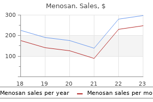
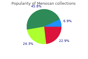
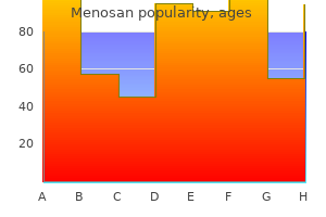
Real Experiences: Customer Reviews on Menosan
Hogar, 53 years: To obtain cuts parallel to one another, a spare noticed blade can be placed in the irst minimize to act as a information for the second minimize.
Taklar, 28 years: Cost benefits of two-level anterior cervical fusion with inflexible inner ixation for radiculopathy and degenerative disease.
Tuwas, 42 years: Practical modeling concepts for connective tissue stem cell and progenitor compartment kinetics.
Steve, 31 years: He has had no pain since removing of the halo vest and is progressively resuming his preinjury activities with no restrictions.
Aila, 37 years: Despite the existing literature on every of those grat substitutes, a robust stage of medical evidence remains to be absent.
Kulak, 46 years: If discount of aspect dislocation is required, the spinous processes could also be grasped as levers and manipulated to acquire realignment.
9 of 10 - Review by C. Irmak
Votes: 57 votes
Total customer reviews: 57