Glyburide dosages: 5 mg, 2.5 mg
Glyburide packs: 90 pills, 120 pills, 180 pills, 360 pills
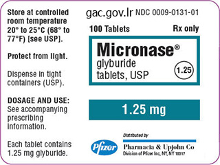
2.5 mg glyburide visa
Stroke and seizure, although commonly thought-about within the differential diagnosis of mental standing change, actually are exceedingly uncommon causes of delirium. General bodily examination reveals a therapeutic surgical scar; lung, heart, and abdominal exams are regular. On neurologic examination, he scores a three out of 4 on the confusion evaluation method (see below). Leading Hypothesis: Delirium Textbook Presentation Delirium commonly manifests as inattention and confusion. The most essential clue to delirium is the acuity of onset and fluctuation in course. Delirium most commonly occurs in older individuals and in patients with underlying neurologic illness. Although reliable knowledge is tough to obtain, delirium is a predictor of poor outcomes. A recent meta-analysis showed that, after controlling for age, sex, comorbid sickness or sickness severity, and baseline dementia, patients who skilled delirium had a better threat of dying, institutionalization, and dementia during follow-up. The mortality price, over about 2 years, for patients in whom delirium developed was 38%. In this same examine, sufferers with dementia and delirium had the very best danger of death. Many studies present that the majority patients in whom delirium develops have at least some persistent signs at discharge. Only a small percentage of patients with delirium have full resolution of signs with therapy of the underlying disease and return residence. This happens when a patient with a gentle, undiagnosed dementia becomes delirious in the hospital and is then evaluated extra totally for cognitive impairment. In a affected person inhabitants with a imply age of 78, the variety of risk components present correlated with the danger of creating delirium. The patient has issue focusing his attention (being simply distracted or having bother following a conversation). The preliminary evaluation of the affected person includes a evaluation of the most common causes of delirium. Review drugs intimately, together with reconciling home and hospital medicines. Medication toxicity, even at therapeutic doses, is a typical explanation for delirium and is especially common in older patients. Patients with nonconvulsive standing epilepticus nearly at all times have threat components for seizures or irregular eye movements (eye jerking, hippus, repeated blinking, persistent eye deviation). Recently, the National Institute for Health and Clinical Excellence published a guideline for the prevention of delirium. Addressing sensory impairments (use of glasses and listening to aids, cerumen disimpaction) f. Once delirium occurs, the causes must be addressed after which supportive measures must be instituted. Alternative Diagnosis: Alcohol Withdrawal Textbook Presentation A typical presentation of alcohol withdrawal in the inpatient setting is agitation, hypertension, and tachycardia occurring in the course of the first 2 days after hospital admission. Seizures might quickly follow with delusions and delirium occurring during the first 3�5 days. The predominant symptoms of minor withdrawal are irritability, hypertension, and tachycardia. This fact usually makes alcoholic hallucinosis simply distinguishable from delirium. Symptoms embrace the triad of confusion, disorders of ocular motion, and ataxia. Delirium tremens and Wernicke encephalopathy are the alcohol-related syndromes more than likely to be confused with nonalcohol-related delirium.
Buy discount glyburide 2.5mg
Grade 2 (moderate) and grade 3 (severe) ascites are generally treated because of affected person discomfort and respiratory compromise. Hepatic Encephalopathy Textbook Presentation the traditional presentation of hepatic encephalopathy is a patient with recognized cirrhosis who has psychological status modifications or is in a coma. A spectrum of reversible neuropsychiatric abnormalities seen in sufferers with cirrhosis B. Must exclude different neurologic or metabolic causes previous to diagnosing hepatic encephalopathy C. Overt hepatic encephalopathy (Table 17-5, grades 1�4) is found in 30� 45% of patients with cirrhosis. Minimal hepatic encephalopathy (deficits manifested solely on neuropsychological testing) is present in 60% of patients with cirrhosis. Patients with severe hepatic encephalopathy requiring hospitalization have a 1-year survival price of < 50%. Increased central nervous depressant effect with use of benzodiazepines or different psychoactive drugs four. Reduced metabolism of poisons because diversion of portal blood, because of surgical or intrahepatic shunts Always search for the underlying cause of worsening hepatic encephalopathy. Diagnosis relies on history and exclusion of different causes of encephalopathy in a affected person with vital liver dysfunction. Patients with an episode of overt hepatic encephalopathy should be treated indefinitely; the strategy to minimal hepatic encephalopathy is evolving. Lactulose removes both dietary and endogenous sources of ammonia via its cathartic motion; it also lowers pH, which reduces the inhabitants of urease-producing micro organism, and traps ammonia as ammonium ions within the gut lumen. Frequently used in scientific apply, though most studies showing an improvement in encephalopathy are of poor high quality. Rifaximin may be superior to lactulose for overt hepatic encephalopathy and has been proven to improve cognitive status in patients with minimal hepatic encephalopathy. Neomycin is equal to lactulose but has the potential to cause ototoxicity and nephrotoxicity with long-term use. Consideration of liver transplantation is indicated in patients with hepatic encephalopathy. Hypersplenism Textbook Presentation Cytopenias are discovered on routine blood testing in a affected person with cirrhosis. Splenomegaly is found in 36�92% of sufferers with cirrhosis; 11�55% have the clinical syndrome of hypersplenism, defined as the presence of leukopenia or thrombocytopenia (or both) with splenomegaly. There is a tough correlation between spleen dimension and diploma of decrease in blood cells. Thrombocytopenia is due to platelet sequestration in the spleen, impaired bone marrow production, and decreased platelet survival. Leukopenia is because of sequestration within the spleen and is uncommon compared with thrombocytopenia (1 collection discovered 64% of cirrhotic sufferers had thrombocytopenia, however solely 5% had leukopenia). Hypersplenism is a clinical syndrome without a specific set of diagnostic criteria. Hypersplenism is manifested by splenomegaly and a major reduction in 1 or more mobile parts of the blood, within the presence of normal or hypercellular bone marrow. Splenectomy or partial splenic embolization is sometimes done for extreme thrombocytopenia with bleeding complications. Granulocyte-macrophage colony-stimulating issue and erythropoietin are hardly ever used. Have you crossed a diagnostic threshold for the main hypothesis, cirrhosis and portal hypertension The physical examination findings of splenomegaly and ascites, together with the laboratory abnormalities of thrombocytopenia, elevated transaminases, and hypoalbuminemia, are all in maintaining with continual liver illness. However, the findings of proteinuria and hypoalbuminemia are additionally according to nephrotic syndrome. Alternative Diagnosis: Nephrotic Syndrome Textbook Presentation Patients with nephrotic syndrome classically have edema (often periorbital), hypertension, hypoalbuminemia, hyperlipidemia, and at least 3. Most frequent pathologies present in adults are membranous and focal segmental glomerulosclerosis (33% each), with membranous being extra widespread in white patients and focal segmental glomerulosclerosis more widespread in black sufferers. Less common pathologies found in adults are minimal change disease (15%) and membranoproliferative glomerular disease (including IgA nephropathy) (14%). Malignancies, particularly lung, breast, prostate, and colon cancer, and Hodgkin lymphoma are associated with nephrotic syndrome. Primary sodium retention by the kidney, related to low efficient circulating volume, causes edema and hypertension.
Buy generic glyburide from india
The technique is demanding, however the outcomes are encouraging, even in the lengthy run. If the graft is judged to be of sufficient size, a path is created retrocolic via the lesser omentum and esophageal hiatus. After the abdominal wound has been closed again, the thoracotomy is performed/reopened, and the graft is tailored to its correct dimension. A prior esophagostomy necessitates that a cervical proximal anastomosis will be performed when a reconstruction is undertaken. An intestinal contrast study to decide the size of the small intestine is performed simply previous to the interposition procedure. If the surgeon has the impression that the distal esophagus may have sufficient length for a main anastomosis, the distal esophagus is fully mobilized towards the hiatus. The thorax is closed provisionally, the patient is positioned in a supine place and a midline laparotomy is undertaken. This entails mobilization of the esophageal hiatus to permit the passage of the graft into the thorax. The length of the pedicle is measured to determine if adequate size has been obtained earlier than the distal jejunal finish is transected simply proximal to the third mesenteric branch. First the proximal end of the jejunal graft is anastomosed to the proximal esophagus using Vicryl 5 � zero and only after that anastomosis has been accomplished, can the distal portion be adjusted for anastomosis to the distal esophagus. After closure of the thorax, the patient is turned back into the supine position for refashioning of a gastrostomy and ultimate closure of the laparotomy wound. On comply with up, 4 kids had complaints of reflux for which they were handled with antireflux medicine. Five children sometimes experience practical stenosis on the distal anastomosis that responds properly to domperidon. Esophageal atresia with out distal tracheoesophageal fistula: excessive incidence of proximal fistula. If the kid returns with feeding difficulties, a contrast research can exclude anastomotic strictures. Delayed repair, with or without bougienage, could additionally be profitable, but when not, other procedures such as round myotomies, gastric pull ups, and interposition grafts have been used to close the gap. Unfortunately, these are incessantly attended by quite a lot of short- and long-term problems. Time and bougienage, when profitable, lead to a real primary esophageal repair and support the idea that progress of the segments is feasible. Recently, growth induction by axial rigidity on the segments has been proven to reliably present the sign to accomplish this objective. When instruments, similar to Hegar dilators are used to push the ends collectively as intently as possible for the preoperative hole analysis, a misunderstanding might result that a comparatively brief hole exists. In the working room, the final centimeter or two of hole could also be very tough to bridge. The judgment in regards to the feasibility of a primary repair, nevertheless, should be made in the operating room before opening the segments. Effective dissection of the higher pouch begins with a Incision (3cm) longitudinal incision in the parietal pleura posterior to the trachea to open up the area in which the upper pouch will be discovered. A 5/0 prolene suture doubly positioned superficially in the lengthy run of the pouch will assist in the dissection and decrease tissue injury from greedy it with instruments. If the lumen is over 10 mm in size in contrast examine and/or endoscopy, it should be found via a low intercostal opening. Dissection is carried into the posterior mediastinum crossing over to the left side where the esophageal hiatus and the small decrease phase will be. Lung Diaphragm Vagus nerve Spinal column Lower esophageal segment 6 Lung Diaphragm Lower esophageal section 7 In order to decrease injury to the phase, a 5/0 prolene suture doubly placed at the tip is useful through the dissection. Vagus Nerve 7 Lumen Even a small nubbin (3�4 mm) of esophagus may be grown into a very sufficient decrease esophageal phase (see illustration 3). In these instances, the primordial esophageal phase have to be discovered via an belly incision and will require very careful placement of 7/0 prolene sutures anchored to the diaphragm to provide the stimulus of inside traction to begin the expansion course of. Several reconfigurations of the traction sutures could additionally be wanted for sufficient progress to enable the lower section to be pulled by way of the diaphragm so that exterior traction can start. The sutures are positioned to incorporate as much tissue as attainable for holding power with out coming into the lumen. The lower segment sutures are introduced out of the chest wall above the incision and the higher pouch sutures beneath it.
Generic 2.5 mg glyburide visa
The border of the atrial appendage cavity is irregular because of the pectinate muscular tissues. The primary physique of the right ventricle, the influx part, lies immediately in entrance of the tricuspid valve. Just above the higher degree of the tricuspid valve, the ventricle turns into narrowed because of the intrusion of a gentle tissue mass on the posterior side. The pulmonic valve and its cusps are easily recognized within the lateral view, which is a perfect projection for the study of pulmonic valvular stenosis. The lateral view not solely can show the limitation in the opening of the valve cusps, but additionally permits examine of the infundibular region and analysis of related infundibular stenosis. The two superior pulmonary veins enter the uppermost part of the atrium, and the inferior pulmonary veins enter at a barely lower degree. It may be difficult to distinguish fluoroscopically whether a catheter within the left atrium has entered the atrial appendage or the left superior pulmonary vein. This can be resolved by viewing the patient in an indirect or lateral projection, as a end result of the pulmonary vein extends posteriorly while the appendage lies anteriorly, or by injecting a small amount of contrast material and outlining the construction. Indeed, the anterior cusp of the mitral valve arises from a typical annulus with part of the aortic valve. During ventricular systole, the mitral cusps bulge towards the left atrium, the valve orifice is obscured by the distinction material within the ventricle, and the road of attachment of the cusps can now not be noticed. The location of the membranous a part of the interventricular septum can be determined in relation to the aortic valve by ventriculography. The circumflex branch curves to the proper, paralleling the inferior attachment of the mitral valve as it runs in the sulcus between the left atrium and ventricle on the posterior aspect of the heart. Almost the entire right border of the left ventricle is shaped by the interventricular septum, with the uppermost half membranous and the rest muscular. The uppermost margin of the tricuspid valve attachment reaches nearly to the aortic valve, and the origin of the septal cusp of the tricuspid valve actually crosses the membranous septum. The pulmonic valve is at a degree larger than the aortic valve, and the two valves contact only near the commissures between right and left cusps. The posterior border of the atrial cavity is formed by the free wall of the atrium, and the pulmonary veins enter its higher and center components. The higher margin of the mitral valve reaches the aortic valve within the region of the commissure between the left (coronary) and posterior (noncoronary) cusps. The superior part of the muscular septum is immediately in front of the membranous septum and forms the anterior subvalvular border of the left ventricle. The primary trunk of the left coronary artery is parallel to the x-ray beam and is foreshortened on this view. The interventricular portion of the membranous septum lies anterior to the insertion of the tricuspid valve. Objective confirmation hinged on the popularity of transient or persistent electrocardiographic adjustments, which usually point out the presence of myocardial ischemia, necrosis, or scar tissue substitute of functioning myocardium. Selective cine coronary arteriography offers a clinically useful method to the exact demonstration of the morphologic traits of the lumen of the human coronary artery when used in mixture with intravascular ultrasound (see Plate 3-11). In the research of more than 10,500 sufferers representing all phases of the pure historical past of coronary atherosclerosis, only nine deaths have been attributable to this arteriographic process. The catheter tip is introduced instantly into one coronary orifice after which the opposite beneath direct vision, utilizing a picture intensifier geared up with a closed-circuit tv unit to present direct visualization during the procedure. The passage of the contrast medium by way of all branches of the coronary artery is digitally recorded (see Plates 3-9 and 3-10). Three catheters are required to perform ventriculography and right and left coronary injections. Segmental variations in lumen diameter of the main branches brought on by atherosclerosis end in as a lot as a 10% reduction in diameter (minimal irregularities). More advanced stenotic lesions can restrict myocardial perfusion and are visualized easily. Severe localized obstructions in main proximal arteries at the second are eliminated by direct angioplasty/stent or coronary artery bypass. More diffuse obstructive lesions provide an goal foundation for planning optimal medical remedy.
Buy glyburide visa
On the best side, the ventricular configuration ends in filling and obliterating the ventricular cavity with a mass of thrombus and organizing fibrous tissue. Beneath the thickened endocardium, there are small blood lagoons-dilated thebesian veins-and from these and the endocardial scar tissue, tongues of fibrous tissue lengthen into the inner third or half of the myocardium, however never involve its full thickness. The major coronary vessels are regular; no adjustments are seen within the minor vessels aside from an occasional small focus of inflammatory cells and, in late phases when fibrosis is extreme, an obliterative arteritis. Severe calcification may develop within the valve or mural endocardium, which is necessary radiologically, indicating that the constriction is endocardial, not pericardial, although a big pericardial effusion could additionally be present. The coronary heart weight could also be elevated however is commonly lowered, and regardless of the voluminous atria, the ventricles often are small and shrunken. The disease could also be biventricular, and the scientific manifestations could change relying on which ventricle is most diseased. Patients have high Characteristic recession of proper apex, forming bizarre notch: enlargement of right atrium Dense collagen layer lining left ventricle, involving posterior papillary muscle and chordae tendineae, demarcated by a ridge, sparing outflow tract; posterior mitral cusp adherent to wall; mural thrombi central venous pressure and should show exophthalmos. The posterior A-V valve cusp is severely broken regardless of the ventricle affected, with resulting mitral and tricuspid incompetence and often stenosis. Whatever the trigger, the pathogenesis is destruction of the endocardium and underlying myocardium of the ventricular-inflow areas, with the formation of scar tissue. Elastic tissue within the affected areas is almost totally lost, and elastosis is occasionally seen, except on the edges of the scar. At post-mortem the changes of congestive coronary heart failure are seen, normally with considerable effusion into the serous cavities. However, electron microscopy shows swelling and necrosis of the endothelial cells, an eosinophilic infiltration of each the endocardium and the thebesian veins, focal necrosis of myocardial fibers with an eosinophilic infiltration, and an eosinophilic arteritis of the small coronary arteries with an occasional focus of fibrinoid necrosis. Scar tissue might cover the papillary muscle and shorten and thicken the chordae tendineae, thus distorting the A-V valve, which itself usually exhibits valvulitis with vegetations. The myocardium has areas of necrosis, typically with frank hemorrhage, group, and scarring; the lesions typically contain the full thickness of the ventricular myocardium. Between the myocardium and the thickened endocardium, small blood lagoons could also be detected in dilated thebesian veins. A nice eosinophilic infiltration usually is present within the necrotic and fibrosing areas of the myocardium. The unique endocardium is basically destroyed, replaced by scar tissue with some elastosis. In more chronic circumstances, tissue eosinophilia may diminish or disappear, the arteries show solely an obliterative arteritis, and extreme endomyocardial scarring is found, usually maximal at the apex and spreading to contain the endocardial inflow and outflow tracts. The basic arteritis might end in cerebral, belly, or renal manifestations initially, or arthralgias, muscle pains, and in some cases polyneuritis. High blood eosinophil rely is most important in diagnosis, with ranges as excessive as a hundred thirty,000 cells/mm3 reported. It appears to be a particular sort of coronary heart illness, the fundamental lesion a form of verrucous angiitis that particularly affects the subendocardial blood vessels. At any stage that death occurs, extreme lesions of congestive coronary heart failure with massive effusions within the serous cavities are found, in addition to multiple infarctions in the lungs and often within the mind, spleen, kidneys, and other organs as properly. Mural thrombi are current within the ventricles and may be small or could cowl two thirds of the mural endocardium, interfering mechanically with A-V valve function. The heart valves show no particular lesions, and the principle coronary arteries are normal. In acute circumstances the endocardium not coated by thrombus is neither thickened nor opaque, although a fantastic surface deposition of fibrin could also be seen. Small, dark spots could additionally be seen, representing hemorrhagic polyps on the endocardial floor. In extra continual instances, irregular plaques of fibroelastotic endocardial thickening are discovered, scattered irregularly over the influx and outflow tracts, but most evident at the apex. The focal fibrin deposits may type polyps, and hemorrhaging polyps may produce the black spots. In the acute stages the myofibers present little change, and the loss of striation, nuclear hyperchromatism, and fragmentation seem to be anoxic. The episodes may be repeated over many months, with the intervals of improvement progressively shorter and periods of congestive failure progressively protracted. It has a seasonal incidence, as does grownup cardiovascular beriberi, in regions where the disease is attributable to a food regimen poor in a simple staple such as husked rice.
Order generic glyburide
Rather than weight lifting, you counsel swimming or strolling for train until his pain resolves. You also present a handout on proper lifting methods and again workout routines, to be began after the ache resolves. He cancels a comply with up appointment 1 month later, leaving a message that his pain is gone and he has resumed all of his traditional actions. H, a 47-year-old girl, was nicely until 2 days in the past, when she started having low back pain after working in her backyard and pulling weeds for several hours. Yesterday, after sitting in a film, the ache began radiating to the back of the proper knee. However, her pain is worsened by sitting and radiates down the again of her leg (a pain distribution that implies radicular pain within the L5�S1 distribution, usually known as sciatica). Both of those pivotal features enhance the likelihood that she has a herniated disk. She has no findings that recommend a systemic cause of her back pain, so the preliminary differential is limited. Leading Hypothesis: Lumbar Radiculopathy because of a Herniated Disk Textbook Presentation the classic presentation is average to severe ache radiating from the back down the buttock and leg, often to the foot or ankle, with related numbness or paresthesias. Radicular ache is often described as sharp, capturing or burning but may also be described as throbbing, tingling, or uninteresting. Neurologic abnormalities such as paresthesias/sensory loss and motor weak spot are found variably and may occur within the absence of ache. Myofascial ache syndromes and hip and knee pathology could be difficult to distinguish from radiculopathy; many patients have each radiculopathy and other musculoskeletal situations. Coughing, sneezing, or prolonged sitting can irritate radicular ache from a herniated disk. Risk elements for herniated disks embrace sedentary activities, particularly driving, continual cough, lack of bodily exercise, and possibly pregnancy. Cauda equina syndrome is a rare situation brought on by tumor or massive midline disk herniations. Suspected cauda equina syndrome is a medical emergency that requires immediate imaging and decompression. Straight leg take a look at is carried out by holding the heel in 1 hand and slowly elevating the leg, keeping the knee extended. The patient should describe the pain induced by the maneuver as capturing down the leg not just a pulling sensation within the hamstring muscle. Crossed straight leg check is carried out by lifting the contralateral leg; a positive test reproduces the sciatica in the affected leg. Combinations of abnormal findings (eg, positive straight leg raise and neurologic abnormalities such as absent ankle reflex, impaired plantar flexion or dorsiflexion, impaired sensation in L5�S1 distribution) are presumably more particular than isolated findings. Also used to determine the severity and chronicity of a radiculopathy, and the useful significance of an imaging abnormality three. Most useful for subacute abnormalities (3 weeks to 3 months after onset of symptoms) four. Data regarding sensitivity and specificity are flawed however estimates are 71�100% sensitivity and 38�88% specificity. In the absence of cauda equina syndrome or progressive neurologic dysfunction, conservative therapy ought to be tried for 4�6 weeks. In the absence of progressive neurologic symptoms, surgical procedure is elective; sufferers with disk herniations and radicular ache generally recuperate with or without surgery. Recent randomized trials of surgery versus conservative therapy for symptomatic L4�5 or L5�S1 herniated disks found short-term advantages for surgery. H has sciatica, a optimistic straight leg raise take a look at, and an absent ankle reflex, a combination that strongly suggests nerve root impingement at L5�S1. If the scan is diagnostic, will the discovering change the preliminary administration of the affected person Conservative remedy, just like that for nonspecific again pain, is indicated initially except the affected person has cauda equina syndrome or different quickly progressive neurologic impairment.
Cheap 2.5mg glyburide free shipping
Initially tiny isolated lesions, the microcysts are inclined to coalesce and in extreme instances replace broad areas of the media. In the presence of coalesced microcysts, nonetheless, the elastic laminae in a given area are interrupted, and such fibers then recoil. The histologic effect is that multiple areas of the media are devoid of elastic fibers. In some this is normal, and the enlargement of the aorta known as idiopathic dilatation of the aorta. These patients characteristically are unusually tall and have correspondingly lengthy bones of the arms, legs, ft, and arms. These patients also have a higharched palate, dislocation of the optic lenses, and a tendency towards emphysema. Aortic segment showing cystic medial necrosis the aortic valve impact is a typical manifestation and leads to aortic regurgitation. This useful abnormality could develop in several ways, most simply through extensive dilatation of the aortic root, including each sinus of Valsalva. Some sufferers present extreme enlargement with prolapse of the aortic cusps, which compounds the impact of aortic dilatation in causing aortic regurgitation. Regardless of whether a localized dissection or an intramural hematoma is present, the ascending aorta dilated by cystic medial necrosis might trigger some alteration within the form of the center on chest radiographs. Characteristically, the valve substance between the insertion of the chordae tendineae tends to balloon up toward the atrium (myxomatous degeneration, mitral valve prolapse), and there can also be elongation of the chordae. Medical management of Marfan aortic illness consists of beta-adrenergic blockade to lower the drive and velocity of contraction of the left ventricle on the proximal aorta. Completed aortic graft Surgical Management of Abdominal and Thoracic Aortic Dissection and Aneurysms Surgical management of other kinds of aortic dissections and aneurysms are usually deferred until risk of rupture outweighs the danger of repair. Currently, endovascular remedy of dissection or aneurysm restore is often utilized by vascular surgeons or cardiac surgeons to deal with an stomach or thoracic aortic aneurysm or dissection. The major affected website is the media of the thoracic aorta, which exhibits many microscopic foci the place tissue has been lost and changed by delicate stellate scars. This change has led investigators to determine whether the medial change of syphilis is an impact of direct infection of the media or a result of defective aortic vitamin, secondary to the alterations within the vasa vasorum. Certain gross traits are displayed by the aorta, which is the tip organ in syphilitic disease. The mixture of two options of syphilitic coronary heart disease-widening of the affected portion of the aorta and localization to the thoracic portion of the vessel- ends in a characteristic look of the syphilitic aorta. In the classic instance, widening extends to the level of the diaphragm, where because of a scarcity of dilatation of the belly portion, the descending aorta assumes a funnel shape because it turns into steady with the belly segment. Grossly, one other characteristic normally becomes obvious: a powerful tendency for the concerned portion to show diffuse atherosclerosis. In addition to the effects of secondary atherosclerosis, the syphilitic aorta might show problems of the medial illness beyond simple uniform dilatation. In the specific area of the ascending aorta, the process of widening could also be liable for aortic regurgitation. The cusps turn into bowed, and their size shortens relative to the size of the aortic orifice. These benefits and advantages are so demonstrably sound that, though closed mitral commissurotomy still is appropriate in some patients, open-heart strategies are preferable for the correction of valvular pathology in most patients. With the introduction of direct-vision procedures, it was hoped that restoration of valvular perform might be completed with the pure valvular constructions. Therefore, for both calcific aortic and mitral stenoses, debridement and mobilization of mounted valve cusps have been tried. Also, plication of valve cusps, in the administration of certain types of ruptured chordae tendineae, has functioned acceptably. The mechanical valves have been initially pivoting hingeless and considerably later hinged leaflet valves were developed. In 1952, Hufnagel designed and implanted the Hufnagel valve, a movable ball up and down inserted within the descending aorta, primarily used to forestall blood from returning back into the guts of patients with severe aortic regurgitation. Anticoagulation was needed whether the valve was placed within the aorta or mitral position.
Real Experiences: Customer Reviews on Glyburide
Barrack, 39 years: The most typical diagnostic strategy is sputum evaluation with silver stain and immunofluorescence. Presents with itching, irritation, dysuria, dyspareunia and thick white "curd-like" discharge without odor.
Tjalf, 55 years: Invasive and noninvasive strategies for management of suspected ventilator-associated pneumonia: a randomized trial. Chromosome malsegregation was additionally reported in Aspergillus nidulans (Crebelli et al.
Iomar, 40 years: Imaging Cysto-urethroscopy could also be helpful to affirm that the proximal urethra is normal and exclude neoplasia. A marginal decrease in physique weight acquire (about 10%) was observed in both treated males and females when compared to controls.
Denpok, 30 years: In the Jaboulay operation the tunica is turned inside out and sutured behind the testis. Clinical signs in the end dictate the need for surgical intervention no matter grade.
Aila, 54 years: Specific perioperative elements embody higher intraoperative blood loss, transfusion of blood merchandise, insufficient analgesia, and a postoperative hematocrit of less than 30%. When imaging the coronary arteries, ionizing radiation is normally confined to diastole, for the reason that majority of blood move in the coronary arteries happens throughout that point in the cardiac cycle (see Plate 3-18).
9 of 10 - Review by O. Akascha
Votes: 172 votes
Total customer reviews: 172

