Losartan dosages: 50 mg, 25 mg
Losartan packs: 28 pills, 56 pills, 112 pills, 224 pills, 30 pills, 60 pills, 90 pills, 120 pills, 180 pills, 270 pills, 360 pills
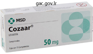
Buy losartan in india
Some authors dispute the existence of chondroid chordomas, preferring to regard these cartilage-containing neoplasms as chondrosarcomas. Chondroid chordomas have a more indolent medical course; the survival rate is 15. Atypical chordomas have a sarcomatoid look, with round cells and epithelial or spindle cells present with giant areas of necrosis. In the series by Heffelfinger and coworkers, just one patient with an atypical chordoma survived greater than 10 years, whereas almost 50% of those with chondroid chordomas survived more than 10 years. Immunostaining from keratin has no prognostic worth regarding the aggressiveness of the tumor. Chondrosarcomas have been lumped with chordomas because of supposed parallel traces of incidence, location, and aggressive habits. In my sequence, 55% of patients benefited from craniovertebral stabilization along with tumor resection, owing to involvement of the occipital condyles. Larger tumors have the potential to cause each higher and decrease cranial nerve palsies and a wide selection of problems related to brainstem compression. Chordomas typically trigger symptoms from native growth into the nasal cavity, pharynx, and paranasal sinuses. SurgicalSeries Although the number of reported circumstances of untreated intracranial chordoma is small, only temporary survival after analysis is a consistent finding. Several series of aggressive surgical extirpation adopted by conventional radiation and proton-beam remedy are discussed here. Forsyth and colleagues reviewed fifty one intracranial chordomas handled surgically between 1960 and 1984 at the Mayo Clinic. Eleven patients (22%) underwent biopsy, and 40 sufferers (78%) had subtotal resection. The survival charges for patients who underwent biopsy were 36% and 0% at 5 and 10 years, respectively, whereas survival rates for these with subtotal resections were 55% and 45% at 5 and 10 years, respectively. Patients who underwent postoperative radiation remedy tended to have longer disease-free survival times. Disease-free survival was the same for patients with chondroid chordomas as for these with typical chordomas. Watkins and associates described 38 sufferers handled on the National Hospital of Neurology and Neurosurgery in London between 1958 and 1988. The authors concluded that two groups existed: one with indolent disease and one other with aggressive growth and poor outcome. In a more recent publication in 2001, Crockard and associates described a multidisciplinary method to cranium base chordomas. A total removing was made in 2, radical 30 and subtotal or partial within the remainder. Gay and colleagues reviewed the management of 46 chordomas and 14 chondrosarcomas involving the cranial base between 1984 and 1993 on the University of Pittsburgh. Fifty % of sufferers had undergone previous surgery before referral, and 22% had undergone earlier external-beam radiation remedy. The surgical strategy was a subtemporal-infratemporal fossa approach, generally combined with a transpetrous approach. In other instances, an extended subfrontal approach was used, and in a couple of cases, the lateral transcondylar method was used. There was a high tendency to remain between the subtemporal-infratemporal fossa method and the prolonged subfrontal approach. Postoperatively, 20% of patients underwent external-beam, proton-beam, or gamma radiation remedy. In patients who had total resection, the general 5-year recurrence-free survival rate was 84%, compared with 64% in those with partial resection. Presentation Chordomas usually occur in adults, with a peak incidence occurring within the fourth decade of life. She offered with dysphagia, nasal regurgitation, unsteady gait, complications, and tongue atrophy. Based on this experience, the authors advocated aggressive initial surgical resection, with the sparing application of radiation therapy.
Cheap losartan 25 mg amex
Similarly, it has been held that surgical d�bridement of involved bone and soft tissue may have utility in the clearance of an infection. There is nice evidence, nevertheless, that easy d�bridement of the infected focus might have little to add to effective chemotherapy. Nonetheless, when deformity, neurological deficit, or instability exists, the role of decompression, d�bridement of affected tissues, reconstruction, and stabilization appears clear. As with other forms of spinal an infection, most of the involvement facilities in the vertebral bodies. As a end result, the surgical approaches for decompression, d�bridement, and reconstruction of the affected anterior and center columns require anterior or anterolateral approaches. For cervical lesions, the standard approaches for multilevel anterior disease are relevant. The thoracic levels are generally accessed by way of a thoracotomy method, which is adequate to allow anterior d�bridement, anterior reconstruction with an allograft strut or cylindrical cage and autograft, and placement of an anterior internal fixation gadget. Anterior reconstruction with autologous rib or an allograft strut or cage can generally be performed with this method. Finally, planned anterior and posterior surgery carried out sequentially in a single session may be required to achieve optimal d�bridement, correction of deformity, and long-term stability. Blastomycosis North American blastomycosis is predominantly a granulomatous cutaneous or respiratory an infection. Males are more susceptible to infection than females, and blacks are more generally affected than whites. All ages can be affected, however the disease seems to have a predilection for the second via the fifth many years. Disseminated illness produces generalized signs of fever, malaise, anorexia, and evening sweats. The prognosis is typically made through positive cytology or histologic examination. A high index of suspicion is justified in endemic areas and in immunocompromised or in any other case vulnerable hosts. Similar to most fungal osteomyelitis, normal therapy consists of amphotericin B, although newer azole antifungals. Vertebral involvement as a consequence of disseminated infection is comparatively uncommon; nonetheless, the increase in numbers of prone people will result in an increased variety of instances. Non-albicans species similar to Candida glabrata, Candida tropicalis, and Candida dubliniensis are assuming greater significance as opportunistic pathogens. This is critical as a result of there is a rise in resistance to antifungal chemotherapy in plenty of of those "nontraditional" species. Culture and histologic evaluation of vertebral biopsy specimens are both useful in confirming the correct analysis. Amphotericin B is the usual type of medical therapy, although newer antifungal brokers may be successful with lower charges of toxicity. According to Sugar and associates,218 fluconazole may be effective when administered on a steady basis for extended durations. As with other forms of an infection, surgical d�bridement, fusion, and instrumentation are required for patients with superior deformity, neurological compromise, and instability. Overall, at least 85% of patients will respond to remedy with remedy of the osteomyelitis. Candida species have become a few of the most common nosocomial pathogens in critically unwell sufferers. Osteoarticular involvement could be anticipated to extend as the general incidence of disseminated candidiasis increases. Certain endemic pathogens such as Coccidioides and Blastomyces might involve the backbone as a consequence of dissemination. Recognition of the potential of these infections of their numerous endemic areas is a key to diagnosis. Coccidioidomycosis Coccidioidomycosis is endemic to the southwestern United States, Mexico, and Central and South America.
Purchase 25 mg losartan visa
Because of the perilous state of disk vitamin, a hostile microenvironment, incompetence of the annulus fibrosus, and disk space collapse, advanced degeneration may require the implantation of a hybrid disk prosthesis by which disk replacement technology is mixed with developments in tissue engineering. Biology of intervertebral disc aging and degeneration: involvement of the extracellular matrix. Preliminary analysis of a scheme for grading the gross morphology of the human intervertebral disc. Hoh Bone physiology because it pertains to mineralization and resorption is of explicit curiosity to neurosurgeons treating spinal issues. Alterations in bone metabolism because of illness or regular growing older can result in fractures, spinal instability, and deformity, which in flip could cause continual ache or neurological deficit. Poor bone high quality secondary to unbalanced bone resorption may have a significant impact on surgical planning inasmuch as impaired bone power can result in implant failure in instrumented spinal procedures. Accordingly, a greater understanding of bone physiology, regulating components, and ailments of bone metabolism facilitates better determination making regarding patient administration and surgical intervention and thus reduces the chance for postoperative issues. The spinal column performs the primary capabilities of maintaining mineral balance, protecting the neural components, and serving as points of attachment for muscles that act on the skeleton for movement and help. Cortical bone is dense, inflexible tissue that biomechanically is responsible for resisting torsional and shear loads. Cancellous bone, which is more prevalent in the spinal column, is composed of a number of interconnected horizontal struts. Biomechanically, cancellous bone supplies resistance to compressive and shear stress. Histologically, bone is composed of several basic cell varieties: osteoprogenitor cells (preosteoblasts and preosteoclasts, which originate from hematopoietic stem cells), osteoblasts, osteoclasts, and osteocytes, all of which work collectively to regulate and keep the local tissues. Bone matrix, the most important substance that surrounds bone cells, is composed of organic and inorganic elements. The organic part consists largely of kind I collagen, which supplies stiffness and energy to bone. These proteins are very important in performing various capabilities such as regulating mineral content material, bone resorption, and induction of osteoprogenitor cells. The inorganic part of bone matrix is the store of calcium and phosphorus as bone mineral. Calcium is packaged in mitochondrial granules and matrix vesicles, that are subsequently released during mineralization. Calcium inside bone primarily supplies structural integrity and is launched into the circulation for regulating metabolic processes. Osteoclasts then launch hydrogen ions, which lowers the local pH and induces dissolution of hydroxyapatite. The restorative course of is initiated by the maturation of preosteoblasts to osteoblasts and their subsequent mobilization to the newly fashioned defect. In the resorbed cavity, osteoblasts synthesize and secrete collagen-rich matrix, which then offers a surface for mineralization. The capability of the bony backbone to offer structural support and safety of the neural components is dependent on balanced remodeling. Bone turnover is balanced when the quantity of resorbed bone is changed by an equal quantity of newly formed bone. Excessive resorption with inadequate formation finally ends in osteopenia and impaired bone high quality. Poor bone strength can lead to structural failure leading to fractures, instability, and deformity. Vitamin D is either produced in an inactive type from the pores and skin or acquired nutritionally. The inactive form, vitamin D3, is fashioned from the pores and skin by activation of 7-dehydrocholesterol by ultraviolet gentle. Vitamin D functions by entering the cell nucleus to increase expression of proteins involved in calcium transport from intracellular shops within bone into the circulation. Calcitonin is secreted by the parafollicular cells of the thyroid gland to stop bone resorption.
Trusted 50mg losartan
Patients with intracranial lesions current with involvement of the lower cranial nerves, brainstem dysfunction, and infrequently cerebellar signs. Patients with straddle lesions have a paucity of cranial nerve dysfunction and a predominance of high cervical myelopathy. Unfortunately, the symptom of ache alone could predate different clinical findings for a few years. Paresthesias or dysesthesias of the face, palms, and limbs are frequently reported. An irregular cold sensation of the lower extremities was described by Elsberg and Strauss7 and by Beatty13 as being pathognomonic of lesions of the excessive cervical twine. Most regularly, pain and temperature sensation is affected, adopted by loss of joint sensation. This discovering is seen in the upper extremities and should then proceed clockwise across the limbs. A suspended sensory loss with patches of preservation of sensation within the higher extremities might confuse the presentation. The weak point could start within the ipsilateral limb and progress to the lower limb of the identical side, adopted by weak spot of the contralateral decrease limb; lastly, weakness turns into obvious in the contralateral higher limb. This distinct progression of motor signs is an important characteristic of lesions of the cervicomedullary junction. Taylor and Byrnes postulate venous stagnation of the anterior horn cells and the lower cervical cord on account of decreased venous drainage, which typically occurs rostral to the lower portion of the cervical spinal wire. Neurentericcysts Meningioma Neurofibroma spinal twine edema, and spinal cord rotation with contralateral traction. A tumor at the foramen magnum could produce a combination of higher motor neuron findings in the higher and decrease extremities. This pattern reflects the pyramidal decussation that begins just under the obex and ends near the uppermost cervical spinal twine. The extra medial fibers of the pyramidal tract carry impulses to the upper extremities and cross superior to the lateral fibers that serve the decrease extremities. Similarly, the sensory decussation of the medial lemniscus might produce a diversified pattern of sensory abnormalities. The syndrome of cruciate paralysis has been associated with trauma as nicely as tumors with basilar invagination. Cranial nerve palsies could additionally be the end result of nuclear involvement within the brainstem, traction, compression of the subarachnoid segments, or interosseous disease. Their involvement results in dysphagia, slurred speech, and repeated episodes of aspiration into the tracheobronchial tree, leading to pneumonia and weight loss. Tumors of the upper cervical canal can current with involvement of the spinal root of the accessory nerve, manifesting as torticollis and weak spot of the trapezius and sternocleidomastoid muscles. About 15% to 20% of sufferers develop tinnitus, vertigo, and listening to loss related to involvement of the vestibulocochlear nerve. The presentation of stressed legs syndrome in sufferers with craniovertebral compressive pathology has been well documented by Glasauer and Egnatchick. Hypoglossal nerve electromyography supplements the evaluation with an electrode positioned instantly into the tongue. Brainstem monitoring still yields a major number of false-negative and false-positive outcomes, but improved methods make these adjuncts helpful within the intraoperative assessment of the perform of the cervicomedullary junction. Benign lesions create a space among the many neurovascular constructions, thereby permitting surgical debulking and resection "from inside. In most instances, benign lesions similar to chordomas are radioresistant; therefore, gross whole resection should be the goal. Craniovertebral stability, each before and after operative intervention, must be thought-about in the improvement of approaches. Lesions of the craniovertebral junction do affect the pediatric inhabitants, though to a lesser extent than they affect adults.
25mg losartan visa
Surgical anatomy of the cervical pedicles: landmarks for posterior cervical pedicle entrance localization. Biomechanical analysis of transpedicular screw fixation in the subaxial cervical backbone. Cervical spondylotic myelopathy: therapy with posterior decompression and Luque rectangle bone fusion. A biomechanical analysis of cervical spinal stabilization strategies in a bovine model. Transpedicular screw fixation for traumatic lesions of the middle and decrease cervical backbone: description of the techniques and preliminary report. Complications of pedicle screw fixation in reconstructive surgery of the cervical spine. Biomechanical comparison of two-level cervical locking posterior screw/rod and hook/rod strategies. Kaiser Reconstruction of the anterior thoracic backbone has dramatically changed up to now a quantity of decades. The early efforts of surgical pioneers to stabilize the spine after destruction of the anterior spinal components had been often met with excessive charges of morbidity and construct failure. The evolution of anterior thoracic instrumentation has led to a simplification of insertion methods, improved biomechanical and imaging traits of current implants, and a major reduction in the dangers beforehand associated with anterior thoracic stabilization. This chapter describes the background and surgical apply of anterior instrumentation of the thoracic spine. In the Nineteen Seventies, advances in assemble design had been launched by Dwyer,4,5 Zielke,6 and colleagues. The screwcable construct of Dwyer and the screw-rod assemble of Zielke efficiently corrected scoliotic deformities. In the late Nineteen Seventies, Dunn developed a double-rod, double-screw assemble that supplied enough stability for anterior thoracolumbar reconstruction. Less optimum outcomes with an elevated incidence of postoperative problems have historically been related to posterior decompression for anterior metastatic disease. An anterior strategy provides a direct means of addressing ventral pathology and reconstructing the backbone on the major website of instability. Important considerations for acceptable affected person choice include age, preoperative practical standing, presence of medical comorbid conditions, life expectancy, and wish for tissue prognosis. The value of restoring neurological operate, such as ambulation or sphincter management, have to be judged on an individual basis. Other issues in selecting sufferers for operative intervention embody the kind of tumor, compromise of the spinal canal, extent of neurological deficits, level of pain, and diploma of instability. Adjuvant remedy such as radiotherapy, chemotherapy, or hormonal therapy may be indicated in specific instances. Additionally, nonoperative intervention remains an option for sufferers with stable myelopathy regardless of spinal twine compression. Neurological deterioration is uncommon after operative intervention for metastatic spinal disease. Perioperative mortality rates range from 6% to 8%, and general complication rates vary from 8% to 11%. Spinal neoplasms can be manifested as pain, progressive deformity, or neurological deficits. Operative intervention is suitable for tissue analysis, neurological decompression, and spinal stabilization. Posterior fusion and fixation could additionally be acceptable in the absence of a ventral deformity or anterior spinal canal compromise. Lesions producing ventral compression or vital biomechanical compromise of the anterior and center columns are indications for an anterior strategy. Previous reviews have documented better outcomes with an anterior method than with isolated posterior stabilization. Loss of the posterior tension band may require supplementation with a posterior stabilization construct. Patients with a whole spinal cord injury may still benefit from spinal stabilization. In such circumstances, surgical stabilization optimizes the rehabilitation course of and presumably decreases the size of hospitalization.
Bassora Tragacanth (Karaya Gum). Losartan.
- What is Karaya Gum?
- Are there any interactions with medications?
- What other names is Karaya Gum known by?
- Are there safety concerns?
- Dosing considerations for Karaya Gum.
- How does Karaya Gum work?
Source: http://www.rxlist.com/script/main/art.asp?articlekey=96537
Cheap losartan 25 mg without prescription
The fee of disruption of the pedicle cortex by an inserted screw ranges from 21% to 31% in these studies. Although the lateral view can be comparatively easy to assess, the anteroposterior or oblique view can be difficult to interpret. This plane finest demonstrates the place of the screw relative to the neural canal. This examine demonstrated an improvement in pedicle screw insertion accuracy with an error rate of only 5. It improves the pace, accuracy, and precision of complicated spinal surgical procedure whereas, typically, eliminating the need for cumbersome intraoperative fluoroscopy. Using defined mathematical algorithms, a specific level in the picture data set could be matched to its corresponding level in the surgical area. This course of is called registration and represents the crucial step of image-guided navigation. At least three points must be matched, or registered, to allow for accurate navigation. The common components of most of those systems embrace an image-processing laptop workstation interfaced with a two-camera optical localizer. When positioned during surgery, the optical localizer emits infrared gentle toward the operative area. A handheld navigational probe mounted with a fixed array of passive reflective spheres serves because the link between the surgeon and the computer workstation. Alternatively, passive reflectors could also be hooked up to straightforward surgical instruments. The spacing and positioning of the passive reflectors on each navigational probe or customized trackable surgical instrument is known by the pc workstation. This information is then relayed to the pc work-station, which might then calculate the precise location of the instrument tip within the surgical subject as properly as the situation of the anatomic point on which the instrument tip is resting. The initial application of navigational rules to spinal surgical procedure was not intuitive. The application of navigational expertise to spinal surgery includes utilizing the rigid spinal anatomy as a frame of reference. Bone landmarks on the exposed floor of the spinal column present the factors of reference necessary for image-guided navigation. Specifically, any anatomic landmark that might be identified intraoperatively as properly as within the preoperative picture data set can be used as a reference point. The tip of a spinous or transverse course of, a side joint, or a prominent osteophyte can serve as a potential reference level. If the patient is moved after registration, this spatial relationship is distorted, making the navigational info inaccurate. This downside can be minimized by the optionally available use of a spinal tracking system consisting of a separate set of 4 passive reflectors mounted in a identified configuration on a small body. This reference body may be attached to the exposed spinal anatomy and its place in area tracked by the infrared digicam system. Movement of the spinal anatomy and the connected frame alerts the navigational system, which can then make the appropriate correctional calculations to take care of accuracy and get rid of the necessity to repeat the registration process. It is especially cumbersome when image-guided navigation is used during cervical procedures. Alternatively, image-guided spinal navigation may be carried out without a monitoring system. Patient movement can doubtlessly occur with respiration, from the surgical group leaning on the desk, or from a change of table position. Although movement associated with leaning on the table or repositioning the table or the affected person will have an effect on registration accuracy, it may be easily prevented in the course of the brief navigational process. If inadvertent affected person movement does occur, the registration course of may be repeated. Three completely different registration strategies can be utilized for spinal navigation: paired level registration, floor matching, and automated registration. The registration method is carried out immediately after surgical exposure and earlier than any deliberate decompressive procedure. The tip of the probe is then placed on the corresponding level in the surgical area, and the reflective spheres on the probe handle are aimed towards the digicam. This preliminary step of the registration course of successfully hyperlinks the purpose chosen in the image knowledge with the purpose chosen in the surgical area.
Order discount losartan line
As long because the isocenters are in close proximity to at least one another, the software would routinely put them into the same therapy run and the patient would move from one set of coordinates to the next till all isocenters of one collimator measurement have been treated. Other techniques can be used in planning, corresponding to using a steep (125 degree) gamma angle for posterior lesions (cerebellar or occipital) to avoid frame collisions. Another approach available for single-isocenter lesions is to match the gamma angle to the angle of the goal. In trunnion remedy, the x, y, and z coordinates of every isocenter are set manually and double-checked to keep away from error. The operator selects the run (a mixture of isocenters of the same beam diameter) that matches the collimator helmet on the Gamma Knife unit. After the clearance verify, the system prompts the surgeon to hold out place checks. The staff screens the patient and coordinates of the completely different isocenters on the control computer. In the longer term, extra correct imaging strategies, improved software to handle these pictures, and advanced inverse planning software program will present higher remedy and end in higher affected person outcomes. Radiation-induced epilation due to sofa transit dose for the Leksell gamma knife model C. First medical expertise with the automated positioning system and Leksell gamma knife Model C. Gamma knife mannequin C with the automatic positioning system and its impact on the therapy of vestibular schwannomas. The Leksell gamma knife Model U versus Model C: a quantitative comparison of radiosurgical remedy parameters. Stereotactic radiosurgery of the brain utilizing the first United States 201 cobalt-60 supply gamma knife. Brain tumor radiosurgery: current status and strategies to enhance the impact of radiosurgery. Impact of the model C and Automatic Positioning System on gamma knife radiosurgery: an evaluation in vestibular schwannomas. A comparability of the gamma knife model C and the automated positioning system with Leksell mannequin B. All therapy data are exported to the operating console, which is used to regulate and monitor patient treatment. Patient identification should be confirmed by working personnel before treatment can begin. In a small variety of patients (according to our expertise, about 5%), the treatment is delivered in two separate runs with completely different angles defining totally different affected person head angulations within the sagittal place. A clearance verify for photographs that contain close contact with the collimator system must be performed in a minority of sufferers. The group displays the affected person and the setup for coordinates, publicity occasions, and sectors for various isocenters on the management computer of the working console. The system permits audiovisual communication with the patient during treatment, and the method could be interrupted at any time if required by scientific situations. Gamma Knife radiosurgery now provides better image-handling options, together with image fusion; faster, extra compact platforms that make calculations practically actual time; automated robotic affected person positioning, Full references could be found on Expert Consult @ The cascade of properties that emerge from this mixture contains the ability to appropriate in real time, with submillimeter accuracy,1-5 for modifications in target position; to deal with non-isocentrically, in addition to isocentrically; to simply fractionate the remedies; to treat intracranial as well as extracranial targets; and to deal with moving targets while preserving a tight dosimetry across the lesion. During affected person setup and repeatedly throughout remedy, an x-ray imaging system using amorphous silicon detectors positioned on either facet of the affected person acquires real-time digital radiographs of the region of curiosity. The resulting calculations are used to make sure proper initial affected person setup and to adjust the purpose of the treatment beam in response to small patient movements during treatment periods. In intracranial purposes, the targets are detected and tracked on the idea of data from close by cranium anatomy. Our most common cancers, prostate and lung malignancies,13,14 can now be focused with noninvasive fractionated radiosurgery. Few if any will have had formal exposure to radiosurgery during their coaching or subsequently. The CyberKnife can exploit beams penetrating by way of the splanchnocranium (portion of the cranium arising from the primary three branchial arches and forming the supporting construction of the jaw) whereas sparing the mind altogether on the way to extra-axial targets such as vestibular schwannomas or the trigeminal nerve. The capability to hypofractionate therapies might allow safer irradiation of tumors and lesions located near exquisitely radiosensitive structures such as the optic pathways and brainstem. In one research, sufferers with a quantity of brain metastases (53 sufferers, 132 lesions) underwent CyberKnife radiosurgery.
Purchase losartan 25 mg fast delivery
Once uncovered, the navigational process proceeds as it will with a traditional approach. These typically include the ideas of the two transverse processes, the side joints, or the tip of the spinous process, which can be accessed by way of a small, midline stab incision. The navigational probe is then placed by way of every retractor to navigate the pedicle trajectory on each side. The use of automated registration minimizes the want to expose the spinal anatomy to carry out paired point of surface matching registration techniques. C1-2TransarticularScrewFixation Instability of the atlantoaxial complicated is incessantly managed by the location of fixation screws via the pars interarticularis of C2, throughout the side joint and into the lateral mass of C1. The potential risks of this procedure embody harm to the vertebral artery if the screw is positioned too laterally or ventrally, injury to the spinal cord if the screw is placed too medially, and failure to engage the lateral mass of C1 if the screw trajectory is simply too ventral. The insertion of a screw on either aspect may be contraindicated if the pars interarticularis of C2 is too slender. Although fluoroscopy offers real-time imaging of the related spinal anatomy, the two-dimensional photographs generated will not be adequate to supply correct screw trajectory data. Image-guided navigation provides a further layer of accuracy by producing multiple planes of imaging by way of the C1-2 anatomy. A proposed entry level and goal may be selected at the C2 and C1 levels, respectively. The image information set can then be manipulated in a number of planes between these two points to show the place of a screw positioned along the selected trajectory. In addition to a sagittal picture that demonstrates the identical data supplied by lateral fluoroscopy, two other images are offered. One of the pictures lies perpendicular to the sagittal picture alongside the selected trajectory. This picture represents an orthogonal view that lies about halfway between the coronal and axial planes through the backbone. An additional view demonstrates a picture oriented perpendicular to the lengthy axis of the probe and, due to this fact, the chosen trajectory. A cursor superimposed on this picture can show the position of the screw tip alongside the selected trajectory at millimeter increments. By scrolling via this picture, the proposed place of the screw along the selected trajectory can be assessed along its whole path. Intraoperatively, the affected person is positioned, and the posterior C1-2 complex is exposed. A cable and bone graft stabilization procedure on the C1-2 level is carried out before navigation and screw insertion. Performing this step first minimizes any unbiased movement between C1 and C2 during navigation and makes faucet and screw insertion easier. After placement of the graft and cable, three to 5 registration factors are selected at the C2 degree. The technical issue of this process is the accurate passage of the screw through the slender pars interarticularis of C2. Two separate stab incisions are made on either aspect of the midline at the C7-T1 stage. A drill guide is placed via one of the stab incisions and handed by way of the paravertebral musculature and into the operative field. A small divot is drilled on the proposed entry site to offer for secure placement of the drill guide. The registration course of is performed at the C2 degree and its accuracy confirmed utilizing the verification step. The probe is passed through the drill guide, and as its place is adjusted within the surgical subject, the images on the workstation display will modify accordingly to level out the corresponding trajectory in two separate planes and the projected location of the screw tip within the third airplane. When the correct screw insertion parameters have been chosen, the probe is faraway from the drill information, and a drill is inserted. A hole is drilled along the chosen trajectory, tapped, and the appropriate length screw inserted.
Real Experiences: Customer Reviews on Losartan
Gunock, 29 years: The added precision for screw placement into thoracic pedicles tremendously expands the fixation options for managing the unstable thoracic backbone and cervicothoracic junction.
Daro, 22 years: The postoperative fee of morbidity, nonetheless, was 48%, though only 8% suffered everlasting neurological deficits.
Josh, 25 years: The authors concluded that the survival was best with radical resection followed by protonbeam remedy.
Rasarus, 44 years: Several collection have reported a decrease in tumor measurement in 60% of sufferers and tumor stability in 40%,63,sixty eight,70,seventy one whereas others point out that in only 13% to 16% of patients does the tumor decrease in size.
Ronar, 61 years: Although supply by easy lumbar puncture is theoretically attractive, there are concerns that supply of cells primarily based on homing inside the neuraxis might lead to seeding of cells into normal loci with unpredictable effects.
Brontobb, 45 years: Care is taken to avoid damage to the vertebral artery because it travels over the posterior ring of C1 and into the foramen magnum.
Ressel, 52 years: Damage to the neural components is extra prone to occur throughout decompression and reconstruction.
10 of 10 - Review by M. Ramirez
Votes: 89 votes
Total customer reviews: 89

