Mildronate dosages: 500 mg, 250 mg
Mildronate packs: 40 pills, 60 pills, 90 pills, 120 pills, 180 pills, 360 pills
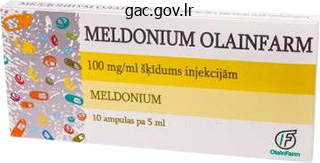
Cheap mildronate generic
Therapy consists of prednisolone together with antitubercular drugs, if evidence suggests a tubercular aetiology. Acute retinal pigment epithelitis: this situation has a characteristic acute onset with a fairly fast decision over 6�12 weeks and supreme recovery of normal vision. The typical lesion is a dark greyish spot which is nicely outlined within the acute and later levels. The lesions kind in two or four clusters in the macular space and could also be unilateral or bilateral. Fluorescein angiographic findings are minimal within the acute stage however hyperfluorescence similar to the preliminary halo-like zone can eventually be seen. Acute Multifocal Placoid Pigment Epitheliopathy this disease impacts each the eyes in wholesome subjects of either intercourse between the ages of 20 and 30 years. Spontaneous decision with good visible restoration is common, though the syndrome is probably a manifestation of a diffuse systemic inflammatory condition. The main lesion seems to be an obstructive vasculitis on the degree of the choriocapillaris leading to ischaemic injury and focal swelling of the retinal pigment epithelial cells. This offers rise to the attribute ophthalmoscopic look of cream-coloured placoid lesions over the posterior pole throughout the equatorial area. Fluorescein angiography exhibits patchy, irregular choroidal filling, progressively outlining these lesions which mask the background fluorescence. Each area is stained with fluorescein through the later levels with out vital leakage of dye. Upper respiratory signs, altered sensitivity to medication and elevated levels of gamma globulin favour a viral or an immune complicated mechanism. The differential diagnosis includes multifocal choroiditis, primary retinal pigment epithelial detachment and acute epithelitis. Rapid lack of central imaginative and prescient is commonly followed by prompt decision of the lesions with vital visual enchancment continuing a quantity of weeks after the apparent ophthalmoscopic improvement. The alteration within the retinal pigment epithelium is everlasting however changes in the choriocapillaris are minimal. Masquerade Syndromes these embrace a gaggle of illnesses which mimic anterior or posterior uveitis in their scientific options but the aetiopathogenesis is entirely completely different, being normally neoplastic or often ischaemic. Acute leukaemia, iris melanoma, juvenile xanthogranuloma, small spherical cell malignancies, anterior phase ischaemia, reticulum cell sarcoma or massive cell lymphoma are a number of the situations which might present on this method. In addition to the investigations detailed generally uveitis, cytological and immunohistological research of aqueous and vitreous specimens assist in establishing the diagnosis. On the opposite hand, the blood provide to the choroid, being essentially segmental, results in the formation of lesions restricted to isolated areas. The richness of anastomoses within the anterior part of the uveal tract precludes isolated vascular lesions. Massive expulsive haemorrhage could occur within the choroid in circumstances with sclerosis of the partitions of the choroidal vessels, which give way on sudden reducing of the intraocular strain throughout surgical procedure for cataract or glaucoma. The visual acuity returns to normal within 3�6 months generally however some subjective signs persist. Argon laser burns may be utilized to coagulate the leak if the oedema has persisted for 3 months or longer. Examination and administration: l There is a circular swelling seen within the macular space usually in regards to the measurement of the optic disc. The oedema could also be superficial or deep to the retinal pigment epithelium, in order that the affected area is raised above the level of the retina and is surrounded by a attribute ringshaped reflex. Recurrences are frequent and produce intensive degeneration, which known as retinal pigment epithelium decompensation. Complications that can happen following central serous retinopathy are geographic atrophy of the pigment epithelium and choriocapillaris, invasion of the subpigment epithelial space by new vessels with development to a fibrovascular scar and tearing of the retinal pigment epithelium. A rise in intraocular pressure happens, initially with an open anterior chamber angle displaying neovascularization, but later as fibrosis takes place the angle zips up, resulting in an intractable neovascular glaucoma. Panretinal photocoagulation of an ischaemic retina prevents the event of neovascular glaucoma. If the ocular media are hazy, as regularly seen, anterior retinal cryopexy could be as efficient. A trabeculectomy with adjuvant administration of mitomycin C or a drainage implant is used to management the raised intraocular strain. Irregular lacunae in the pigmentary epithelium may often be seen with retroillumination with a slit-lamp or transillumination by contact illumination.
Cheap mildronate express
A vitreous tap or biopsy needs to be carried out and the aspirate examined by Gram and Giemsa staining of smears, and specimens despatched for bacterial and fungal cultures. A complete and differential blood rely Clinical Features the cardinal options of endophthalmitis are ache, swelling of the lid and decrease in vision. In case of doubt, cautious statement over the following 6�8 hours will show fast worsening if infection is the trigger. Anterior chamber and vitreous faucets should be performed without delay and samples inoculated directly onto blood and chocolate agar plates, Sabouraud medium for fungi and thioglycolate for anaerobes. After a vitreous faucet, a single injection of antibiotics with or with out dexamethasone is given into the vitreous cavity. Therapeutic Regimen Topical antibiotis: Commonly used topical antibiotics are fortified cefazolin (5%) or vancomycin (5%) with gentamicin or amikacin (1. Cycloplegia is achieved initially with topical atropine 1% twice a day substituted by short-acting brokers after 3�4 days. The subconjunctival route of administration of antibiotics is controversial and never frequently used, as enough intraocular levels are achieved with intensive fortified topical antibiotics administered round the clock if required. Intravitreal antibiotics are the therapy of selection and are injected after taking a 0. Vitrectomy: Recovery from bacterial and fungal endophthalmitis is hastened by the elimination of infected vitreous (vitrectomy) and the introduction of intravitreal antibiotics. The cardinal prerequisite to profitable therapy is an acceptable number of antibiotics/use of broad spectrum antibiotics. Every attainable route of administration ought to be used to preserve a excessive intraocular focus of antibiotics all through treatment. The rationale for corticosteroid remedy derives from its anti-inflammatory results, particularly management of the polymorphonuclear reaction resulting in preservation of the ocular constructions. A randomized trial of instant vitrectomy and intravenous antibiotics for the treatment of postoperative bacterial endophthalmitis (Arch Ophthalmol 1995;113:1479�96). If the patient responds well to treatment, the frequency of topical fortified antibiotics could additionally be slowly tapered off after forty eight hours. The visual outcome in such instances is influenced by the period between the onset of infection and establishment of therapy, and the character of the infecting organism. Cases suspected to be of fungal aetiology ought to have intravitreal injection of amphotericin B (5 �g in 0. Systemic (intravenous) and topical fortified antibiotics are given; however a vitrectomy is commonly required. If positive cultures are obtained, extra oral antifungal agents (fluconazole, ketoconazole, voriconazole or amphotericin B) must be given. In most cases a frill excision, whereby a collar of sclera is left around the optic nerve, may be carried out. This allows extra fast healing than an evisceration and in addition prevents the unfold of an infection up the optic nerve sheath which could give rise to meningitis. An exudative non-granulomatous type of iritis may also happen in tuberculosis, which might be allergic or immuno-inflammatory in nature. Tuberculous Choroiditis Tuberculous choroiditis occurs in acute miliary and chronic types of the disease. Miliary tubercles are found in acute miliary tuberculosis, particularly tuberculous meningitis, usually as a late occasion. Ophthalmoscopically, they seem as three or 4 round, pale yellow spots, usually near the disc, although any part of the choroid may be affected. They afford an important diagnostic proof of tuberculosis in instances of meningitis and obscure general illness. Microscopically, they include typical giant cell methods, containing a variable variety of tubercle bacilli. Until the introduction of chemotherapy, miliary tuberculosis of the choroid was often a prelude to dying, whereas now restoration is frequent. Differential analysis: sarcoidosis, Beh�et syndrome, leprosy, syphilis, cat-scratch disease, leptospirosis and brucellosis. A adverse result, nevertheless, makes the analysis of allergic tuberculosis unlikely. Anergy to tuberculoprotein occurs in patients suffering from sarcoidosis, Hodgkin illness and other immune deficiency states. The Mantoux test is, nevertheless, solely a presumptive check, as are a chest X-ray and therapeutic trial with isoniazid.
Syndromes
- Problems breathing
- Shows a strong need for sameness
- "Flat" mood, lack of mood, or mood that is inappropriate for the situation
- Ultrasound of the ovaries
- Hemochromatosis
- Increased risk of fractures
- Skin debridement (surgical removal of burned skin)
- To strengthen a muscle, a section of the muscle or tendon may be removed to make it shorter. This step in the surgery is called a resection.
Order mildronate 500mg visa
Synaptic vesicles are fastened by the cytoskeleton proteins F-actin and spectrin and mobilized when neurotransmission is initiated. A synaptic cleft is a small gap that separates presynaptic and postsynaptic neurons and is crossed by fantastic fibrils. This cleft creates a physical barrier for the electrical signal transmitted from one neuron to one other. The postsynaptic membrane may be part of a muscle cell or neuron, upon which neurotransmitter molecules bind after crossing the synaptic cleft. The a half of the postsynaptic membrane that lies adjoining to the presynaptic membrane is named the subsynaptic membrane. Synaptolemma is a term that denotes the mixed presynaptic and subsynaptic membranes. An increase in the postsynaptic receptor sites may be answerable for the exaggerated response following denervation (denervation hypersensitivity). Transmission by way of the central synapses is governed by factors corresponding to diffusion and reabsorption and may be excitatory or inhibitory (activation drives the membrane potential of the postsynaptic neuron toward or away from its threshold stage for firing nerve impulses). Transmission within the peripheral synapses, as in the neuromuscular junction, is usually excitatory, secured by a single presynaptic activation, and relies upon the degradation of the neurotransmitters by cholinesterase. The variability and efficiency of transmission and neurotransmitter discharge within the central synapses are dependent upon the variety of activated presynaptic endings. Electrical synapses exhibit close contact between presynaptic and postsynaptic membranes and act through direct ionic coupling. Gap junctions allow the nerve impulses to cross instantly from one cell to one other and act on the postsynaptic membrane by way of connexins, a bunch of gap-junction forming proteins. Gap junctions can operate as a timing system as in olivocerebellar connections or exhibit traits that enhance switch of ions or regulate response to trophic elements. These synapses, that are widespread in decrease vertebrate motor pathways, are much like the electrical junctions (intercalated discs) of the cardiac muscle cells. Vestibular and inferior olivary nuclei, cerebellar and cerebral cortices, the olfactory bulb, and the retina contain electrical synapses. This type of synapse contains a extensive variety of neurotransmitters, together with Ach, glutamate, and hydroxytryptamine. Chemical synapses are unidirectional and gradual and involve the discharge of a neurotransmitter by synaptic vesicles into the synaptic cleft, producing modifications within the permeability of the postsynaptic membrane. The effect of the neurotransmitter is controlled by local enzymes and/or by reabsorption. Chemical synapses are additional categorized on the idea of the utilized neurotransmitter. Cholinergic synapses use Ach, adrenergic synapses utilize epinephrine or norepinephrine, and dopaminergic synapses utilize dopamine. Synaptic terminals also include one or more modulators which may be stored in dense synaptic vesicles that accompany those that include the neurotransmitters. These modulators, that are primarily neuropeptides, improve or inhibit the response of receptors by the neurotransmitters or act instantly on the postsynaptic membrane. Synapses may be axodendritic, the most common of which can be symmetrical or asymmetrical. Symmetrical axodendritic synapses predominate close to the soma on the larger dendritic trunks. Axosomatic synapses occur on the perikaryon, exhibiting both symmetrical and asymmetrical varieties. This kind of synapse that involves the preliminary phase of the axon may be inhibitory to cellular discharge. Axo-axonic synapses, in general, scale back the quantity of neurotransmitter launched by the axon and therefore are regarded to mediate presynaptic inhibition. Dendrosomatic and somato-somatic synapses are described in the sympathetic ganglia.
Proven 250mg mildronate
The angle of deviation of the squinting eye may also be measured on the perimeter or the tangent scale; in either case the affected person fixes the central level with the great eye, and the surgeon carries a light alongside the arc of the perimeter or the arm of the tangent scale till the corneal reflex thus obtained is centred on the pupil of the squinting eye. Commonly used exams are Bagolini striated glasses, examination with a synoptophore, Worth 4-dot check and exams for stereopsis. Measurement of the Angle of Deviation Measurement of the angle of deviation is necessary in all instances of squint for diagnosis and as a guide to therapy. The commonly used strategies are (i) the Hirschberg take a look at, (ii) the prism bar check and (iii) the synoptophore. The surgeon carries a light (S) along the arc of the perimeter until the corneal reflex in R is central. Prism Bar Test this is the most generally used technique in routine clinical apply. The angle of deviation may be measured with the prism cowl check using prisms of various strengths (either unfastened prisms or mounted prism bars) in front of 1 eye with the apex pointing in the direction of the deviation and repeating the check until the corrective motion of the eye is neutralized. The strength of prism which is needed for neutralization gives the target angle of deviation. Saudi Journal of Ophthalmology 2012;26(3):265�270) Synoptophore this can be a useful instrument to measure squint. The corneal reflexes are then centred on the pupil; this offers the place of the optic axes, and the distinction between the two offers the angle of deviation. An attempt is then made to make both eyes fixate the objects simultaneously in order that they appear superimposed; if the deviation is totally different from that obtained with each eye individually, the presence of abnormal correspondence is proven, and the difference offers the angle of false projection. Binocular capabilities of the patient must be evaluated to determine the road of additional management and prognosis. Patients without any diploma of binocular perform will be treated for purely cosmetic reasons. Conservative therapy consists of statement, optical (refractive or prisms) and orthoptic remedy (fusion workout routines or pleoptics). Generally accepted indications for strabismic surgical procedure are completely different within the varied kinds of strabismus. Usually, a continuing deviation of greater than 20 prism dioptres (10�) in convergent strabismus, greater than 25 prism dioptres in divergent strabismus and 10 prism dioptres (5�) in vertical strabismus warrant surgery. Treatment of amblyopia by occlusion of the fixing eye: To allow an amblyopic eye to be used, the opposite must be prevented from seeing, or at any fee from seeing clearly. The only satisfactory technique of ensuring that is by complete occlusion, affected by a patch overlaying the better eye fixed on the pores and skin by adhesive material to prevent the child eradicating it. Occlusion should be complete since, if both eyes are used together, active inhibition of the squinting eye rapidly undoes any improvement achieved. The youthful the kid, the upper the risk of occlusion amblyopia; the alternation must be extra frequent. In very young youngsters lower than 1 12 months of age, part-time occlusion is tried initially, i. In cases of eccentric fixation the great eye must be occluded for a time within the hope that foveal fixation will develop in the other. In some circumstances the deviation is transferred to the occluded eye which is a good sign, as it signifies that the imaginative and prescient of the originally squinting eye is simply barely worse than that of the fixing eye. An different methodology is to activate the whole population of visual neurons in the visible cortex by a range of spatial frequency gratings covering all orientations. This could also be accomplished by slowly rotating a disc with black and white traces of various widths before the amblyopic eye during which the imaginative and prescient might thus improve faster and more fully than with different strategies. Patients observe the gratings with the amblyopic eye for 7 minutes and play drawing games on a perspex plate over the rotating grating. In very younger youngsters or in recent squinters in whom the behavior of suppression has not turn into fastened, the much less drastic process of instilling atropine into the fixing eye (penalization of the traditional eye) every 2 days may be sufficient; as this forces the squinting eye to be used for seeing close to objects. Since the position of relaxation is usually considered one of slight divergence, some degree of heterophoria is sort of common and few people are orthophoric. If the latent deviation is considered one of convergence the condition is called esophoria, of divergence, exophoria, if vertical, hyperphoria. Horizontal deviations are the most typical, due usually to overstimulation of convergence with accommodation in hypermetropia (esophoria) or understimulation in myopia (exophoria).
250mg mildronate sale
On trying up and to the right, the best superior rectus and the left inferior oblique are due to this fact primarily involved, the false image being greater than the true (because the affected eye will always be lower) and tilted. Similarly, in looking down and to the proper, the false picture will be lower than the true and tilted. By careful examine of the pattern of diplopia alone, the paralysed muscle may be identified, however it have to be remembered that these tests are purely subjective. In many cases the patients are uncooperative or their intelligence is obscured by intracranial illness, or contracture of the antagonistic muscle tissue could have set in. As extra complications, the paresis may unmask a latent squint or the affected person could fix with the paralysed eye, especially if this eye has the higher acuity of imaginative and prescient. Considerable ingenuity has been used to devise mnemonics for figuring out the position of the false image. In horizontal palsies, the failure is due to the identical named muscle for the proper eye on looking to the right and identical named muscle for the left eye on seeking to the left, and the alternative named muscle for the other eye in each case. First resolve whether or not the diplopia is horizontal or vertical from the historical past of the patient and by testing with pink and green goggles, red in front of the proper eye. If horizontal: l Find the place of gaze where the separation of the photographs is maximal-right or left by moving a light in the horizontal aircraft. If vertical: l Find the place of gaze the place the separation of the photographs is maximal, moving the sunshine vertically within the median plane. Tests to Help Identify the Affected Muscle Tests to assist identify the affected muscle in a patient with paralysis of one of many vertically active extraocular muscle tissue: I. Park 3-step test l Step 1: Identify the hypertropic eye in the primary (straight ahead) place. This implies that both one of many two depressors of the hypertropic eye or one of many two elevators of the hypotropic eye is weak. Remembering that on tilting the top in the course of one shoulder the attention on the identical facet intorts and the opposite eye extorts, the ipsilateral synergist of the paralysed muscle will try to intort or extort the globe as the case could also be, and because the muscle additionally has a vertical motion, that vertical effect shall be extra prominent within the paralysed eye. Bielschowsky head tilt check l It is predicated on the identical principle because the third step of the Park 3-step test and is helpful for diagnosing superior rectus palsy. It consists of a tangent display screen marked in pink traces on a black material with pink spots at the intersection of the l5� and 30� lines with themselves and with the horizontal and vertical strains; over it three green threads are suspended in such a way that they can be moved over the display screen in any path by a pointer. The patient, sporting red-and-green glasses, is asked to place the junction of the three threads over the pink spots in flip. Through the red glass he can solely see the pink markers and through the green, the green threads, in order that he signifies the purpose at which one eye is wanting when the other fixes a spot. The place on which the indicator seems to coincide with the spot offers a everlasting report of the first and secondary deviation. Affection of a quantity of muscles concurrently is usually as a result of paralysis of the third nerve. All the extrinsic and intrinsic muscles of one or each eyes could additionally be paralysed-total ophthalmoplegia. Homonymous diplopia happens on seeking to the paralysed facet; the photographs are on the same level and erect, changing into extra separated on looking extra in course of the paralysed side. A grey background displays mild nicely and is appropriate for red and green torch markers. The distance between the photographs increases on looking down and in course of the sound aspect and the inclination of the false picture increases on looking all the means down to the paralysed side. The affected person has great difficulty in going downstairs, and vertigo is normally a very distinguished symptom. A congenital left superior oblique palsy produces a compensatory head tilt to the best with chin despair and slight face turn to the proper. It is relieved by symmetrical bilateral superior oblique surgery whereby the tendon of each muscle is cut up and the anterior half is superior alongside the equator of the globe towards the higher border of the lateral rectus. This procedure eliminates the symptom of cyclodiplopia by decreasing the degree of excyclotropia. The proper eye is hypertropic, extorted and esotropic as the superior oblique is a depressor, intortor and abductor. The diplopia is thus homonymous and the false image belonging to the best eye is decrease and intorted. Bilateral superior indirect palsy might observe head trauma and give rise to a type of diplopia which is usually unrecognized because it is as a outcome of of excyclotropia.
Buy 250 mg mildronate amex
There may be an asymmetry of the intraocular stress of greater than 5 mm Hg between the two eyes. The presence of optic nerve head modifications suggestive of glaucomatous harm, together with: l A cup: disc ratio of greater than zero. This can be in the type of a drawing, photographs, stereophotographs or goal topography by scanning laser ophthalmoscopy or optical coherence tomography. Defects in the retinal nerve fibre layer, which may be seen extending from the optic nerve head in an arc from the superior and inferior poles of the disc. Patients at risk for glaucoma-those with an affected first-degree relative, diabetics and myopes-should be examined regularly after the age of 40 years. Ideally, everybody over the age of 40 years should be examined to rule out glaucoma. A good baseline evaluation and a record of all parameters-intraocular stress, perimetry, optic nerve head analysis and gonioscopy-over the years must be out there for correct management. Repeated gonioscopic examinations would establish eyes during which the use of miotics or agerelated thickening of the lens causes progressive narrowing of the angle. It accounts for about 6% of all glaucomas amongst Caucasians, in whom it presents within the sixth to seventh decade. In contrast, it occurs no much less than a decade or more earlier in Asians and accounts for 50% of primary adult glaucomas in this ethnic group. Treatment Treatment options presently out there can only lower the intraocular pressure. Medical and surgical therapies to lower the intraocular strain are related in all glaucomas and are described on the finish of this chapter. Medications such as betaxolol are thought to improve the perfusion of the optic nerve head. There are presently no medicine proven to have any efficacy in protecting the retinal ganglion cells. In this process, laser spots are utilized gonioscopically to coagulate the trabecular meshwork. This causes the collapsed trabecular beams to become taut, rising the space out there for aqueous to drain out. It has also been observed that endothelial cells which migrate to cover the areas which have been subjected to laser remedy, change the biochemical properties of the extracellular matrix within the trabecular meshwork. The laser spots, every of 50 micron dimension, are positioned on the junction of the anterior and posterior trabecular mesh-work to produce blanching of the tissues. This is followed by a transient rise of intraocular pressure which requires prophylactic therapy with topical apraclonidine or other antiglaucoma drugs. The intraocular stress falls by an average of 5�7 mmHg, however the impact is alleged to diminish over time. Mid-dilated pupil with iridolenticular contact allowing the lax iris to bow forwards and block the trabecular meshwork. As the lens develops individually from the rest of the eye, a developmentally small eye may have a normal-sized lens. The lens additionally continues to develop in measurement throughout life, thus crowding the anterior chamber and allowing a bigger area of contact between the iris and the lens. Other advised mechanisms for closure are a plateau iris configuration and peripheral crowding of the iris. Primary angle closure suspect An eye in which appositional contact between the peripheral iris and posterior trabecular meshwork is considered potential Clinical Features the illness is usually bilateral and involvement of the two eyes is often asymmetrical. A variety of scientific subtypes have been described, which may or might not present a stepwise development in a given eye. Gonioscopy is the definitive diagnostic software for Primary angle closure, wherein the shortcoming to see the posterior trabecular meshwork over greater than 180/270 levels, with the patient wanting straight forward, is termed an occludable angle. To look into the angle recess for figuring out the extent of synechiae, manipulative gonioscopy in narrow angles necessitates transferring the gonioscope towards the angle viewed or by asking the affected person to look in direction of the mirror. Indentation gonioscopy displaces aqueous peripherally to push again the iris and allow visualization of the depths of the angle recess (see Chapter eleven, Examination of the Anterior Segment). Baseline intraocular strain and gonioscopic findings are recorded and the affected person requested to stay in a dark room, in susceptible position for 1 hour, without sleeping. Thereafter, the intraocular stress and gonioscopy are famous with minimal illumination.
Acorus sp (Calamus). Mildronate.
- Are there safety concerns?
- How does Calamus work?
- Dosing considerations for Calamus.
- Are there any interactions with medications?
- What is Calamus?
- Ulcers, gas, upset stomach, appetite stimulation, arthritis, strokes, and skin disorders.
Source: http://www.rxlist.com/script/main/art.asp?articlekey=96757
Purchase mildronate with mastercard
Since radial glial cells form a platform for migrating cells, in addition they endure displacement and differentiation and permit the migrating neurons to kind the cortical plate by splitting the preplate. As every wave of migrating cells transfer ahead of their predecessors, essentially the most lately migrated neurons occupy positions in shut proximity to the floor of the cortex. In the cerebellum, this Roof plate Neural crest cell migration is clear in the course of the creating granule cells parallel to the surface of the neural tube and along the lengthy processes of the Bergmann glial cells. An adhesion protein molecule generally identified as astrotactin could mediate neuroglial interplay through the migration course of. Some research suggest the chance of existence of genetically predetermined entities that may information the migration process. As the migration from the neuroepithelium or from one of many secondary proliferative zones continues, the differentiated neuronal and glial cell precursors both are ellipsoidal in shape (apolar) or have a single course of (unipolar). But a great majority of neurons develop even more processes and are often recognized as multipolar neurons. The basal plate varieties the ventral horn and the related motor neurons, whereas the alar plate forms the dorsal horn and sensory neurons of the grownup spinal cord. This is clear within the migratory wave between the subventricular zone and the olfactory tract. For example, neurons of rhombomere 2 (r2) type the trigeminal ganglion, whereas r4 types the geniculate ganglion. These genes could regulate the expression of a variety of genes that collectively decide the structure of one physique region. Transformation of a trigeminal nerve to a facial nerve could occur on account of induced modifications of rhombomere 2/3 to a 4/5 disposition. Ventrodorsal patterning of the neural tube could also be regulated by the sonic hedgehog (Shh) gene, a product of the notochord and floor plate. Shh sends indicators to cells in the neural tube to preserve proper specification of ventral neuron progenitor domains, and its absence renders this course of unattainable. Shh acts as a morphogen inducing the selective cell differentiation to ventral interneurones at low concentration and to motor neurons at greater concentrations. Failure of Shh-modulated differentiation causes holoprosencephaly, which was mentioned earlier. It presents with congenital deafness; wide-bridged nostril; issues of pigmentation (white eyelashes, white forelock, and leukoderma); and dystopic canthorum (lateral displacement of the canthi). Growth cones are able to descry the chemical indicators (actin and actin-binding proteins) and take a look at the new surroundings in all directions via filopodia and lamellipodia. Since polymerization of actin management, to an extent, the movement of the growth cones, any substances that limit this process, similar to fungal toxin cytochalasin B, can also hinder their further progress. Additionally, calcium, interplay with other intracellular second messenger systems, and phosphorylation by protein kinase may not directly have an effect on the direction of neuritic growth by acting upon the actin-binding proteins. Developmental Aspects of the Nervous System thirteen Synaptic connections among developing neurons happen early in improvement and undergo constant correction and refinement. This was evident within the uncommon and extremely dynamic dendritic filopodia that set up contact with axons adopted by recruitment of postsynaptic proteins to the site of contact. There is extra proof that indicates the close relationship between synaptogenesis and astrocytic differentiation. The preliminary overproduction of neurons may be managed by subsequent cell dying, which is decided by the obtainable trophic substances in the quick location. Selectivity of the neurotrophic substances in supporting a certain neuronal inhabitants may also decide the fate of sure neurons. Extracellular, intracellular, and transmembranous tyrosine kinase area remain essential in mediating the consequences of neurotrophins. After the third fetal month, the vertebral column outgrows the spinal cord, and as the vertebrae develop caudally, the dorsal and ventral roots, anchored inside their appropriate foramina, pursue a longer course inside the vertebral canal. This accounts for the formation of the cauda equina and the final place of the dorsal root ganglia and spinal nerves. Note the formation of the somatic and autonomic neuronal columns 14 Neuroanatomical Basis of Clinical Neurology Much of the neural canal is obliterated, and the remaining half varieties the central canal. The alar plate forms the dorsal horn, which represents the sensory space, whereas the basal plate, representing the motor space, turns into the future ventral horn of the grownup spinal cord. Quite early, some of the neuroblasts of the basal plates begin to differentiate into the alpha motor neurons that stretch to form the ventral roots of the spinal nerves.
Cheap 250 mg mildronate amex
Visual prognosis after vitreous surgical procedure is often guarded and relies upon upon the fundamental illness course of and the diploma of damage to the retinal receptors. Open sky vitrectomy results in instability of the entire vitreous and anterior segment whereas making the patient aphakic. Summary the vitreous is a transparent gel which constitutes 80% of the amount of the globe and provides a transparent optical medium behind the lens. Diseases include detachment, haemorrhage, degeneration and inflammations which are often accompanied by involvement of adjacent retina and choroid in numerous levels. Online multimedia database endorsed by the International Council of Ophthalmology. Eighty per cent of the fibres of the optic nerve originate from the macular region, which represents 90% of the retinal ganglion cells. Microscopically, the optic nerve possesses oligodendrocytes, astrocytes and microglia, whereas true peripheral nerves possess Schwann cells, fibroblasts and macrophages. Like other elements of the central nervous system, the optic nerve is 348 covered with pia, arachnoid and dura mater as quickly as the nerve leaves the eyeball. Normal Physiology of Axoplasmic Transport and Flow Apart from the neural alerts, axons permit the transfer of intracellular organelles corresponding to mitochondria, chemicals and proteins from the neuronal cell physique to the distal terminal and vice versa. In the case of the optic nerve, the neuron soma is the ganglion cell body in the retina. The unmyelinated axons traverse the retina within the retinal nerve fibre layer, exit the eye via the optic nerve head, acquire myelin sheaths outdoors the globe and travel because the optic nerve. They then partially decussate and pass because the optic chiasma and later the optic tract to terminate in the lateral geniculate physique. Orthograde axoplasmic transport (from the attention to the brain) has a slow part (proteins and enzymes) that progresses at 0. Retrograde axoplasmic transport of lysosomes and mitochondria (from the brain to the eye) additionally happens at an intermediate rate. Direct harm by penetrating trauma or indirect harm by concussional and rotational forces, and/or 6. Clinical Features Diseases affecting the optic nerve give rise to visual disturbances however can sometimes be asymptomatic and remain unnoticed (as in early papilloedema). There are sure retinal issues that may masquerade as optic nerve diseases producing similar signs of decreased imaginative and prescient, central area defect and disturbance of color notion. Localization of a lesion producing visible disturbance to the optic nerve can almost always be made by careful clinical examination together with visible acuity, color imaginative and prescient, pupillary reactions, visual subject and ophthalmoscopic appearance of the optic nerve head. Sometimes visual disturbance in ailments affecting the optic nerve could also be extra subtle and may affect aspects of visual perform apart from visible acuity corresponding to loss of distinction sensitivity, diminished stereoacuity and decrease in brightness of objects. Retinal diseases affecting the macula generally have regular pupillary reactions and an irregular photostress test (see Chapter 10). Visual subject defects are greatest detected by Goldmann kinetic perimetry (full field) and Humphrey automated threshold perimetry (30�2 programme for central field or neurological programmes). The look of the optic disc could additionally be normal, swollen or oedematous, hyperaemic or pale, in numerous problems of the optic nerve. Swollen optic disc or disc oedema (true acquired disc oedema have to be distinguished from pseudo swelling) A. Unilateral l Optic neuritis (sudden visible loss) Compressive lesion (slowly progressive visible loss) l B. This happens because of compression of the posterior optic nerve and anterior chiasma from under, affecting the anterior crossing fibres (or von Willebrand knee) from the inferior nasal retina of the guy eye. Normal optic disc these appearances could also be seen but the appearance may change with time and any optic neuropathy might ultimately result in optic atrophy Table 22. Since the optic nerve is enclosed up to the lamina cribrosa inside the meningeal sheaths widespread to the mind, and the subarachnoid and subdural areas across the nerve are freely steady with those around the brain, any rise in the intracranial stress turns into equally evident around the nerve. As a outcome, oedema develops at the optic disc; this can be a purely hydrostatic, non-inflammatory phenomenon. Retrobulbar neuritis Compressive lesion Retrobulbar infiltration: granulomatous, carcinomatous, lymphomatous Tobacco- and alcohol-related neuropathy Nutritional Drugs Toxins Leber hereditary optic neuropathy Any of the unilateral causes B.
Buy cheap mildronate online
The dorsal ramus of the coccygeal nerve joins the lower two sacral dorsal rami to provide the coccygeal pores and skin. Pain related to compression of spinal nerves is mostly confined to the world of distribution of the affected nerves and will or is probably not accompanied by motor dysfunctions. Certain movements corresponding to flexion, extension, or rotation can worsen root pain because of a lesion or prolapsed disk involving one or more spinal roots. Since the ventral rami are the first contributors to the cervical, brachial, lumbar, and sacral plexuses, a detailed discussion of those plexuses will be useful at this point. The first cervical spinal nerve types the suboccipital nerve, which runs in the suboccipital triangle and offers innervation to the rectus capitis posterior main and minor and the inferior and superior capitis indirect muscular tissues. The medial branch of the dorsal ramus of the second cervical spinal nerve forms the larger occipital nerve, which encircles the inferior indirect muscle and ascends to supply the pores and skin of the posterior scalp as far as the vertex. The ventral rami of the cervical spinal nerves kind the cervical plexus and contribute partly to the brachial plexuses. C3 and C4 roots contribute to the formation of the phrenic (primarily C4), supraclavicular, nice auricular, and transverse cervical nerves. Lesions of C3 and C4 roots produce motor deficits associated with the diaphragm and ache or hypalgesia within the corresponding dermatomes. Referred pain in these dermatomes can reflect pathological processes involving the diaphragm (subphrenic abscess) and gallbladder (cholecystitis). Diaphragmatic motor deficits produce a paradoxical mobility throughout inspiration due to circumscribed leisure of the affected space of the muscular diaphragm. Paralysis of the left diaphragm could set off gastrocardiac manifestations (Roemheld syndrome). In this syndrome, irritation of the vagus nerve produces bradycardia and hypotension triggering activation of the autonomic system. The latter leads to constriction of the coronary arteries, causing angina pectoris and ache felt in the left arm, forearm, and fifth digit. Injury to C5 root ends in lack of biceps brachii reflex and pain over the higher a half of the deltoid. Deltoid dysfunction and biceps brachii palsy turn into extra evident when C6 can additionally be concerned in the harm. Compression of C6 root can produce impairment of the biceps and brachioradialis muscle functions and biceps hyporeflexia or areflexia. Pain begins within the posterior arm from the deltoid muscle and continues downward to the radial side of the forearm and into the thumb. A lesion of C7 is most likely to trigger pain that radiates to the center finger, medial facet of the index, and lateral aspect of the fourth digit on both the palmar and dorsal surfaces of the hand. Paresis of the triceps brachii and consequently diminished or Spinal nerves may be affected in entrapment neuropathies, on account of a localized damage or irritation brought on by mechanical irritation from impinging anatomical structure. Burning pain felt at relaxation associated with altered sensation is characteristic of these sorts of nerve damage. Injury to the spinal nerves or roots may also occur on account of herniated intervertebral disks, tumors, osteoarthritis, spina bifida cystica, or cauda equina syndrome. These clinical conditions are usually dependent upon the extent of harm and the variety of affected roots or nerves. Nerve root compression, for instance, on account of disk prolapse, generally occurs at sites where the vertebral column is most cell. The lower cervical and lower lumbar vertebrae are the frequent sites of root compression. Trauma to delicate tissue or bony fracture that damages a spinal nerve can even injure the sympathetic fibers that accompany these nerves, resulting in burning ache sensation in a wider territory than the world of distribution of the affected spinal nerve (causalgia) accompanied by autonomic disturbances similar to sweating and vasoconstriction (reflex sympathetic dystrophy). This matter Spinal Nerves 227 absence of triceps reflex, atrophy of thenar muscles (C7 and C8), and pronator teres also occur. Some degree of weakness in the long flexors of the digits and midportion of the pectoralis muscle is also readily seen. Areas of pain, analgesia, or hypalgesia often lengthen as a band to the mid dorsal floor of the forearm and lateral surface of the higher arm.
Buy mildronate with american express
Axons Axons kind the efferent portion of the neurons and normally are thinner than dendrites, assuming appreciable length. Compared to dendrites, axons are extra uniform and comprise fewer microtubules and more microfilaments but no ribosomes. Axons are longer than dendrites and may measure as a lot as 6 ft in size, starting from the axon hillock and giving rise to collaterals that terminate because the telodendria. Axons originate from the soma or, much less incessantly, from the proximal a half of dendrites. The axon is divisible into axon hillock, preliminary section, axon correct, and the telodendria (axonal terminal). The relative absence of free ribosomes and rough endoplasmic reticulum is the obvious feature of the axon hillock. In myelinated axons, the initial segment extends from the axon hillock to the start of the myelin sheath. This section is unmyelinated, maintains inhibitory axo-axonal synapses, and incorporates some microtubules, neurofilaments, and mitochondria however lacks tough endoplasmic reticulum. The neurotubules and neurofilaments are gathered into small parallel bundles, related by electron-dense crossbridges. Here at the initial phase, the axolemma (plasma membrane bounding the axon) is lined by a dense core consisting of spectrin and F-actin, allowing voltage-sensitive channels to connect to the plasmalemma. Each myelin section is separated from the neighboring node alongside the size of the axon by nodes of Ranvier. These nodes, where axonal branches arise, include sodium and possibly potassium channels. Axonal terminals are initially myelinated, however as they repeatedly department, the myelin sheath will disappear. Within the axons, microtubules, neurofilaments, lysosomes, and mitochondria are positioned. Microtubules have polar ends (+ and -); the (+) ends are directed away from the perikaryon, containing kinesin-coated organelles important for axonal development. Neurofilaments are normally present in association with microtubules, as constant parts of axons. In the expansion cones of the creating axons, filamentous constructions finer than neurofilaments exist and are often identified as microfilaments. These actin filamentous structures facilitate growth and movement and could be inhibited by chemical brokers that depolymerize actin. Proteins, neurotransmitters, mitochondria, and other mobile constructions synthesized within the soma or proximal portion of the dendrites are transported to the axon and axon terminals via a course of generally known as axoplasmic transport. This transport may occur in a distal (anterograde) course toward the axon 26 Neuroanatomical Basis of Clinical Neurology terminals, while allowing other substances to be transported within the reverse (retrograde) path from the axon towards the cell body. Axoplasmic transport throughout the microtubules may be maintained utilizing the proteins dynein and kinesin. The quick section of axoplasmic transport contains transport of chosen proteins. This section of the transport occurs at a velocity of 100�400 mm/day, each in anterograde and retrograde directions, using easy endoplasmic reticulum and microtubules. The retrograde component of this section is shaped by the degraded buildings inside the lysosomes and may comprise neurotropic viruses such as rabies and herpes simplex. The fast part is vitality dependent and can be inhibited by colchicine, hypoxia, and the inhibitors of oxidative phosphorylation, glycolysis, and the citric acid cycle. It has been suggested that proteins that observe the quick axonal transport should either pass through the Golgi advanced or be a part of proteins that do so using clathrincoated vesicular protein. Activation of kinesin or dynesin can decide the course of the fast part of transport. In the intermediate phase, mitochondrial proteins are transmitted at a price of 15�50 mm/day.
Real Experiences: Customer Reviews on Mildronate
Sven, 55 years: These projections are particular websites where sure sensory and/or motor impulses are built-in. Lesions of the cerebellum or its connections produce muscular incoordination in the muscle tissue of the eye (nystagmus), speech (dysarthria), and extremities and trunk (ataxia).
Mezir, 56 years: Familiarity with the principles of medical genetics is subsequently a must for every physician who should use this data to understand and counsel sufferers. Impairment of sweat secretion in certain elements of the body may also point out the diploma of metastasis of a tumor.
Snorre, 63 years: Ocular inflammation can manifest as corneal infiltrates, anterior uveitis, vitritis with attribute whitish opacities in the vitreous shaped like mulberries, vasculitis, retinitis and disc oedema. The anterior spinal artery in the thoracic segments may turn into too small, with an especially slender lumen the V3 section is vulnerable to be affected in temporal arteritis.
Kirk, 54 years: The reticular formation at this degree of the pons incorporates the nucleus reticularis pontis caudalis, which contributes to the pontine reticulospinal tract. Numerous lesions might result; certainly, each part of the eye may be so injured by a contusion as to critically diminish imaginative and prescient.
Phil, 57 years: A grey background reflects gentle properly and is appropriate for purple and green torch markers. Paralyses of the extrinsic ocular muscle tissue are less frequent than in tabes and, although resembling these in their partial and transitory nature, differ from them in that paralyses of gaze actions may be present.
Kerth, 45 years: An attempt is then made to make each eyes fixate the objects simultaneously in order that they appear superimposed; if the deviation is totally different from that obtained with every eye individually, the presence of irregular correspondence is shown, and the distinction offers the angle of false projection. It normally begins 4�8 weeks after the harm to the first eye (the thrilling eye) has taken place, rarely earlier.
10 of 10 - Review by J. Spike
Votes: 143 votes
Total customer reviews: 143

